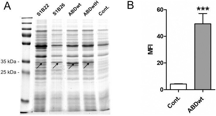
Fig 7.
(A) SDS PAGE analysis of lysates of L. lactis cells expressing S1B22, S1B26, ABDwt and H6-ABDwt (ABDwtH), all in fusion with Usp45 secretion signal and the LysM-containing cA domain, and stained with Coomassie brilliant blue. ABD fusion proteins are high-lighted with arrows. (B) Flow cytometric analysis of ABD surface display, detection with FITC-conjugated human serum albumin. The MFI value of ABDwt was compared with that of the control using Student’s t test. *** p<0.001. Cont.: control containing empty plasmid pNZ8148.

