

Abstract
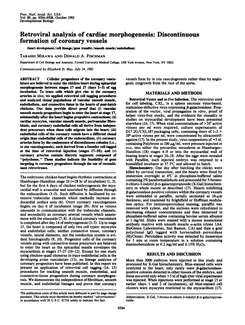
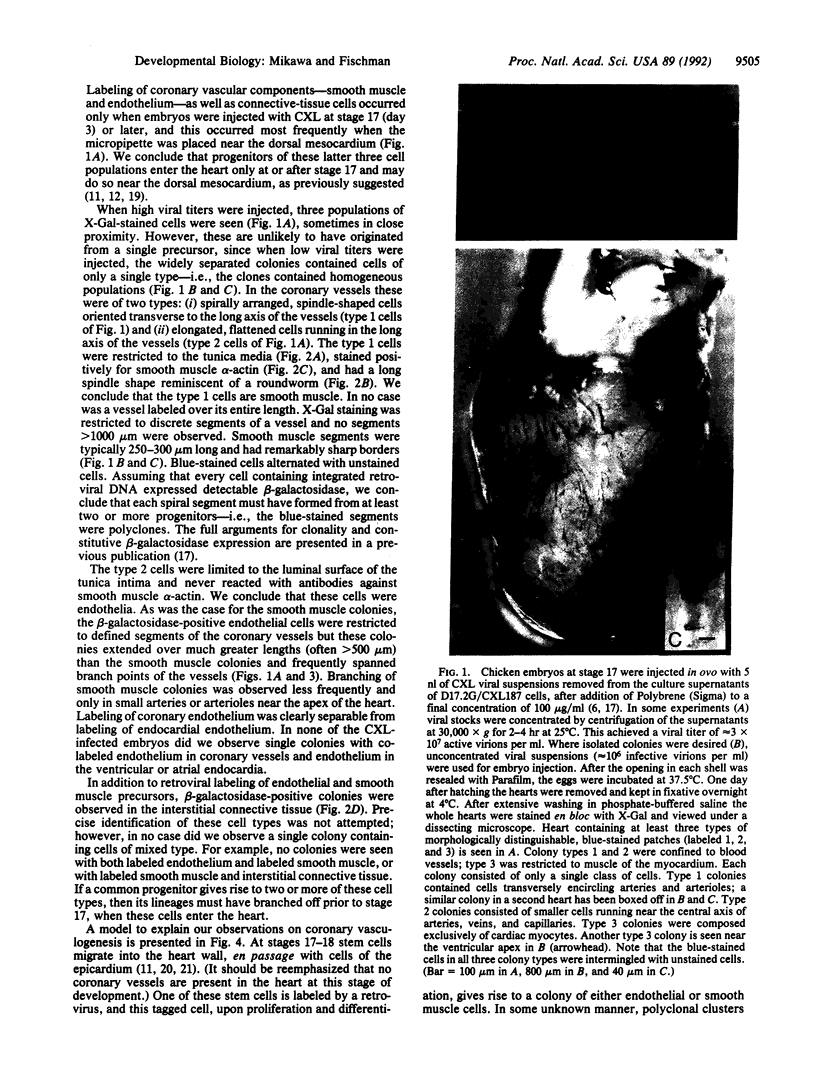
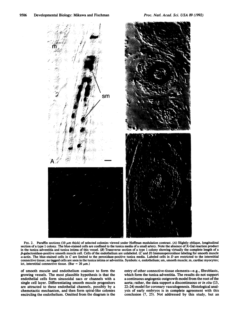
Cellular progenitors of the coronary vasculature are believed to enter the chicken heart during epicardial morphogenesis between stages 17 and 27 (days 3-5) of egg incubation. To trace cells which give rise to the coronary arteries in vivo, we applied retroviral cell tagging procedures and analyzed clonal populations of vascular smooth muscle, endothelium, and connective tissue in the hearts of post-hatch chickens. Our data provide direct proof that (i) vascular smooth muscle progenitors begin to enter the heart at stage 17, substantially after the heart begins propulsive contractions; (ii) cardiac myocytes, vascular smooth muscle, perivascular fibroblasts, and coronary endothelial cells all derive from independent precursors when these cells migrate into the heart; (iii) endothelial cells of the coronary vessels have a different clonal origin than endothelial cells of the endocardium; (iv) coronary arteries form by the coalescence of discontinuous colonies (i.e., in situ vasculogenesis), each derived from a founder cell tagged at the time of retroviral injection (stages 17-18); and (v) coronary arteries contain discrete segments composed of "polyclones." These studies indicate the feasibility of gene targeting to coronary progenitors through the use of recombinant retroviruses.
Full text
PDF
Images in this article

Selected References
These references are in PubMed. This may not be the complete list of references from this article.
- Benditt E. P., Gown A. M. Atheroma: the artery wall and the environment. Int Rev Exp Pathol. 1980;21:55–118. [PubMed] [Google Scholar]
- Bogers A. J., Gittenberger-de Groot A. C., Dubbeldam J. A., Huysmans H. A. Scanning electron microscopy substantiates histology in showing the inadequacy of the existing theories on the development of the proximal coronary arteries and their connections with the arterial trunks. Acta Morphol Neerl Scand. 1988;26(4):225–237. [PubMed] [Google Scholar]
- Bogers A. J., Gittenberger-de Groot A. C., Poelmann R. E., Péault B. M., Huysmans H. A. Development of the origin of the coronary arteries, a matter of ingrowth or outgrowth? Anat Embryol (Berl) 1989;180(5):437–441. doi: 10.1007/BF00305118. [DOI] [PubMed] [Google Scholar]
- Cepko C. Retrovirus vectors and their applications in neurobiology. Neuron. 1988 Jul;1(5):345–353. doi: 10.1016/0896-6273(88)90184-5. [DOI] [PubMed] [Google Scholar]
- Coffin J. D., Poole T. J. Embryonic vascular development: immunohistochemical identification of the origin and subsequent morphogenesis of the major vessel primordia in quail embryos. Development. 1988 Apr;102(4):735–748. doi: 10.1242/dev.102.4.735. [DOI] [PubMed] [Google Scholar]
- Coffin J. D., Poole T. J. Endothelial cell origin and migration in embryonic heart and cranial blood vessel development. Anat Rec. 1991 Nov;231(3):383–395. doi: 10.1002/ar.1092310312. [DOI] [PubMed] [Google Scholar]
- Hiruma T., Hirakow R. Epicardial formation in embryonic chick heart: computer-aided reconstruction, scanning, and transmission electron microscopic studies. Am J Anat. 1989 Feb;184(2):129–138. doi: 10.1002/aja.1001840204. [DOI] [PubMed] [Google Scholar]
- Ho E., Shimada Y. Formation of the epicardium studied with the scanning electron microscope. Dev Biol. 1978 Oct;66(2):579–585. doi: 10.1016/0012-1606(78)90263-4. [DOI] [PubMed] [Google Scholar]
- Manasek F. J. Embryonic development of the heart. I. A light and electron microscopic study of myocardial development in the early chick embryo. J Morphol. 1968 Jul;125(3):329–365. doi: 10.1002/jmor.1051250306. [DOI] [PubMed] [Google Scholar]
- Manasek F. J. Embryonic development of the heart. II. Formation of the epicardium. J Embryol Exp Morphol. 1969 Nov;22(3):333–348. [PubMed] [Google Scholar]
- Manasek F. J. Histogenesis of the embryonic myocardium. Am J Cardiol. 1970 Feb;25(2):149–168. doi: 10.1016/0002-9149(70)90576-x. [DOI] [PubMed] [Google Scholar]
- Manasek F. J. The ultrastructure of embryonic myocardial blood vessels. Dev Biol. 1971 Sep;26(1):42–54. doi: 10.1016/0012-1606(71)90106-0. [DOI] [PubMed] [Google Scholar]
- Mikawa T., Borisov A., Brown A. M., Fischman D. A. Clonal analysis of cardiac morphogenesis in the chicken embryo using a replication-defective retrovirus: I. Formation of the ventricular myocardium. Dev Dyn. 1992 Jan;193(1):11–23. doi: 10.1002/aja.1001930104. [DOI] [PubMed] [Google Scholar]
- Mikawa T., Fischman D. A., Dougherty J. P., Brown A. M. In vivo analysis of a new lacZ retrovirus vector suitable for cell lineage marking in avian and other species. Exp Cell Res. 1991 Aug;195(2):516–523. doi: 10.1016/0014-4827(91)90404-i. [DOI] [PubMed] [Google Scholar]
- Noden D. M. Embryonic origins and assembly of blood vessels. Am Rev Respir Dis. 1989 Oct;140(4):1097–1103. doi: 10.1164/ajrccm/140.4.1097. [DOI] [PubMed] [Google Scholar]
- Noden D. M. Origins and assembly of avian embryonic blood vessels. Ann N Y Acad Sci. 1990;588:236–249. doi: 10.1111/j.1749-6632.1990.tb13214.x. [DOI] [PubMed] [Google Scholar]
- Rychter Z., Jelínek R. Progress of vascularization of the ventricular myocardium in the chick embryo. Physiol Bohemoslov. 1971;20(2):131–138. [PubMed] [Google Scholar]
- Rychter Z., Ostádal B. Fate of "sinusoidal" intertrabecular spaces of the cardiac wall after development of the coronary vascular bed in chick embryo. Folia Morphol (Praha) 1971;19(1):31–44. [PubMed] [Google Scholar]
- Rychter Z., Ostádal B. Mechanism of the development of coronary arteries in chick embryo. Folia Morphol (Praha) 1971;19(2):113–124. [PubMed] [Google Scholar]
- Rychterová V. Principle of growth in thickness of the heart ventricular wall in the chick embryo. Folia Morphol (Praha) 1971 Aug;19(3):262–272. [PubMed] [Google Scholar]
- Sanes J. R. Analysing cell lineage with a recombinant retrovirus. Trends Neurosci. 1989 Jan;12(1):21–28. doi: 10.1016/0166-2236(89)90152-5. [DOI] [PubMed] [Google Scholar]
- Schwartz S. M., Heimark R. L., Majesky M. W. Developmental mechanisms underlying pathology of arteries. Physiol Rev. 1990 Oct;70(4):1177–1209. doi: 10.1152/physrev.1990.70.4.1177. [DOI] [PubMed] [Google Scholar]
- Van Mierop L. H. Location of pacemaker in chick embryo heart at the time of initiation of heartbeat. Am J Physiol. 1967 Feb;212(2):407–415. doi: 10.1152/ajplegacy.1967.212.2.407. [DOI] [PubMed] [Google Scholar]
- Waldo K. L., Willner W., Kirby M. L. Origin of the proximal coronary artery stems and a review of ventricular vascularization in the chick embryo. Am J Anat. 1990 Jun;188(2):109–120. doi: 10.1002/aja.1001880202. [DOI] [PubMed] [Google Scholar]