Abstract
Purpose
Seroma is among the most common complications of laparoscopic total extraperitoneal (TEP) for especially large indirect inguinal hernia, and may be regarded as a recurrence by some patients. A potential area localized behind the mesh and extending from the inguinal cord into the scrotum may be one of the major etiological factors of this complication. Our aim is to describe a novel technique in preventing pseudorecurrence by using fibrin sealant to close that potential dead space.
Methods
Forty male patients who underwent laparoscopic TEP for indirect inguinal hernia with at least 100-mL volume were included in this prospective clinical study. While fibrin sealant was used to close the potential dead space in the study group, nothing was used in the control group. The volume of postoperative fluid collection on ultrasound was compared between the groups.
Results
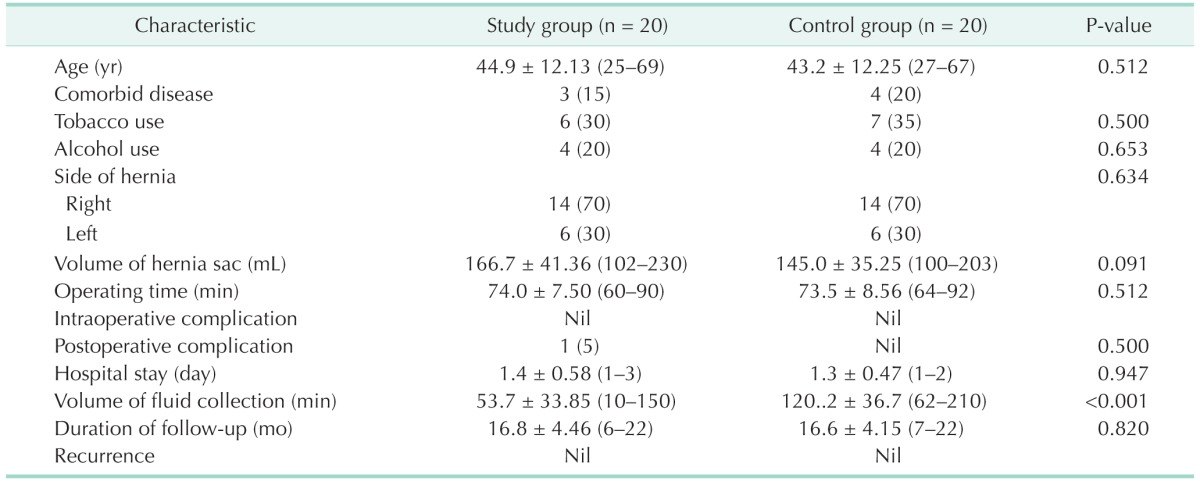
Patient characteristics and the volumes of hernia sac were similar between the 2 groups. The mean volume of postoperative fluid collection was found as 120.2 mL in the control group and 53.7 mL in the study group, indicating a statistical significance (P < 0.001).
Conclusion
Minimizing the potential dead space with a fibrin sealant can reduce the amount of postoperative fluid collection, namely the incidence of pseudorecurrence.
Keywords: Fibrin sealant, Inguinal hernia, Laparoscopic total extraperitoneal hernia repair, Seroma, Pseudorecurrence
INTRODUCTION
Inguinal hernia repair is one the most frequently performed surgical procedures worldwide. Although many surgical techniques have been described to date, laparoscopic procedures have been increasingly used over the past 2 decades, with favorable clinical outcomes such as less postoperative pain, faster recovery of routine physical activity and highly acceptable cosmetic results [1,2,3]. Today, totally extraperitoneal (TEP) and transabdominal preperitoneal (TAPP) techniques are the 2 most widely adopted laparoscopic procedures. However, laparoscopic methods have some potential complications such as suture/staple-related chronic groin pain, surgical site infection, hematoma, seroma, and recurrence [4,5,6]. Among those, seroma in the deep inguinal ring or scrotum is a common complication following TEP, with a reported incidence of up to 15% [7]. It is fact that a swelling at the surgical site due to the seroma may be sometimes regarded as a recurrence by patients. This undesirable complication, also called pseudorecurrence, has prompted us to use an alternative and atraumatic method of fixation of deep inguinal rings with an adhesive material.
The aim of this study is to evaluate the efficacy of a fibrin sealant (Tisseel LYO, Baxter Healthcare, DeerWeld, IL, USA) in preventing pseudorecurrence in laparoscopic TEP repair of inguinal hernia, in comparison to routine mechanical stapling.
METHODS
Patients
Between 2012 and 2014, a total of 40 consecutive male patients who underwent laparoscopic TEP hernioplasty for indirect and large inguinal hernia were included in this prospective study. All patients had a hernia sac with at least 100-mL volume on preoperative ultrasonography (US), which indicated a high risk of postoperative pseudorecurrence. The patients were randomized into 2 groups: the study group (n = 20, used fibrin sealant) and the control group (n = 20, not used fibrin sealant). Other types of groin hernia such as direct and femoral hernia, urgent and recurrent cases, American Society of Anesthesiologists 3–4 scores, and anticoagulant drug use were the exclusion criteria. All operations were carried out by the same surgical team, and all the sonographic examinations of the patients were performed by a single experienced radiologist. The volumes of indirect hernia sac and postoperative fluid collection were automatically calculated on US. The sonographic measurement of hernia sac was done while the patient was in the normal anatomical position (standing) without any procedure such as the Valsalva maneuver or coughing. Demographic and clinical data of the patients, including age, comorbid diseases, tobacco and alcohol use, side of hernia, operative time, intraoperative and postoperative complications, were also recorded.
Each patient was informed about the surgical procedures and possible perioperative complications. This study was conducted in accordance with the principles outlined in the Declaration of Helsinki. Written informed consent forms were obtained from all patients before the operation.
Surgical technique
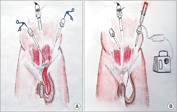
All patients were operated on in a supine Trendelenburg's position under general anesthesia. Antibiotic prophylaxis with 1-g cephalosporin was given intravenously on induction of anesthesia. A standard three-port technique was used. After disinfection of the surgical area with bovidine iodine, a transverse 2-cm infraumbilical incision was made, and the subcutaneous tissue was dissected down to the anterior sheath of rectus abdominis muscle. The anterior sheath was opened, and a spacemaker balloon (Covidien, Mansfield, MA, USA) was inserted between the muscle and the posterior sheath to open a preperitoneal space. It was then removed and replaced with a 12-mm structural balloon trocar (in the form of crow's feet). A 30-degree camera was inserted through this trocar, and carbon dioxide was insufflated at a pressure of 12 mmHg. Then, 2 other trocars were inserted at standard points (5-mm trocar approximately 8–10 cm proximal to the symphysis pubis in each group, another 5-mm trocar about 3 cm proximal to the right or left anterior superior iliac spine in study group and about 3-cm left or right to the symphysis pubis in control group) (Fig. 1A). After dissecting the extraperitoneal space by using endoscissors and diathermy, the indirect hernia sac was reduced and the spermatic cord was parietalized for a length of 3–4 cm. In the study group, the fibrin sealant (8 mL) was sprayed into the inner surfaces of the potential dead space located behind the mesh and extending along the inguinal canal into the scrotum by using a laparoscopic applicator (Duplocath 35 M.I.C, Baxter AG, Vienna, Austria) through the trocar near the anterior superior iliac spine (in order to provide a suitable angle for the application of fibrin sealant) (Fig. 1B). On the other hand, no adhesive material and/or mechanical devices were used for the dead space in the control group. A standard 10 cm × 15 cm polypropylene mesh (ProLite, Atrium Medical, Hudson, NH, USA) was introduced to cover the posterior wall of the inguinal canal, deep inguinal ring, and femoral ring on each side. Titanium tacks (ProTack, Covidien) were used for the fixation of mesh in each group. After bleeding control, the wound was closed without using a drain. Finally, compression was applied to the wound by using a 5-kg sandbag for a duration of 6 hours.
Fig. 1. (A) A drawing showing the placement of the trocars in a patient of study group. (B) Application of fibrin sealant into the potential dead space by using a laparoscopic applicator through the trocar near the anterior superior iliac spine.
Postoperative period
Routine analgesics including diclofenac sodium and paracetamol were used for postoperative analgesia. All patients were uneventfully discharged within 1–3 days after the operation. Two weeks after surgery, volumes of seroma at the surgical site were calculated on US. All patients were also evaluated at regular intervals in terms of pseudorecurrence and other possible complications. No recurrence was observed during the mean follow-up period of 16.7 months.
Statistical analysis
The IBM SPSS Statistics ver. 21.0 (IBM Co., Armonk, NY, USA) was used for data analyses. The results of descriptive analysis for demographic and clinical variables were presented as mean ± standard deviation/percentages for continuous variables, and number/percentage for categorical variables. Fisher exact test and Mann-Whitney U-test were used to test for the significance of association between the 2 groups, in terms of all dependent variables. Significance level was accepted as P < 0.05.
RESULTS
A total of 40 male patients (20 patients in study group, 20 patients in control group) with a mean age of 44 years underwent laparoscopic TEP for inguinal hernia. All patients had large indirect inguinal or scrotal hernia. There were no significant differences between the 2 groups in terms of basic patient characteristics. The volumes of hernia sac on US were also similar between the study and control groups (P = 0.091).
Laparoscopic TEP hernioplasty was successfully performed in all patients. There were no conversions to open surgery or TAPP hernia repair. The mean operative time was 74 minutes and 73.5 minutes in the study and control groups, respectively (P = 0.512). No intraoperative complication or hospital mortality was observed. Postoperative hematoma was developed in one patient of study group, which was easily brought under control with compression to the surgical region.
On US at the second week of surgery, mean volume of fluid collection at the surgical site was found to be 120.2 mL in the control group while the patients in the study group had a mean volume of fluid collection of 53.7 mL. Statistically, the volume of fluid collection was shown to be significantly decreased in the patients of fibrin sealant group in comparison to the patients of non-fibrin sealant group (P < 0.001).
During the postoperative period, clinically apparent seroma was found in 3 patients (15%) in the control group while only 1 patient (5%) in the study group had a clinically apparent seroma. Multiple aspirations with compression on the surgical site were performed to reduce fluid collection. All seromas disappeared after the aspirations. All demographic data, clinical characteristics, and intraoperative and postoperative findings of the patients in the 2 groups were presented in the Table 1.
Table 1. The comparison of all demographic, clinical and perioperative findings of the 2 groups.

Values are presented as mean±standard deviation (range) or number (%).
Nil, 0.
DISCUSSION
Today, using prosthetic mesh has become the standard strategy in laparoscopic hernia surgery due to its effectiveness in preventing recurrence. Although use of various tissue-penetrating devices such as sutures and staples is still common in routine practice, these techniques have been showed to be associated with an increase of several complications including hematoma and postoperative pain. While hematoma mainly develops due to the vascular injury caused by sutures or staples, postoperative pain is caused by nerve compression, and may lead to big socioeconomic problems and severe limitation to routine daily activities [8,9]. Due to such concerns, many atraumatic and sutureless mesh fixation methods have been developed in recent years. Since the first work on application of an adhesive tissue sealant for mesh fixation in a swine model by Katkhouda et al. in 2001, many clinical studies on this issue have been reported [10,11,12]. In fact, 2 major tissue sealant groups, those containing human fibrinogen as the main adhesive sealant and those that primarily consist of polyethylene glycol, are still available. However, fibrin sealants are more frequently used than the other adhesive materials, and comprise of both fibrinogen and thrombin, which are converted to fibrin to form an adhesive. It is well known that fibrin is a protein with adhesive properties involved in the final steps of coagulation cascade. Its adhesive and hemostatic properties have been demonstrated in a number of clinical and experimental studies [13,14]. Fibrin sealants were primarily used as a hemostatic agent in various operations, particularly in cardiovascular surgery [15]. It is also indicated for prevention of leaks at bowel anastomoses [16]. In addition, its effectiveness has been shown in wound healing [17].
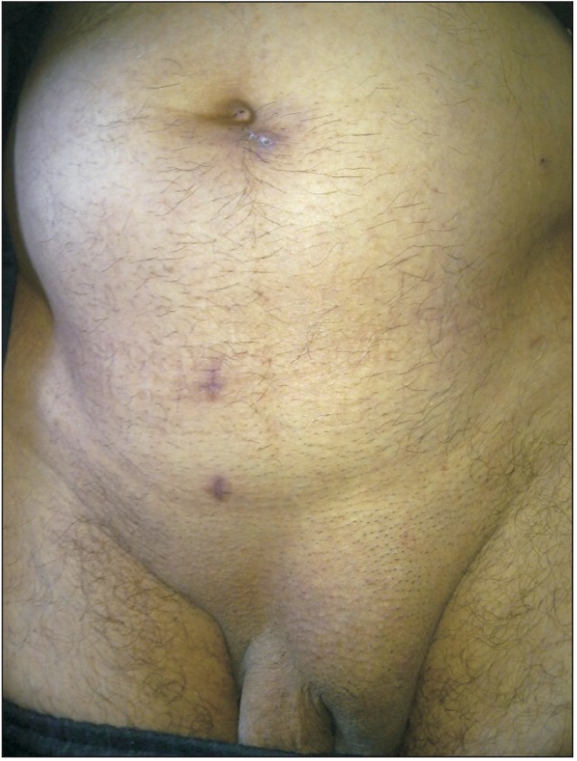
Although the first report on using fibrin sealants for mesh fixation had a primary aim of reducing the recurrence rate [18], minimizing bleeding complications and postoperative chronic groin pain were other primary endpoints in subsequent studies [11,19,20,21,22]. However, postoperative seroma, in other words pseudorecurrence, is also an important and common complication of laparoscopic large hernia repair (Fig. 2). It is well known that fluid collection at the surgical site is a normal process of wound healing depending on inflammatory response directly related to various surgical applications such as cutting, catheterization, suturation, etc. The extent of dissecting area, being old or recurrent hernia, and use of prosthetic patch also contribute to the development of fluid collection. The effectiveness of an adhesive material for mesh fixation in inguinal hernia repair upon seroma formation has not been clearly demonstrated. Although some authors suggested otherwise, many clinical studies on this issue showed that seroma formation is less frequently seen with use of an adhesive material for mesh fixation [12,20,23]. However, in those studies, the effectiveness of the adhesive materials was only evaluated in fixation of the prosthetic patch to the underlying tissues, instead of staples, tacks, or sutures. It is fact that a potential dead space located behind the mesh and extending along the inguinal canal into the scrotum occurs after retraction of indirect hernia sac towards the abdomen and placement of mesh during laparoscopic hernia repair. In addition, depending on our experience in laparoscopic surgery, we suggest that the size of this potential dead space takes an important place in the occurrence of pseudorecurrence, and therefore minimizing its size can reduce the amount of fluid collection. As far as we know, there is no clinical study regarding any intervention of the dead space. Hence, our work is the first report on this topic in the literature. It should be noted here that fibrin sealant was only used to close the dead space, and the fixation of the mesh to the standard points was performed by tacks. The study was designed in this form in order to determine the effect of fibrin sealant alone on the development of pseudorecurrence.
Fig. 2. The image of a pseudorecurrence following laparoscopic total extraperitoneal for left scrotal hernia in a patient of control group.

Although the present study clearly showed the success of the fibrin sealant in preventing pseudorecurrence and high satisfaction of the patients from the procedure, it has also some limitations. First of all, although sufficient for statistical assessment, it is a pilot study with small number of cases, and large-scale studies may be needed for confirmation. Secondly, only the patients who had an indirect hernia sac greater than 100-mL volume were included in the study. This was because the hospital in which the study was carried out is a high-volume center on hernia surgery, and according to our experience, clinically apparent seroma regarded as a pseudorecurrence by some patients usually occurs in patients with indirect inguinal hernia higher than that size.
At this point, we should state that the patients with direct inguinal hernia were not included in the present study. It is well known that pseudorecurrence is hardly ever seen after laparoscopic hernia repair for large direct inguinal hernia. This is due to the potential space behind the prosthetic patch being easily inverted into retzius and closed to cooper ligament by sutures or staples. On the other hand, this is not possible for large indirect or scrotal hernias by routine methods. Therefore, unfortunately, the risk of pseudorecurrence is high following routine TEP for these kinds of hernias. Additionally, aspiration is the single therapeutic approach for pseudorecurrence, and carries a significant risk of infection and hematoma. Also, multiple aspirations are usually needed in the majority of patients with pseudorecurrence, which have negative impacts on patient satisfaction and are associated with increased hematoma and infection risks. Considering all these unwanted situations, our technique can be an effective and alternative method to reduce the risk of pseudorecurrence following TEP for large indirect inguinal hernias.
In conclusion, pseudorecurrence is one of the most annoying complications of TEP, and especially occurs in patients with large indirect or scrotal inguinal hernia. In addition to the other factors in its etiology, the potential space localized behind the mesh and extending from the inguinal cord into the scrotum is one of the main causes of this desired complication. Minimization of this dead space with a fibrin sealant can reduce the incidence of pseudorecurrence.
Footnotes
CONFLICTS OF INTEREST: No potential conflict of interest relevant to this article was reported.
References
- 1.Bringman S, Ramel S, Heikkinen TJ, Englund T, Westman B, Anderberg B. Tension-free inguinal hernia repair: TEP versus mesh-plug versus Lichtenstein: a prospective randomized controlled trial. Ann Surg. 2003;237:142–147. doi: 10.1097/00000658-200301000-00020. [DOI] [PMC free article] [PubMed] [Google Scholar]
- 2.Griffin KJ, Harris S, Tang TY, Skelton N, Reed JB, Harris AM. Incidence of contralateral occult inguinal hernia found at the time of laparoscopic trans-abdominal pre-peritoneal (TAPP) repair. Hernia. 2010;14:345–349. doi: 10.1007/s10029-010-0651-6. [DOI] [PubMed] [Google Scholar]
- 3.Park BS, Ryu DY, Son GM, Cho YH. Factors influencing on difficulty with laparoscopic total extraperitoneal repair according to learning period. Ann Surg Treat Res. 2014;87:203–208. doi: 10.4174/astr.2014.87.4.203. [DOI] [PMC free article] [PubMed] [Google Scholar]
- 4.Franneby U, Sandblom G, Nordin P, Nyren O, Gunnarsson U. Risk factors for long-term pain after hernia surgery. Ann Surg. 2006;244:212–219. doi: 10.1097/01.sla.0000218081.53940.01. [DOI] [PMC free article] [PubMed] [Google Scholar]
- 5.Bansal VK, Misra MC, Babu D, Victor J, Kumar S, Sagar R, et al. A prospective, randomized comparison of long-term outcomes: chronic groin pain and quality of life following totally extraperitoneal (TEP) and transabdominal preperitoneal (TAPP) laparoscopic inguinal hernia repair. Surg Endosc. 2013;27:2373–2382. doi: 10.1007/s00464-013-2797-7. [DOI] [PubMed] [Google Scholar]
- 6.Pahwa HS, Kumar A, Agarwal P, Agarwal AA. Current trends in laparoscopic groin hernia repair: A review. World J Clin Cases. 2015;3:789–792. doi: 10.12998/wjcc.v3.i9.789. [DOI] [PMC free article] [PubMed] [Google Scholar]
- 7.Wang WJ, Chen JZ, Fang Q, Li JF, Jin PF, Li ZT. Comparison of the effects of laparoscopic hernia repair and Lichtenstein tension-free hernia repair. J Laparoendosc Adv Surg Tech A. 2013;23:301–305. doi: 10.1089/lap.2012.0217. [DOI] [PubMed] [Google Scholar]
- 8.McCormack K, Scott NW, Go PM, Ross S, Grant AM EU Hernia Trialists Collaboration. Laparoscopic techniques versus open techniques for inguinal hernia repair. Cochrane Database Syst Rev. 2003;(1):CD001785. doi: 10.1002/14651858.CD001785. [DOI] [PMC free article] [PubMed] [Google Scholar]
- 9.O'Rourke MG, O'Rourke TR. Inguinal hernia: aetiology, diagnosis, post-repair pain and compensation. ANZ J Surg. 2012;82:201–206. doi: 10.1111/j.1445-2197.2011.05755.x. [DOI] [PubMed] [Google Scholar]
- 10.Katkhouda N, Mavor E, Friedlander MH, Mason RJ, Kiyabu M, Grant SW, et al. Use of fibrin sealant for prosthetic mesh fixation in laparoscopic extraperitoneal inguinal hernia repair. Ann Surg. 2001;233:18–25. doi: 10.1097/00000658-200101000-00004. [DOI] [PMC free article] [PubMed] [Google Scholar]
- 11.Lovisetto F, Zonta S, Rota E, Mazzilli M, Bardone M, Bottero L, et al. Use of human fibrin glue (Tissucol) versus staples for mesh fixation in laparoscopic transabdominal preperitoneal hernioplasty: a prospective, randomized study. Ann Surg. 2007;245:222–231. doi: 10.1097/01.sla.0000245832.59478.c6. [DOI] [PMC free article] [PubMed] [Google Scholar]
- 12.Descottes B, Bagot d'Arc M. Fibrin sealant in inguinal hernioplasty: an observational multicentre study in 1,201 patients. Hernia. 2009;13:505–510. doi: 10.1007/s10029-009-0524-z. [DOI] [PMC free article] [PubMed] [Google Scholar]
- 13.Canonico S. The use of human fibrin glue in the surgical operations. Acta Biomed. 2003;74(Suppl 2):21–25. [PubMed] [Google Scholar]
- 14.Zieren J, Castenholz E, Baumgart E, Müller JM. Effects of fibrin glue and growth factors released from platelets on abdominal hernia repair with a resorbable PGA mesh: experimental study. J Surg Res. 1999;85:267–272. doi: 10.1006/jsre.1999.5608. [DOI] [PubMed] [Google Scholar]
- 15.Schenk WG, 3rd, Burks SG, Gagne PJ, Kagan SA, Lawson JH, Spotnitz WD. Fibrin sealant improves hemostasis in peripheral vascular surgery: a randomized prospective trial. Ann Surg. 2003;237:871–876. doi: 10.1097/01.SLA.0000071565.02994.DA. [DOI] [PMC free article] [PubMed] [Google Scholar]
- 16.Jenkins ED, Lerdsirisopon S, Costello KP, Melman L, Greco SC, Frisella MM, et al. Laparoscopic fixation of biologic mesh at the hiatus with fibrin or polyethylene glycol sealant in a porcine model. Surg Endosc. 2011;25:3405–3413. doi: 10.1007/s00464-011-1741-y. [DOI] [PMC free article] [PubMed] [Google Scholar]
- 17.Wilson C, Robinson S, French J, White S. Strategies to reduce pancreatic stump complications after open or laparoscopic distal pancreatectomy. Surg Laparosc Endosc Percutan Tech. 2014;24:109–117. doi: 10.1097/SLE.0b013e3182a2f07a. [DOI] [PubMed] [Google Scholar]
- 18.Chevrel JP, Rath AM. The use of fibrin glues in the surgical treatment of incisional hernias. Hernia. 1997;1:9–14. [Google Scholar]
- 19.Canonico S, Sciaudone G, Pacifico F, Santoriello A. Inguinal hernia repair in patients with coagulation problems: prevention of postoperative bleeding with human fibrin glue. Surgery. 1999;125:315–317. [PubMed] [Google Scholar]
- 20.Lau H. Fibrin sealant versus mechanical stapling for mesh fixation during endoscopic extraperitoneal inguinal hernioplasty: a randomized prospective trial. Ann Surg. 2005;242:670–675. doi: 10.1097/01.sla.0000186440.02977.de. [DOI] [PMC free article] [PubMed] [Google Scholar]
- 21.Ceccarelli G, Casciola L, Pisanelli MC, Bartoli A, Di Zitti L, Spaziani A, et al. Comparing fibrin sealant with staples for mesh fixation in laparoscopic transabdominal hernia repair: a case control-study. Surg Endosc. 2008;22:668–673. doi: 10.1007/s00464-007-9458-7. [DOI] [PubMed] [Google Scholar]
- 22.Chan MS, Teoh AY, Chan KW, Tang YC, Ng EK, Leong HT. Randomized double-blinded prospective trial of fibrin sealant spray versus mechanical stapling in laparoscopic total extraperitoneal hernioplasty. Ann Surg. 2014;259:432–437. doi: 10.1097/SLA.0b013e3182a6c513. [DOI] [PubMed] [Google Scholar]
- 23.Damiano G, Gioviale MC, Palumbo VD, Spinelli G, Buscemi S, Ficarella S, et al. Human fibrin glue sealing versus suture polypropylene fixation in Lichtenstein inguinal herniorrhaphy: a prospective observational study. Chirurgia (Bucur) 2014;109:660–663. [PubMed] [Google Scholar]


