Abstract
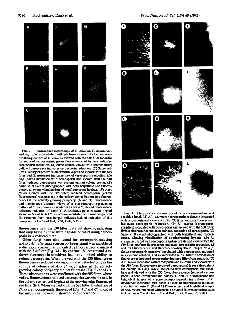
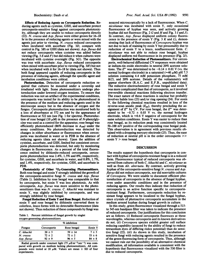
Fungi that are resistant or sensitive to the singlet oxygen-generating toxin cercosporin were assayed for their ability to detoxify it by reduction. Cercosporin reduction was assayed microscopically by using bandpass filters to differentiate between fluorescence emission from cercosporin and reduced cercosporin. Hyphae of the resistant Cercospora and Alternaria species emitted a green fluorescence, indicative of reduced cercosporin. Hyphae of nonviable cultures and of cercosporin-sensitive fungi did not reduce cercosporin. Sensitive fungi occasionally reduced cercosporin when incubated with reducing agents that protect against cercosporin toxicity. Cercosporin could not be efficiently photoreduced in the absence of the fungus. Cercospora species were also resistant to eosin Y but were sensitive to rose bengal. Microscopic observation demonstrated that Cercospora species were not capable of reducing rose bengal but were capable of reducing eosin Y. These observations were supported by in vitro electrochemical measurements that revealed the following order with respect to ease of reduction: cercosporin >> eosin Y > rose bengal. The formal redox potential (E 0') of cercosporin at pH 7.5 was found to be -0.14 V vs. the normal hydrogen electrode. We conclude that Cercospora species protect themselves against cercosporin by the reduction and detoxification of the toxin molecule.
Full text
PDF
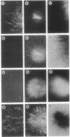
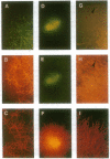
Images in this article
Selected References
These references are in PubMed. This may not be the complete list of references from this article.
- Daub M. E., Briggs S. P. Changes in tobacco cell membrane composition and structure caused by cercosporin. Plant Physiol. 1983 Apr;71(4):763–766. doi: 10.1104/pp.71.4.763. [DOI] [PMC free article] [PubMed] [Google Scholar]
- Daub M. E., Hangarter R. P. Light-induced production of singlet oxygen and superoxide by the fungal toxin, cercosporin. Plant Physiol. 1983 Nov;73(3):855–857. doi: 10.1104/pp.73.3.855. [DOI] [PMC free article] [PubMed] [Google Scholar]
- Goldenberg H. Plasma membrane redox activities. Biochim Biophys Acta. 1982 Oct 20;694(2):203–223. doi: 10.1016/0304-4157(82)90025-9. [DOI] [PubMed] [Google Scholar]
- Hartman P. E., Dixon W. J., Dahl T. A., Daub M. E. Multiple modes of photodynamic action by cercosporin. Photochem Photobiol. 1988 May;47(5):699–703. doi: 10.1111/j.1751-1097.1988.tb02767.x. [DOI] [PubMed] [Google Scholar]
- Hartman P. E. Ergothioneine as antioxidant. Methods Enzymol. 1990;186:310–318. doi: 10.1016/0076-6879(90)86124-e. [DOI] [PubMed] [Google Scholar]
- Meister A., Anderson M. E. Glutathione. Annu Rev Biochem. 1983;52:711–760. doi: 10.1146/annurev.bi.52.070183.003431. [DOI] [PubMed] [Google Scholar]
- Rougee M., Bensasson R. V., Land E. J., Pariente R. Deactivation of singlet molecular oxygen by thiols and related compounds, possible protectors against skin photosensitivity. Photochem Photobiol. 1988 Apr;47(4):485–489. doi: 10.1111/j.1751-1097.1988.tb08835.x. [DOI] [PubMed] [Google Scholar]
- Sollod C. C., Jenns A. E., Daub M. E. Cell surface redox potential as a mechanism of defense against photosensitizers in fungi. Appl Environ Microbiol. 1992 Feb;58(2):444–449. doi: 10.1128/aem.58.2.444-449.1992. [DOI] [PMC free article] [PubMed] [Google Scholar]
- Upchurch R. G., Walker D. C., Rollins J. A., Ehrenshaft M., Daub M. E. Mutants of Cercospora kikuchii Altered in Cercosporin Synthesis and Pathogenicity. Appl Environ Microbiol. 1991 Oct;57(10):2940–2945. doi: 10.1128/aem.57.10.2940-2945.1991. [DOI] [PMC free article] [PubMed] [Google Scholar]


