Case Report
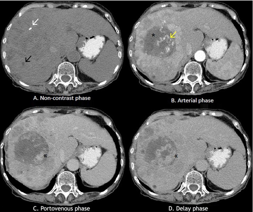
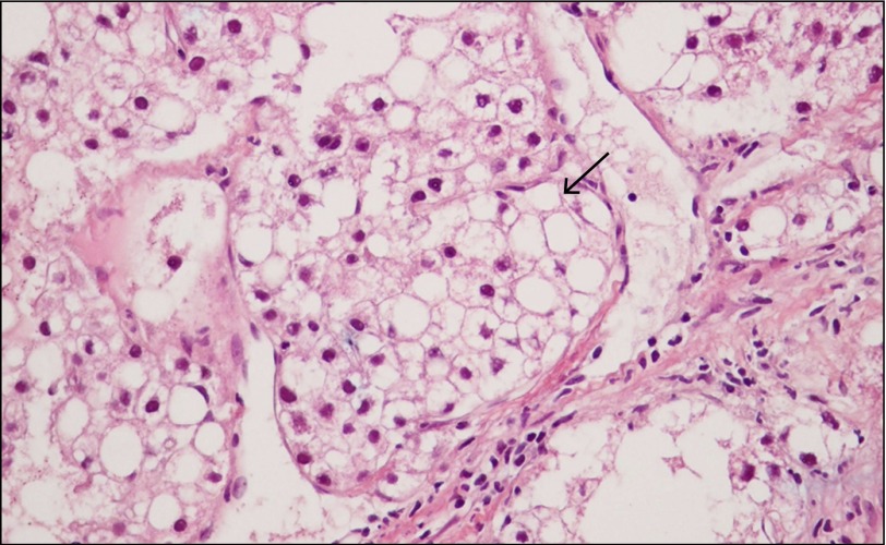
A 77-year-old woman presented with progressive right upper quadrant abdominal pain and 33-pound weight loss over 3 months. Physical examination revealed an enlarged liver with a span of 14 cm. Laboratory results showed: aspartate aminotransferase, 126 IU/L; alanine aminotransferase, 50 IU/L; total bilirubin, 2.8 mg/dL; alkaline phosphatase, 213 IU/L; α-fetoprotein, 11 IU/mL, and hepatitis B surface antigen was positive. Computed tomography (CT) demonstrated liver cirrhosis with a huge heterogeneous mass in the right hepatic lobe. It exhibited peripheral nodular enhancement in arterial phase with centripetal fill-in through portovenous and delay phase (Figure 1), mimicking radiologic characteristics of hemangioma, the most common primary liver tumor. Other parts of the mass also showed hyperattenuation in arterial phase with rapid washout in portovenous and delay phase (Figure 2), representing typical radiologic characteristics of hepatocellular carcinoma (HCC).1 Liver biopsy showed dysplastic small cells with polymorphic nuclei (Figure 3), thus confirming the diagnosis of HCC. Fatty change was also noted (Figure 4).
Figure 1.
Dynamic CT showing atypical imaging of HCC with (A) focal bright hyperdensity (white arrow) and low attenuated area (black arrow) representing calcification and fat tissue, respectively, (B) nonenhancing area (star) and focal arterial enhancing area (arrow) representing necrotic area and peliotic change, respectively, and (C and D) delayed enhancing area (double arrowhead) representing fibrosis.
Figure 2.
(A and B) CT showing heterogenous arterial enhancing mass with rapid washout in (C) portovenous and (D) delay phase, representing radiologic features of HCC.
Figure 3.
Histopathology showing dysplastic small cells with polymorphic nuclei and increased nuclear to cytoplasmic ratio in nests of thick trabeculae (arrow), confirming diagnosis of HCC.
Figure 4.
Histopathology showing part of clear cells with vacuoles pushing the nucleus to the periphery of the cell representing macrovesicular steatosis/fatty change (arrow).
The diagnosis of HCC is generally made by classical enhancement pattern of dynamic contrast enhanced imaging. However, large HCC may have atypical enhancement patterns, due to calcification, fatty change, tumor necrosis, peliotic change,2 or fibrosis that mimics other benign or malignant hepatic masses, including hemangioma,3 large regenerative nodules, abscess, or angiosarcoma. As most of blood supply to HCC is from branches of hepatic artery, HCC has early enhancement in arterial phase and “washout” in venous and delay phase. Hemangioma also receives blood supply from branches of hepatic artery; however, it contains multiple cavernous venous spaces. This leads to radiologic features of peripheral nodular enhancement, which represents venous spaces, in arterial phase, and “progressive” enhancement through venous and delay phase. In summary, HCC and hemangioma are sometimes difficult to differentiate radiographically, particularly in the setting of fatty liver or cirrhosis. A biopsy with a histopathological diagnosis is required in such setting.
Disclosures
Author contribution: P. Aumpansub, R. Chaiteerakij, A. Sanpavat, and B. Chaopathomkul generated the data. P. Aumpansub and R. Chaiteerakij drafted the manuscript. R. Chaiteerakij, K. Thanapirom, N. Geratikornsupuk, S. Treeprasertsuk, A. Sanpavat, and B. Chaopathomkul reviewed the manuscript. R. Chaiteerakij is the article guarantor.
Financial disclosure: None.
Informed consent was obtained for this publication.
References
- 1.Sherman M. The radiological diagnosis of hepatocellular carcinoma. Am J Gastroenterol. 2010; 105:610–12. [DOI] [PubMed] [Google Scholar]
- 2.Hoshimoto S, Morise Z, Suzuki K, et al. Hepatocellular carcinoma with extensive peliotic change. J Hepatobiliary Pancreat Surg. 2009; 16(4):566–70. [DOI] [PubMed] [Google Scholar]
- 3.Kawasaki T, Kudo M, Inui K, Ogawa C, Chung H, Minami Y. Hepatocellular carcinoma mimicking carvenous hemangioma on angiography and contrast enhanced harmonic ultrasonography, a case report. Hepatol Res.2003; 25:202–12. [DOI] [PubMed] [Google Scholar]


