Abstract
Purpose
This study aims to compare the dosimetric data of local tumor's bed dose escalation (boost) with photon beams (external beam radiation therapy – EBRT) versus high-dose-rate interstitial brachytherapy (HDR-BT) after breast-conserving treatment in women with early-stage breast cancer.
Material and methods
We analyzed the treatment planning data of 136 irradiated patients, treated between 2006 and 2013, who underwent breast-conserving surgery and adjuvant whole breast irradiation (WBI; 50.4 Gy) and boost (HDR-BT: 10 Gy in one fraction [n = 36]; EBRT: 10 Gy in five fractions [n = 100]). Organs at risk (OAR; heart, ipsilateral lung, skin, most exposed rib segment) were delineated. Dosimetric parameters were calculated with the aid of dose-volume histograms (DVH). A non-parametric test was performed to compare the two different boost forms.
Results
There was no difference for left-sided cancers regarding the maximum dose to the heart (HDR-BT 29.8% vs. EBRT 29.95%, p = 0.34). The maximum doses to the other OAR were significantly lower for HDR-BT (Dmax lung 47.12% vs. 87.7%, p < 0.01; rib 61.17% vs. 98.5%, p < 0.01; skin 57.1% vs. 94.75%, p < 0.01; in the case of right-sided breast irradiation, dose of the heart 6.00% vs. 16.75%, p < 0.01).
Conclusions
Compared to EBRT, local dose escalation with HDR-BT presented a significant dose reduction to the investigated OAR. Only left-sided irradiation showed no difference regarding the maximum dose to the heart. Reducing irradiation exposure to OAR could result in a reduction of long-term side effects. Therefore, from a dosimetric point of view, an interstitial boost complementary to WBI via EBRT seems to be more advantageous in the adjuvant radiotherapy of breast cancer.
Keywords: adjuvant radiotherapy, breast cancer, dosimetry, EBRT, HDR brachytherapy (HDR-BT)
Purpose
Breast cancer is the most common form of cancer among women in the western world. The current treatment for early-stage breast cancer includes breast-conserving surgery, whole-breast irradiation, and an additional local radiation dose to the tumor bed (boost). Randomized studies have shown the benefit of boosting in order to minimize the local recurrence rate [1, 2, 3, 4]. However, the radiation doses delivered to organs at risk (heart, lung, skin, rib segment) can increase long-term risks of normal tissue side effects. A recent study has shown an increase of 7.4% per Gy in the risk of ischemic heart events [5]. The risk of pneumonitis correlates with the average dose to the lung [6], and a reduction of the dose delivered to the skin shows a better cosmetic outcome [4, 7, 8].
There are different techniques for delivering the boost: via photons, electrons, and interstitial implants. Limited data exist on the advantages of the different boost forms [3, 9]. This study has aimed to compare the dosimetric parameters of a multicatheter single-shoot interstitial tumor-bed boost with high-dose-rate brachytherapy (HDR-BT) to those of a photon boost via external beam radiotherapy (EBRT). Data on incidental irradiation to organs at risk when boosting the tumor bed in women with early-stage breast cancer have been analyzed.
Material and methods
One hundred thirty-six patients with early-stage breast cancer who underwent breast-conserving surgery and whole breast irradiation (WBI; 50.4 Gy) between January 2006 and December 2013 were enrolled in this retrospective investigation. Patients signed a general consent form before treatment. Table 1 summarizes patients and tumor characteristics. Thirty-six patients received an interstitial HDR-BT boost (10 Gy in 1 fraction). For a valid statistical comparison, 100 patients were randomly selected from the institutional EBRT boost group of patients (photon-boost: 10 Gy in 5 fractions). For the EBRT boost, a three-field three-dimensional conformal radiation therapy (3D-CRT) technique was used.
Table 1.
Patient and tumor characteristics
| Characteristic | HDR-BT (n = 36) | EBRT (n = 100) |
|---|---|---|
| Age in years | ||
| Median | 58 | 62 |
| Range* | 54.75-65.25 | 50-69 |
| Breast volume [cc] | ||
| Median | 1102.6 | 1179.3 |
| Range | 874-1380.75 | 871.9-1491.2 |
| Side of the tumor | ||
| Right-sided | 21 | 46 |
| Left-sided | 15 | 54 |
| Localization of tumor | ||
| Upper outer quadrant | 21 | 58 |
| Upper inner quadrant | 4 | 10 |
| Lower outer quadrant | 2 | 17 |
| Lower inner quadrant | 4 | 8 |
| Other | 5 | 7 |
| Size of the tumor [cm] | ||
| Median | 18 | 16 |
| Range | 11-29.25 | 11-23 |
HDR-BT – high-dose-rate interstitial brachytherapy, EBRT – external beam radiotherapy
Range: 25th quartile – 75th quartile
Patients at higher risk for local recurrence were recommended to receive an HDR-BT boost. If a patient refused to undergo a brachytherapy treatment, she received an EBRT boost. For each patient, the best treatment was planned individually. The target volume was defined as the former site of the tumour (clip marked and correlated to preoperative mammography images), plus a 2-3 cm safety margin in all directions. The HDR-BT clinical target volume encompassed the tumour bed and an at least 10 mm safety margin in all directions, and it was equal to the planning target volume (PTV). The minimum quality parameters were D90 ≥ 90% (dose delivered to 90% of the PTV), Dskin < 50 Gy (maximal dose delivered to the skin), V150 < 70 cm3 (volume receiving 150% of the reference dose). The dose non-uniformity ratio (DNR) should be < 30% [10].
Contouring
Organs at risk (heart, ipsilateral lung, skin, most exposed rib segment) were contoured on the planning computed tomography (CT) with the treatment planning systems (BrachyVision and Eclipse/VARIAN, Palo Alto Ca, USA; OncentraBrachy/Nucletron, an Elekta company, Elekta AB, Stockholm, Sweden) according to the existing contouring atlases [11, 12]. Dosimetric characteristics were obtained from dose-volume histograms (DVH). The DNR was calculated (DNR = v150/v100) as a quality parameter of interstitial implants. The absolute volumes in cubic centimeters (cc) of the organs at risk were reported. The organ-specific description is as follows.
Heart
The whole heart was delineated with pericardium and fatty tissue. This includes the part inferior to the left pulmonary artery down to the diaphragma [11]. The dosimetric parameter was Dmax (%) (maximum dose given in percentage), as high maximum dose values can cause atherogenic, as well as perfusion defects, and can lead to cardiovascular events [13]. Due to the reduced number of slices of the planning CT to minimize radiation exposure and the lack of contrast agents, it was not possible to delineate correctly the entire left anterior descending coronary artery (LAD) in the planning CT. The results of the radiation exposure to the LAD would have been flawed.
Lung
The ipsilateral lung was delineated excluding the large bronchi. The Dmean (Gy) and Dmax (%) were reported for each patient. Dmean (Gy) is the mean dose in Gy to the lung – it seems to correlate best with the risk of pneumonitis [6, 14, 15]. Furthermore, the maximum dose to the lung can cause secondary malignancies (Dmax [%]) [16, 17, 18, 19].
Most exposed rib segment
The most exposed rib segment was identified with the aid of consecutive point dose measurements on the planning CT. The maximum dose (Dmax [%]) and the dose in Gy that irradiates 1 cc of the rib (d1cc) were reported.
Skin
The skin was defined as a thick layer of 5 mm underneath the skin surface, as, according to the data in the literature, irradiation of the vessels located in the superficial and mid-dermis can cause telangiectasia [20]. The maximum dose to the skin (Dmax [%]) and the dose in Gy that irradiates 1cc of the skin (d1cc [Gy]) were analyzed for each patient.
Statistical analysis
IBM SPSS Statistics (Version 22.0.0.0, Chicago, IL, USA) was used for statistical analysis. Medians and quartiles were distinguished and non-parametric test (the Mann-Whitney-U Test) was performed on the data. The Mann-Whitney-U Test was used to control for discordant values in this small database. Rank-based box plots illustrated the findings of the tests. The doses of the HDR-BT boost were compared to those of the EBRT boost. Differences in the irradiation of a right- or left-sided tumor were accessed for doses to the heart. For all other organs at risk, it was assumed that there was no difference in the doses caused by the localization of the tumor. The null hypothesis (no difference between the HDR-BT and EBRT boost irradiation) was rejected at a p-value less than 0.01.
Due to different treatment schedules, the isoeffective dose can differ. We therefore calculated the biological effective dose (BED) for a better comparison between HDR-BT and EBRT. The values for the α/β-ratio were chosen to be 3 Gy (heart, lung, most exposed rib), and 2.8 Gy (skin), as values for late-responding normal tissues [21, 22].
Results
There were 36 eligible interstitial HDR-BT cases, from which 21 patients received right-sided irradiation and 15 patients left-sided irradiation. In the EBRT group, there were 46 right-sided and 54 left-sided tumors. Patient and tumor characteristics are given in Table 1.
The reference dose-line was the 100% dose-line on the planning CTs. The results are listed as medians. The range indicates values in the 25th quartile and the 75th quartile, given in brackets.
The DNR, as a parameter for quality and a prognostic factor for acceptable toxicity, for interstitial implants should be below 0.35 [23]. In this study, the mean DNR was 0.3 (0.23-0.35), which shows an excellent result and well-calculated interstitial HDR-BT plans. The characteristics of radiotherapy are given in Table 2.
Table 2.
Characteristics of radiotherapy
| Characteristic | HDR-BT (n = 36) | EBRT (n = 100) |
|---|---|---|
| Whole breast irradiation [Gy] | ||
| Median dose | 50.4 | 50.4 |
| Range* | 50-50.4 | 50.4-50.4 |
| Boost [Gy] | ||
| Median dose | 10 | 10 |
| Range | 8-10 | 10-10 |
| Target volume [cc] | ||
| Median | 103.61 | 131.75 |
| Range | 65.54-121.66 | 95-178.18 |
| DNR | ||
| Median | 0.3 | |
| Range | 0.23-0.35 | |
| Number of fractions (boost) | ||
| Median | 1 | 5 |
| Range | 1-1.5 | 5-5 |
| Number of needles (HDR-BT) | ||
| Median | 15 | |
| Range | 12-18 | |
| Number of planes (HDR-BT) | ||
| Median | 3 | |
| Range | 3-4 | |
| Number of fields (EBRT) | ||
| Median | 3 | |
| Range | 3-3 | |
HDR-BT – high-dose-rate interstitial brachytherapy, EBRT – external beam radiotherapy, Gy – Gray, cc – cubic centimeter, DNR – dose non-uniformity ratio
Range: 25th quartile – 75th quartile
Heart
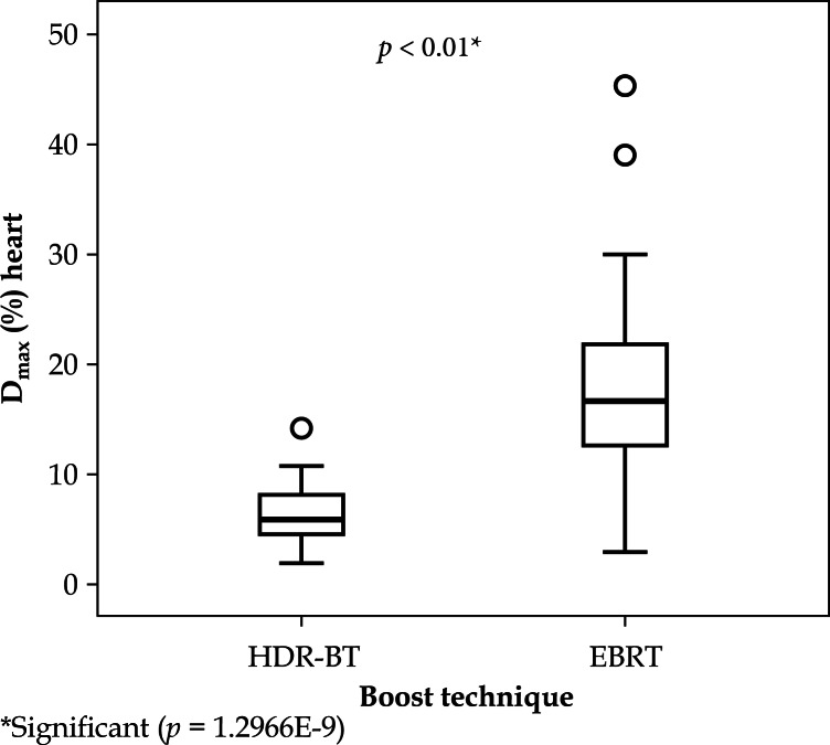
Tumors located on the left side led to a higher maximum dose of irradiation to the heart than tumors located on the right side: HDR-BT left-sided irradiation 29.8% (19.58-32.68%) vs. right-sided irradiation 6.00% (4.79-8.07%); left-sided EBRT 29.95% (21.80-49.98%) vs. right-sided EBRT 16.75% (12.67-22.28%).
Our study has shown no significant difference between the two local dose escalation techniques in the heart dose reduction regarding left-sided tumors (p ≈ 0.34). Radiation of right-sided cancer with interstitial implants showed less radiation exposure to the heart compared to EBRT (p < 0.01; Figure 1).
Fig. 1.
Box plot of irradiation of the heart (right-sided breast cancer): there is a significant difference between external beam radiotherapy (EBRT) and high-dose-rate interstitial brachytherapy (HDR-BT)
The mean dose to the heart was 0.27 Gy (0.19-0.59 Gy) for HDR-BT patients. In patients with left-sided breast cancer, the heart received 0.67 Gy (0.39-1.80 Gy); the mean heart dose of patients with right-sided irradiation was 0.24 Gy (0.17-0.27 Gy).
For EBRT patients, the mean heart dose was 0.37 Gy (0.16-0.68 Gy). Regarding the left-sided irradiation, the heart received 0.50 Gy (0.29-0.86 Gy) vs. 0.23 Gy (0.1-0.47 Gy) for a right-sided tumor.
Lung
The ipsilateral lung volume of the interstitial irradiated patients was 1245.95 cc, while, for the EBRT patients it was 1235.35 cc.
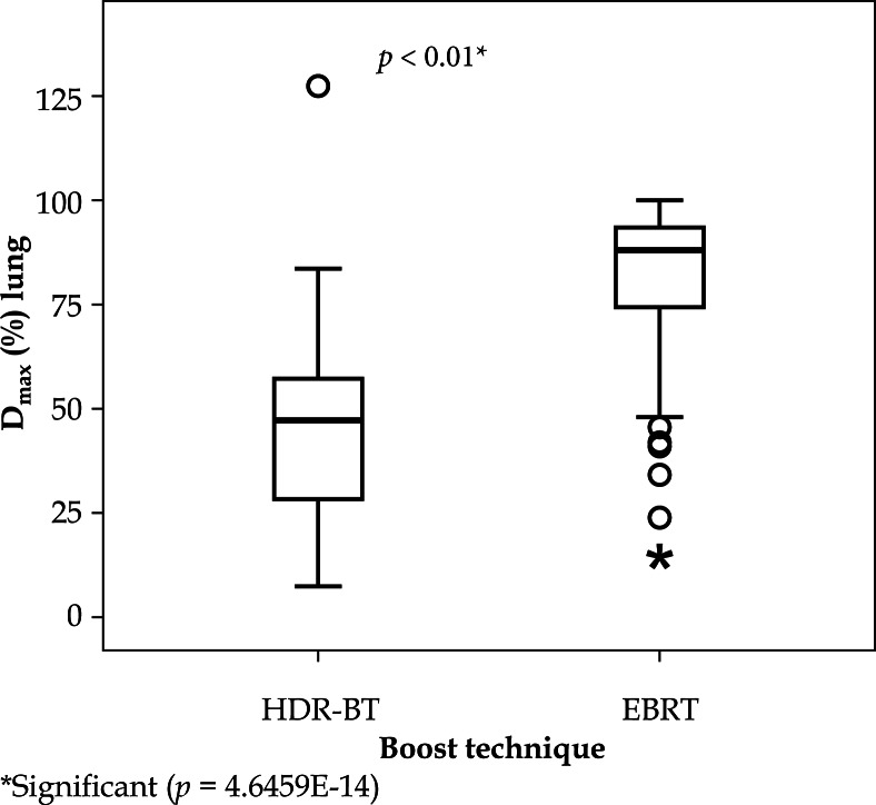
With HDR-BT, the lung received maximum doses of 3.25 Gy (1.99-4.68 Gy), and with EBRT, 8.74 Gy (7.42-9.34 Gy). The difference is significant (p < 0.01, Figure 2).
Fig. 2.
Box plot of irradiation of the lung. It shows a significant difference between the two techniques in favour of high-dose-rate interstitial brachytherapy (HDR-BT)
Mean doses to the lung are understood as markers for pneumonitis risk. A higher mean dose indicates higher pneumonitis risk. Our data show a significant reduction of the mean dose with interstitial implants: 0.40 Gy (0.27-0.57 Gy) vs. 0.79 Gy (0.52-0.99 Gy), p < 0.01.
Most exposed rib segment
The volume of the most exposed rib segment was 19.25 cc (HDR-BT) vs. 22.8 cc (EBRT). The maximum dose was calculated at 4.29 Gy (2.76-6.27 Gy) for HDR-BT and 9.84 Gy (9.55-10.05 Gy) for EBRT.
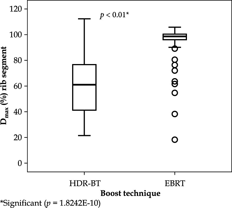
The maximum dose (Dmax [%]) of EBRT given in the DVH is significantly higher than the dose of interstitial implants: 98.5% (96.11-100.4%) vs. 61.17% (41.24-77.64%), p < 0.01 (Figure 3). Additionally, 1 cc of rib received less radiation with an HDR-BT boost: 3.37 Gy (1.85-4.59 Gy) vs. 9.27 Gy (8.52-9.70 Gy), p < 0.01.
Fig. 3.
Box plot of irradiation of the most exposed rib segment. There is a significant difference between the two treatment modalities
Skin
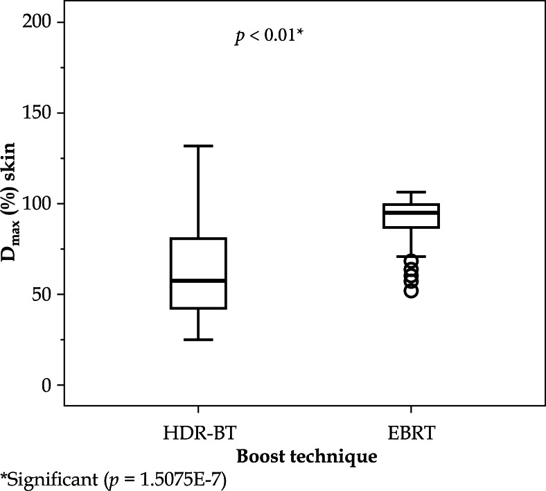
The HDR-BT technique reduces the maximum dose to the skin compared to EBRT. The maximum dose was calculated to be 4.01 Gy (2.28-6.72 Gy) with HDR-BT and 9.44 Gy (8.54-9.92 Gy) with EBRT. The same effect is visible when comparing the percentage maximum doses: HDR-BT 57.1% (41.71-81.15%) vs. EBRT 94.75% (86.17-98.94%); p < 0.01 (Figure 4). One cubic centimeters of skin is significantly less irradiated with the HDR-BT boost than with EBRT: 3.23 Gy (1.86-4.90 Gy) vs. 8.57 Gy (7.59-9.28 Gy); p < 0.01.
Fig. 4.
Box plot of irradiation of the skin. There is a significant difference between the two treatment modalities
Biological effective dose
There are differences in the treatment regimens between the HDR-BT and EBRT. Even after adjusting the doses using the BED equation, OAR received significantly less radiation with HDR-BT. Table 3 lists the adjusted BED values.
Table 3.
Biological effective dose (BED) values for heart, lung, most exposed rib segment, and skin
| HDR-BT [Gy] | EBRT [Gy] | p-value | |
|---|---|---|---|
| Heart Dmax | 5.37 (4.00-7.76) | 10.73 (8.49-16.36) | < 0.01 |
| Heart Dmax left-sided irradiation (α/β = 3) | 8.83 (5.90-13.30) | 14.47 (10.4-21.37) | < 0.01 |
| Heart Dmax right-sided irradiation (α/β = 3) | 4.29 (3.85-5.20) | 9.27 (7.64-10.94) | < 0.01 |
| Lung Dmax (α/β = 3) | 12.61 (8.70-17.00) | 32.47 (27.64-34.46) | < 0.01 |
| Lung Dmean (α/β = 3) | 4.24 (3.80-5.10) | 5.92 (5.05-6.63) | < 0.01 |
| Rib Dmax (α/β = 3) | 15.92 (10.80-21.52) | 36.12 (35.18-36.83) | < 0.01 |
| Skin Dmax (α/β = 2.8) | 16.20 (11.37-27.17) | 37.27 (33.36-39.01) | < 0.01 |
HDR-BT – high-dose-rate interstitial brachytherapy, EBRT – external beam radiotherapy, Dmax – maximum dose [Gy], Dmean – mean dose [Gy]
Range: 25th quartile – 75th quartile
Discussion
There are only a few published studies that compare dosimetric characteristics between the different local dose escalation modalities. Our study has been able to record characteristics of maximum doses to the heart, and thus provide insight into the evaluated forms of irradiation. High-dose-rate brachytherapy plans for right-sided breast cancer have shown a reduction of the maximum doses to the heart. Nonetheless, it is important to keep in mind that the maximum doses to the heart depend on the anatomy of the chest and on the inter-/intra-fraction movements of the heart [24, 25, 26,27].
In 2013, a Korean study compared electron and X-ray beams for tumor bed boost irradiation in 131 patients with left-sided breast cancer [28]. The maximum dose to the left ventricle was 3.66 ± 2.70 Gy (average ± standard deviation). We calculated the maximum dose of the photon boost to be 3.34 Gy, which lies within the same range as the results of Park and Kim. In our study, the HDR-BT boost reduced the maximum doses to the heart (maximum dose 1.65 Gy).
In regard to the treatment of a left-sided tumor, there were no significant differences between the two techniques in terms of Dmax (%). One limitation of the evaluation presented is that we did not consider the localization or depth of the tumor bed. Tumors located deeper in the breast are closer to the heart and lead to a higher maximum dose to the heart. It is possible that our study could have analyzed more deeply-located tumors, and thus produced erroneously high results.
The DVH contains no information on the exact localization of the maximum dose. It makes a difference if muscle or coronary arteries are irradiated. Irradiation of vessels can lead to arteriosclerosis and can cause a myocardial infarction [13].
A study on pneumonitis risk due to irradiation of breast cancer has shown a reduction of the risk by reducing the high dose volume of the lung [29]. The pneumonitis risk is proportional to the mean dose to the lung [6, 14, 15, 29].
In the study presented, the HDR-BT boost showed a reduction of the mean dose and of the maximum dose to the lung compared to the EBRT boost (mean dose 0.40 Gy vs. 0.79 Gy; maximum dose 47.12% vs. 87.7%). Van Parijs published average dose values for the ipsilateral lung between 0.47 Gy and 1.09 Gy [30]. Our study has been able to confirm the reported doses by Van Parijs.
Another long-term risk is the development of secondary malignancies after irradiation. Women who received radiation therapy as part of their breast cancer treatment are at higher risk for secondary malignancies of the lung after 15 years [17]. Grantzau reported that the risk for lung cancer after irradiation increases linearly with every Gray delivered to the lung, by 8.3%. The risk for smokers is even higher (17.3% per Gy) [18]. The reduction of the maximum lung dose decreases the risk of secondary cancer. According to our results, the HDR-BT boost seems to deliver fewer maximum doses to the lung. Randomized studies with larger cohorts are needed to confirm these findings.
Only limited data exist on the dosimetric characteristics of irradiated ribs. Long-term risks to ribs have scarcely been documented. There is one study that published dosimetric data on the rib with a HDR-BT boost [31]. In that study, the most exposed rib received 24.5% of the prescribed dose. Potentially, the variation in the depth of the tumor could explain the different results in their study as compared to our study. More studies on the doses delivered to the rib are necessary to obtain valid data.
Many publications have shown that minimizing the dose to the skin leads to a better cosmetic outcome [4, 7, 28, 32, 33, 34, 35].
Our results show a dose reduction with the HDR-BT boost. When evaluating the results, one has to keep in mind that the outcomes depend on the localization and the depth of the tumor. The more superficial the tumor, the higher the maximum dose to the skin. It is also important to consider the skin-delineation technique for treatment planning. The detected doses to the skin change when defining a different width of the skin [24].
At the moment, there is no consistent recommendation regarding skin delineation for radiation treatment planning [26, 36]. It is difficult to compare our findings for the maximum doses delivered to the skin to other results. Without knowing the exact localization of the tumor, it would also not be valid to compare the results.
Limitations
The retrospective design is a further limitation of our investigation. Women were not randomized equally for either the HDR-BT boost or EBRT boost. A retrospective design cannot control bias, because of different prognostic factors. Additionally, it is important to stress the individual anatomical differences between women. It is disputable whether or not the women can be generally compared. Another limitation is the missing documentation of the exact localization and depth of the tumor.
Our presented cohort contains only a low number of cases – a higher number of cases would lead to more reliable results. Furthermore, the study only examined the dosimetric characteristics, but not the clinical results of the irradiation. Last but not least, we have neither assessed the recurrence rate nor the cosmetic outcome after treatment. Further studies with larger cohorts and long-term follow-up are necessary in the future.
Another limitation is the fact that we only examined 3D-planned photon boost cases in the EBRT group. We excluded data on electron boosts for technical reasons. Prospective studies on the comparison between electron, photon, and interstitial boosts are needed.
Studies have also shown that the contouring and the delineation processes depend on the examiner. Hence, the results can vary widely [37, 38]. An advantage of this presentation is that the same person performed delineation on all the subjects, and the contouring atlases were considered.
Multiple new irradiation techniques are emerging (intraoperative radiotherapy – IORT), intensity-modulated radiotherapy (IMRT), irradiation in prone position, deep inspiration breath-hold techniques (DIBH), and need to be assessed. Long-term data are still lacking, but early findings have shown a benefit of these methods [39, 40]. More studies with prospectively collected data are now in progress and need to be evaluated in the future.
Conclusions
In this limited retrospective study, we were able to demonstrate a reduction of doses delivered to organs at risk with the use of an HDR-BT boost, following WBI via EBRT in breast-conserving treatments in patients with early-stage breast cancer. We have provided data on dosimetric characteristics, and stress the importance of reducing the delivered dose to organs at risk in order to minimize long-term side effects.
Disclosure
Authors report no conflict of interest.
References
- 1.Bartelink H, Horiot JC, Poortmans PM, et al. Recurrence rates after treatment of breast cancer with standard radiotherapy with or without additional radiation. N Engl J Med. 2001;345:1378–1387. doi: 10.1056/NEJMoa010874. [DOI] [PubMed] [Google Scholar]
- 2.Bartelink H, Horiot JC, Poortmans PM, et al. Impact of a higher radiation dose on local control and survival in breast-cancer: 10-year results of the randomized boost versus no boost EORTC 22881-10882 trial. J Clin Oncol. 2007;25:3259–3265. doi: 10.1200/JCO.2007.11.4991. [DOI] [PubMed] [Google Scholar]
- 3.Polgár C, Fodor J, Orosz Z, et al. Electron and high-dose-rate brachytherapy boost in the conservative treatment of stage I-II breast cancer: first results of the randomized Budapest boost trial. Strahlenther Onkol. 2002;178:615–623. doi: 10.1007/s00066-002-1053-1. [DOI] [PubMed] [Google Scholar]
- 4.Poortmans PM, Colette L, Horiot JC, et al. Impact of the boost dose of 10 Gy versus 26 Gy in patients with early stage breast cancer after a microscopically incomplete lumpectomy: 10-year results of the randomized EORTC boost trial. Radiother Oncol. 2009;90:80–85. doi: 10.1016/j.radonc.2008.07.011. [DOI] [PubMed] [Google Scholar]
- 5.Darby SC, Ewertz M, McGale P, et al. Risk of ischemic heart disease in women after radiotherapy for breast cancer. N Engl J Med. 2013;368:987–998. doi: 10.1056/NEJMoa1209825. [DOI] [PubMed] [Google Scholar]
- 6.Seppenwoolde Y, Lebesque JV, de Jaeger K, et al. Comparing different NTCP models that predict the incidence of radiation pneumonitis. Normal tissue complication probability. Int J Radiat Oncol Biol Phys. 2003;55:724–735. doi: 10.1016/s0360-3016(02)03986-x. [DOI] [PubMed] [Google Scholar]
- 7.Collette S, Collette L, Budiharto T, et al. Predictors of the risk of fibrosis at 10 years after breast conserving therapy for early breast cancer: a study based on the EORTC trial 2281-10882 “boost versus no boost”. Eur J Cancer. 2008;44:2587–2599. doi: 10.1016/j.ejca.2008.07.032. [DOI] [PubMed] [Google Scholar]
- 8.Taylor ME, Perez CA, Halverson KJ, et al. Factors influencing cosmetic results after conservation therapy for breast cancer. Int J Radiat Oncol Biol Phys. 1995;31:753–764. doi: 10.1016/0360-3016(94)00480-3. [DOI] [PubMed] [Google Scholar]
- 9.Poortmans P, Bartelink H, Horiot JC, et al. The influence of the boost technique on local control in breast conserving treatment in the EORTC “boost versus no boost” randomized trial. Radiother Oncol. 2004;72:25–33. doi: 10.1016/j.radonc.2004.03.007. [DOI] [PubMed] [Google Scholar]
- 10.Strnad V, Pötter R, Kovács G. Practical Handbook of Brachy-therapy. Bremen: UNI-MED; 2014. pp. 184–189. [Google Scholar]
- 11.Feng M, Moran JM, Koelling T, et al. Development and validation of a heart atlas to study cardiac exposure to radiation following treatment for breast cancer. Int J Radiat Oncol Biol Phys. 2011;79:10–18. doi: 10.1016/j.ijrobp.2009.10.058. [DOI] [PMC free article] [PubMed] [Google Scholar]
- 12.Kong FM, Ritter T, Quint DJ, et al. Consideration of dose limits for organs at risk of thoracic radiotherapy: atlas for lung, proximal bronchial tree, esophagus, spinal cord, ribs, and brachial plexus. Int J Radiat Oncol Biol Phys. 2011;81:1442–1457. doi: 10.1016/j.ijrobp.2010.07.1977. [DOI] [PMC free article] [PubMed] [Google Scholar]
- 13.Lee MS, Finch W, Mahmud E. Cardiovascular complications of radiotherapy. Am J Cardiol. 2013;112:1688–1696. doi: 10.1016/j.amjcard.2013.07.031. [DOI] [PubMed] [Google Scholar]
- 14.Kwa SLS, Lebesque JV, Theuws JCM, et al. Radiation pneumonitis as a function of mean lung dose: an analysis of pooled data of 540 patients. Int J Radiat Oncol Biol Phys. 1998;42:1–9. doi: 10.1016/s0360-3016(98)00196-5. [DOI] [PubMed] [Google Scholar]
- 15.Kahán Z, Csenki M, Varga Z, et al. The risk of early and late lung sequelae after conformal radiotherapy in breast cancer patients. Int J Radiat Oncol Biol Phys. 2007;68:673–681. doi: 10.1016/j.ijrobp.2006.12.016. [DOI] [PubMed] [Google Scholar]
- 16.Deutsch M, Land SR, Begovic M, et al. The Incidence of Lung Carcinoma after Surgery for Breast Carcinoma with and without Postoperative Radiotherapy. Results of National Surgical Adjuvant Breast and Bowel Project (NSABP) Clinical Trials B-04 and B-06. Cancer. 2003;98:1362–1368. doi: 10.1002/cncr.11655. [DOI] [PubMed] [Google Scholar]
- 17.Grantzau T, Mellemkjær L, Overgaard J. Second primary cancers after adjuvant radiotherapy in early breast cancer patients: A national population based study under the Danish Breast Cancer Cooperative Group (DBCG) Radiother Oncol. 2013;106:42–49. doi: 10.1016/j.radonc.2013.01.002. [DOI] [PubMed] [Google Scholar]
- 18.Grantzau T, Thomsen MS, Vaeth M, et al. Risk of second primary lung cancer in women after radiotherapy for breast cancer. Radiother Oncol. 2014;111:366–373. doi: 10.1016/j.radonc.2014.05.004. [DOI] [PubMed] [Google Scholar]
- 19.Roychoudhuri R, Evans H, Robinson D, et al. Radiation-induced malignancies following radiotherapy for breast cancer. Br J Cancer. 2004;91:868–872. doi: 10.1038/sj.bjc.6602084. [DOI] [PMC free article] [PubMed] [Google Scholar]
- 20.Van Limbergen E, Briot E, Drijkoningen M. The source-skin distance measuring bridge: A method to avoid radiation telangiectasia in the skin after interstitial therapy for breast cancer. Int J Radiat Oncol Biol Phys. 1990;18:1239–1244. doi: 10.1016/0360-3016(90)90464-u. [DOI] [PubMed] [Google Scholar]
- 21.Joiner M, Kogel A. Basic clinical radiobiology. London: Hodder Arnold; 2009. pp. 102–134. [Google Scholar]
- 22.Fowler JF. 21 years of Biologically Effective Dose. Br J Radiol. 2010:554–568. doi: 10.1259/bjr/31372149. [DOI] [PMC free article] [PubMed] [Google Scholar]
- 23.Dutta S, Sharma SC, Sigh R, et al. Quality Assessment of Interstitial Implants in High-Dose-Rate Brachytherapy After Lumpectomy in Patients of Early Stage Breast Cancer. Indian J Surg Oncol. 2010;1:294–302. doi: 10.1007/s13193-011-0059-5. [DOI] [PMC free article] [PubMed] [Google Scholar]
- 24.Berger D, Kauer-Dorner D, Seitz W, et al. Concepts for critical organ dosimetry in three-dimensional image-based breast brachytherapy. Brachytherapy. 2008;7:320–326. doi: 10.1016/j.brachy.2008.08.002. [DOI] [PubMed] [Google Scholar]
- 25.Duma MN, Molls M, Trott KR. From heart to heart for breast cancer patients – cardiovascular toxicities in breast cancer radiotherapy. Strahlenther Onkol. 2014;190:5–7. doi: 10.1007/s00066-013-0465-4. [DOI] [PubMed] [Google Scholar]
- 26.Major T, Polgár C, Lövey K, et al. Dosimetric characteristics of accelerated partial breast irradiation with CT image-based multicatheter interstitial brachytherapy: A single institution's experience. Brachytherapy. 2011;10:421–426. doi: 10.1016/j.brachy.2010.12.006. [DOI] [PubMed] [Google Scholar]
- 27.Taylor CW, Nisbet A, McGale P, et al. Cardiac exposures in breast cancer radiotherapy: 1950s-1990s. Int J Radiat Oncol Biol Phys. 2007;69:1484–1495. doi: 10.1016/j.ijrobp.2007.05.034. [DOI] [PubMed] [Google Scholar]
- 28.Park SH, Kim JC. Comparison of electron an x-ray beams for tumour bed boost irradiation in breast-conserving treatment. J Breast Cancer. 2013;16:300–307. doi: 10.4048/jbc.2013.16.3.300. [DOI] [PMC free article] [PubMed] [Google Scholar]
- 29.Willner J, Jost A, Baier K, et al. A Little to a Lot or a Lot to a Little? An Analysis of Pneumonitis Risk from Dose-Volume Histogram Parameters of the Lung in Patients with Lung Cancer Treated with 3-D Conformal Radiotherapy. Strahlenther Onkol. 2003;179:548–556. doi: 10.1007/s00066-003-1078-0. [DOI] [PubMed] [Google Scholar]
- 30.Van Parijs H, Reynders T, Heuninckx K, et al. Breast conserving treatment for breast cancer: dosimetric comparison of different non-invasive techniques for additional boost delivery. Radiat Oncol. 2014;9:36. doi: 10.1186/1748-717X-9-36. [DOI] [PMC free article] [PubMed] [Google Scholar]
- 31.Wiercinska J, Wronczewska A, Kabacinska R, et al. Transition from Paris dosimetry system to 3D image-guided planning in interstitial breast brachytherapy. J Contemp Brachytherapy. 2015;6:479–484. doi: 10.5114/jcb.2015.56492. [DOI] [PMC free article] [PubMed] [Google Scholar]
- 32.Dodwell DJ, Povall J, Gerrard G, et al. Skin teleangiectasia: the influence of radiation dose delivery parameters in the conservative management of early breast cancer. Clin Oncol. 1995;7:248–250. doi: 10.1016/s0936-6555(05)80613-2. [DOI] [PubMed] [Google Scholar]
- 33.Ott OJ, Lotter M, Sauer R, et al. Accelerated partial-breast irradiation with interstitial implants – the clinical relevance of the calculation of skin doses. Strahlenther Onkol. 2007;183:426–431. doi: 10.1007/s00066-007-1738-6. [DOI] [PubMed] [Google Scholar]
- 34.Perera F, Chisela F, Stitt L, et al. TLD skin dose measurements and acute and late effects after lumpectomy and high-dose-rate brachytherapy only for early breast cancer. Int J Radiat Oncol Biol Phys. 2005;62:1283–1290. doi: 10.1016/j.ijrobp.2005.01.007. [DOI] [PubMed] [Google Scholar]
- 35.Vrieling C, Collette L, Fourquet A, et al. The influence of the boost in breast-conserving therapy on cosmetic outcome in the EORTC “boost versus no boost” trial. Int J Radiat Oncol Biol Phys. 1999;45:677–685. doi: 10.1016/s0360-3016(99)00211-4. [DOI] [PubMed] [Google Scholar]
- 36.Lasota J, Kabacinska R, Makarewicz R. Dose estimation for different skin models in interstitial breast brachytherapy. J Contemp Brachytherapy. 2014;6:200–207. doi: 10.5114/jcb.2014.43167. [DOI] [PMC free article] [PubMed] [Google Scholar]
- 37.Hurkmans CW, Borger JH, Pieters BR, et al. Variability in target volume delineation on CT scans of the breast. Int J Radiat Oncol Biol Phys. 2001;50:1366–1372. doi: 10.1016/s0360-3016(01)01635-2. [DOI] [PubMed] [Google Scholar]
- 38.Li XA, Tai A, Arthur DW, et al. Variability of target and normal structure delineation for breast cancer radiotherapy: an RTOG multi-institutional and multiobserver study. Int J Radiat Oncol Biol Phys. 2009;73:944–951. doi: 10.1016/j.ijrobp.2008.10.034. [DOI] [PMC free article] [PubMed] [Google Scholar]
- 39.Bruzzaniti V, Abate A, Pinnarò O, et al. Dosimetric and clinical advantages of deep inspiration breath-hold (DIBH) during radiotherapy of breast cancer. J Exp Clin Cancer Res. 2013;32:88. doi: 10.1186/1756-9966-32-88. [DOI] [PMC free article] [PubMed] [Google Scholar]
- 40.Varga Z, Cserháti A, Rárosi F, et al. Individualized positioning for maximum heart protection during breast irradiation. Acta Oncol. 2014;53:58–64. doi: 10.3109/0284186X.2013.781674. [DOI] [PubMed] [Google Scholar]


