Key Clinical Message
A case of parvovirus B19‐induced pure red cell aplasia occurring in a heart transplant recipient is reported. The diagnosis of this rare but clinically important complication can be suspected on the basis of the pathognomonic morphological features of the bone marrow.
Keywords: Organ transplant, parvovirus B19, pure red cell aplasia
Case Report
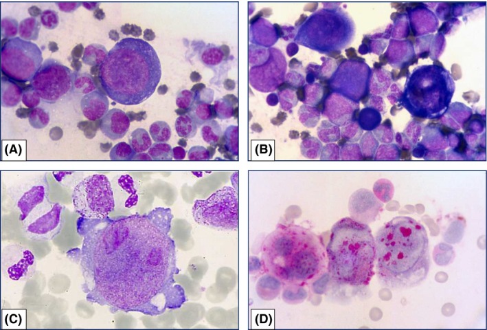
A 47‐year‐old male, heart transplant recipient, treated with immunosuppressive therapy, presented with severe normochromic normocytic anemia (Hb 6.7 g/dL, MCV 94 fL) and reticulocytopenia (reticulocytes 1 × 10⁹/L), normal leukocyte, differential and platelet count, and high levels of erythropoietin (420 U/mL). Vitamin B12, folate, iron, and ferritin levels were normal. Bone marrow smears showed good cellularity with marked hypoplasia of the erythroblastic lineage and almost complete lack of maturing erythroblasts, normal granulopoiesis, and megakaryopoiesis. There were hyperbasophilic giant cells with prominent nuclear inclusions resembling large nucleoli, cytoplasmic vacuoles and blebs (Fig. 1A and B, 1000x, Fig. 1C, 1600x). These cells showed very strong positivity to PAS reaction with coarse blocks of glycogen that looked different from the type of reactivity present in normal megakaryocytes (Fig. 1D, 1000x). They were giant proerythroblasts with morphological features suggestive for parvovirus B19 infection.
Figure 1.

(A–C) Bone marrow smear showing normal granulopoiesis, lack of maturing erythroblasts, giant proerythroblasts with hyperbasophilic cytoplasm (MGG, 1A,B, x1000), cytoplasmic vacuoles and blebs, and very large nuclear inclusions (MGG, 1C, x1600). (D) Two erythroblasts (right) showing strong PAS positivity with coarse blocks of glycogen; this positivity pattern is different from that observed in the megakaryocyte (left) (PAS reaction, x1000).
The diagnosis of parvovirus B19‐induced pure red cell aplasia was confirmed by immunocytochemical and molecular tests. Viral capsid antigens were detected on bone marrow smears by an immunoperoxidase technique, and parvovirus B19 DNA was revealed in the peripheral blood by polymerase chain reaction (PCR). Parvovirus B19 infection was confirmed also by serology showing parvovirus B19‐specific IgM.
High dose i.v. immunoglobulin treatment allowed a prompt recovery.
Discussion
Pure red cell aplasia caused by parvovirus B19 infection is a rare but clinically important complication that may arise in the period after transplantation 1, 2, 3, 4. The diagnosis can be suspected on the basis of a careful examination of the bone marrow, showing pathognomonic morphological features of early erythroblasts. Giant proerythroblasts have nuclear viral inclusions resembling large nucleoli. Their monstrous feature can give rise to problems of differential diagnosis with hematologic malignancies as myelodysplastic syndrome or acute erythroid leukemia, and also with bone marrow infiltration by cancer cells. Parvovirus infection is usually confirmed by serology but, in immunocompromised patients, immunohistochemical or molecular techniques may be necessary to confirm diagnosis.
Conflict of Interest
None declared.
References
- 1. Eid, A. J. , Brown R. A., Patel R., and Razonable R. R.. 2006. Parvovirus B19 infection after transplantation: a review of 98 cases. Clin. Infect. Dis. 43:40–48. [DOI] [PubMed] [Google Scholar]
- 2. Gosset, C. , Viglietti D., Hue K., Antoine C., Glotz D., and Pillebout E.. 2012. How many times can parvovirus B19‐related anemia recur in solid organ transplant recipients? Transpl. Infect. Dis. 14:E64–E70. [DOI] [PubMed] [Google Scholar]
- 3. Crabol, Y. , Terrier B., Rozenberg F., Pestre V., Legendre C., Hermine O., et al. 2013. Intravenous immunoglobulin therapy for pure red cell aplasia related to human parvovirus B19 infection: a retrospective study of 10 patients and review of the literature. Clin. Infect. Dis. 56:968–977. [DOI] [PubMed] [Google Scholar]
- 4. Kelleher, E. , McMahon C., and McMahon C. J.. 2015. A case of parvovirus B19‐induced pure red cell aplasia in a child following heart transplant. Cardiol. Young 25:373–375. [DOI] [PubMed] [Google Scholar]


