Abstract
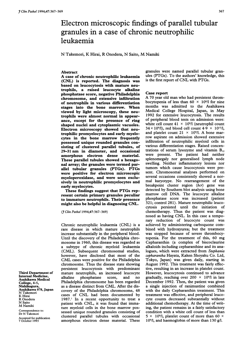
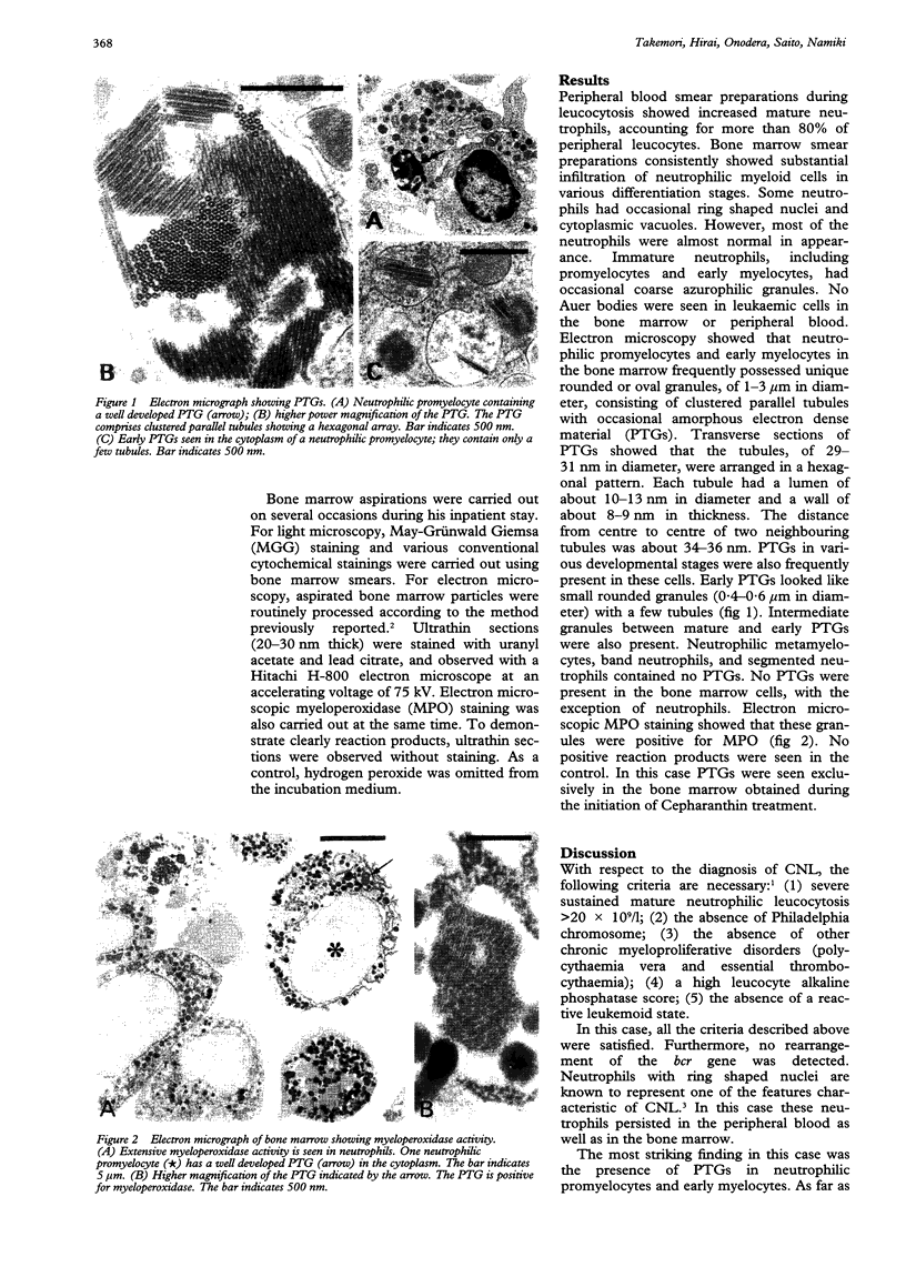
A case of chronic neutrophilic leukaemia (CNL) is reported. The diagnosis was based on leucocytosis with mature neutrophils, a raised leucocyte alkaline phosphatase score, negative Philadelphia chromosome, and extensive infiltration of neutrophils in various differentiation stages into the bone marrow. When viewed by light microscopy, these neutrophils were almost normal in appearance, except for the presence of ring shaped nuclei and cytoplasmic vacuoles. Electron microscopy showed that neutrophilic promyelocytes and early myelocytes in the bone marrow frequently possessed unique rounded granules consisting of clustered parallel tubules, of 29-31 nm in diameter, and occasional amorphous electron dense material. These parallel tubules showed a hexagonal array; the granules were termed parallel tubular granules (PTGs). PTGs were positive for electron microscopic myeloperoxidase, and were seen exclusively in neutrophilic promyelocytes and early myelocytes. These findings suggest that PTGs represent certain primary granules peculiar to immature neutrophils. Their presence might also be helpful in diagnosing CNL.
Full text
PDF


Images in this article
Selected References
These references are in PubMed. This may not be the complete list of references from this article.
- Dixon B. R., Mukherjee T. M., Ho J. Q. The ultrastructural identification of Auer body precursors in a case of acute promyelocytic leukemia using high-angle specimen tilt. Am J Clin Pathol. 1984 Jan;81(1):132–137. doi: 10.1093/ajcp/81.1.132. [DOI] [PubMed] [Google Scholar]
- Gorius J. B., Houssay D. Auer bodies in acute promyelocytic leukemia. Demonstration of their fine structure and peroxidase localization. Lab Invest. 1973 Feb;28(2):135–141. [PubMed] [Google Scholar]
- Kanoh T., Saigo K., Yamagishi M. Neutrophils with ring-shaped nuclei in chronic neutrophilic leukemia. Am J Clin Pathol. 1986 Dec;86(6):748–751. doi: 10.1093/ajcp/86.6.748. [DOI] [PubMed] [Google Scholar]
- Kobayashi M., Katayama T., Ochiai S., Yoshida M., Kaito K., Masuoka H., Shimada T., Nishiwaki K., Sakai O. [High-dose cepharanthin therapy of idiopathic thrombocytopenic purpura]. Rinsho Ketsueki. 1992 Mar;33(3):405–407. [PubMed] [Google Scholar]
- Miura A. B., Takahashi T., Nishinari T. [Chronic neutrophilic leukemia]. Rinsho Ketsueki. 1987 Apr;28(4):505–512. [PubMed] [Google Scholar]
- Takemori N., Hirai K., Onodera R., Uenishi H., Saito N., Takasugi Y., Namiki M., Muraoka S. Disseminated intravascular coagulation in a patient with acute myeloid leukemia. Ultrastructural evidence of hypercoagulation in bone marrow. Am J Clin Pathol. 1993 Jun;99(6):695–701. doi: 10.1093/ajcp/99.6.695. [DOI] [PubMed] [Google Scholar]
- Tulliez M., Breton-Gorius J. Three types of Auer bodies in acute leukemia. Visualization of their protein by negative contrast after peroxidase cytochemistry. Lab Invest. 1979 Nov;41(5):419–426. [PubMed] [Google Scholar]


