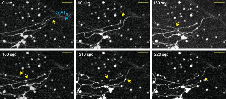
Figure 3. Real-time disassembly of an ER network in the presence of cytATL.
A network was formed from a crude Xenopus egg extract in the presence of the dye DiIC18 in a computer-controlled microfluidics device. CytATL-GFP (5 µM) in cytosol containing an energy regenerating system was then slowly perfused into the chamber at a total laminar flow rate of 0.5 µL/min from multiple ports (one is indicated by a blue arrow). The arrival of cytATL- GFP arrival in the chamber was monitored by GFP fluorescence (not shown). Images represent snapshots of a real-time video (see Video 3). Yellow arrows point to the detachment or breakage of tubules. Scale bar = 10 µm.

