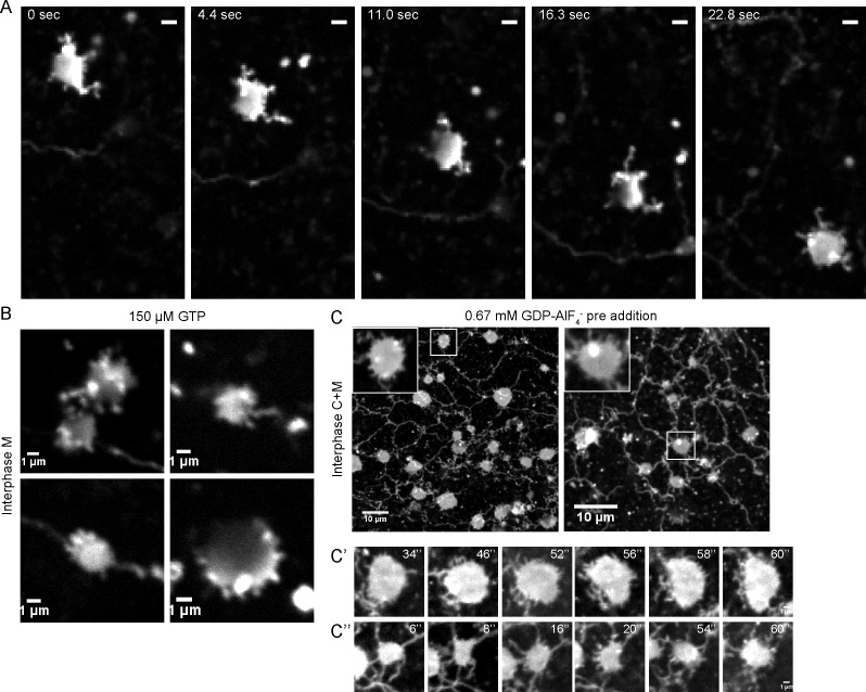
Figure 4. Intermediates during in vitro ER network formation.
(A) DiIC18-prelabeled light membranes were mixed with buffer and an energy regenerating system. The sample was imaged immediately by confocal microscopy. Scale bar = 2 µm. (B) Xenopus egg light membranes were incubated in the presence of 150 µM GTP and octadecyl rhodamine. Scale bar = 1 µm. (C) Interphase Xenopus egg cytosol, light membranes, 0.67 mM GDP-AlF4-, and an energy-regenerating system were incubated for 30 min. The membranes were stained with octadecyl rhodamine. Scale bars = 10 µm. Insets and images in C’ and C’’ show magnified views of small sheets from which short tubules emanate. Scale bars = 1 µm.

