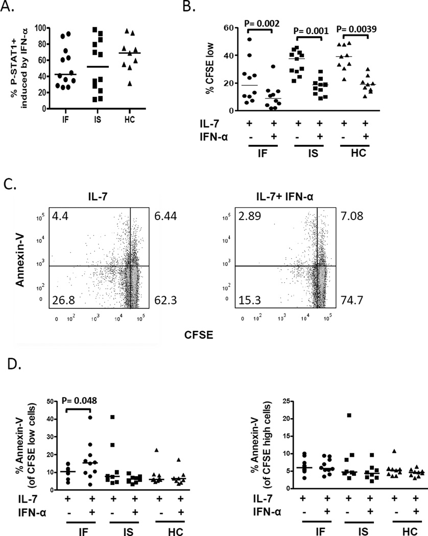
Fig. 4. CD4+ T cell responses to IFN-α were not diminished in IF subjects. PBMCs from subjects were incubated in the presence or absence of IFN-α.
All data shown are gated from CD3+CD4+ cells. (A) P-STAT1 induction following 15 minutes of IFN-α treatment (IF n= 12, IS n= 12, HC n= 9). (B-D) PBMCs were CFSE stained and assessed for proliferation and cell death in cells treated with IL-7 in the absence or presence of IFN-α for 7 days. (B) Compares %CFSE low (IF n= 10, IS n= 11, HC n= 9) and (C) are representative dot plots comparing cell death as measured by annexin-V binding CD4+ T cells that were not excluded by viability dye. (D) Summary data of cell death in divided cells (%CFSE low) and undivided cells (CFSE high) (IF n= 10, IS n= 8, HC n= 9). P-values were obtained by Wilcoxon signed rank test. Abbreviations: immune failure, IF; immune success, IS; healthy control, HC.

