

Abstract
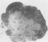
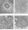
AIM--To describe the clinical, histological and immunohistochemical features in four cases of an uncommon benign lymph node lesion which may mimic a neoplastic process. METHODS--Four cases of inflammatory pseudotumour of lymph nodes were studied using conventional staining (haematoxylin and eosin, PAS, Gordon and Sweets reticulin stain, and the Ziehl-Neelsen stain) and with immunohistochemical techniques using a variety of antibodies (CD3, L26, CD15, CD21, CD30, KP1, MAC 387, vimentin, alpha SMA, HHF-35, D33, CD34, and S100). RESULTS--The lesion comprises a proliferation of spindle cells expanding the connective tissue framework of lymph nodes and is associated with a plasma cell and small lymphocyte infiltrate. There are variable numbers of macrophages, neutrophils and eosinophils, and varying degrees of fibrosis. Vascular changes are common but vary in degree and type. CONCLUSIONS--Inflammatory pseudotumour of lymph nodes is an uncommon benign reaction pattern which may be misdiagnosed as a neoplastic or even a malignant process. Increased awareness of its histological features should help prevent such misdiagnoses.
Full text
PDF


Images in this article

Selected References
These references are in PubMed. This may not be the complete list of references from this article.
- Chen K. T. Mycobacterial spindle cell pseudotumor of lymph nodes. Am J Surg Pathol. 1992 Mar;16(3):276–281. doi: 10.1097/00000478-199203000-00008. [DOI] [PubMed] [Google Scholar]
- Davis R. E., Warnke R. A., Dorfman R. F. Inflammatory pseudotumor of lymph nodes. Additional observations and evidence for an inflammatory etiology. Am J Surg Pathol. 1991 Aug;15(8):744–756. doi: 10.1097/00000478-199108000-00004. [DOI] [PubMed] [Google Scholar]
- Dorfman R. F., Berry G. J. Kikuchi's histiocytic necrotizing lymphadenitis: an analysis of 108 cases with emphasis on differential diagnosis. Semin Diagn Pathol. 1988 Nov;5(4):329–345. [PubMed] [Google Scholar]
- Facchetti F., De Wolf Peeters C., De Wever I., Frizzera G. Inflammatory pseudotumor of lymph nodes. Immunohistochemical evidence for its fibrohistiocytic nature. Am J Pathol. 1990 Aug;137(2):281–289. [PMC free article] [PubMed] [Google Scholar]
- Hui P. K., Chan J. K., Ng C. S., Kung I. T., Gwi E. Lymphadenopathy of Kimura's disease. Am J Surg Pathol. 1989 Mar;13(3):177–186. doi: 10.1097/00000478-198903000-00001. [DOI] [PubMed] [Google Scholar]
- Hurt M. A., Santa Cruz D. J. Cutaneous inflammatory pseudotumor. Lesions resembling "inflammatory pseudotumors" or "plasma cell granulomas" of extracutaneous sites. Am J Surg Pathol. 1990 Aug;14(8):764–773. [PubMed] [Google Scholar]
- Kemper C. A., Davis R. E., Deresinski S. C., Dorfmann R. F. Inflammatory pseudotumor of intra-abdominal lymph nodes manifesting as recurrent fever of unknown origin: a case report. Am J Med. 1991 Apr;90(4):519–523. [PubMed] [Google Scholar]
- Perrone T., De Wolf-Peeters C., Frizzera G. Inflammatory pseudotumor of lymph nodes. A distinctive pattern of nodal reaction. Am J Surg Pathol. 1988 May;12(5):351–361. doi: 10.1097/00000478-198805000-00003. [DOI] [PubMed] [Google Scholar]
- Ramani P., Bradley N. J., Fletcher C. D. QBEND/10, a new monoclonal antibody to endothelium: assessment of its diagnostic utility in paraffin sections. Histopathology. 1990 Sep;17(3):237–242. doi: 10.1111/j.1365-2559.1990.tb00713.x. [DOI] [PubMed] [Google Scholar]
- Suster S. Nodal angiolymphoid hyperplasia with eosinophilia. Am J Clin Pathol. 1987 Aug;88(2):236–239. doi: 10.1093/ajcp/88.2.236. [DOI] [PubMed] [Google Scholar]