Abstract
Methicillin-resistant Staphylococcus aureus (MRSA), an important human pathogen, has evolved an inducible mechanism for resistance to β-lactam antibiotics. We report herein that the integral membrane protein BlaR1, the β-lactam sensor/signal transducer protein, is phosphorylated on exposure to β-lactam antibiotics. This event is critical to the onset of the induction of antibiotic resistance. Furthermore, we document that BlaR1 phosphorylation and the antibiotic-resistance phenotype are both reversed in the presence of synthetic protein kinase inhibitors of our design, restoring susceptibility of the organism to a penicillin, resurrecting it from obsolescence in treatment of these intransigent bacteria.
Keywords: BlaR1, phosphorylation, MRSA, kinase inhibitor, Stk1
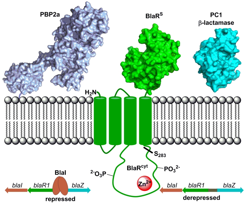
Staphylococcus aureus is a broadly antibiotic-resistant Gram-positive bacterium. β-Lactam antibiotics were the drugs of choice for treatment of infection by S. aureus, but a variant of this organism, methicillin-resistant Staphylococcus aureus (MRSA) emerged in 1961, which exhibits resistance to the entire class of β-lactams. This organism has been a global clinical problem for over half a century. The molecular basis for the broad resistance of MRSA to β-lactams, which is incidentally inducible, was traced to a set of genes within the bla and mec operons. The BlaR1 (or the cognate MecR1) protein is a β-lactam antibiotic sensor/signal transducer, which communicates the presence of the antibiotic in the milieu to the cytoplasm in a process that is largely not understood (Fig. 1).1-3 Signal transduction leads to activation of the cytoplasmic domain of BlaR1 (or MecR1), a zinc protease,2,4 which turns over the gene repressor BlaI (or MecI) in derepressing transcriptional events that result in expression of antibiotic-resistance determinants, the class A β-lactamase PC1 and/or the penicillin-binding protein 2a (PBP2a; Figure 1).5,6
Figure 1.
The bla system includes the β-lactam-antibiotic sensor/signal transducer protein BlaR1, which is acylated by the β-lactam antibiotics in the extracellular sensor domain (BlaRS). This initiates signal transduction through the membrane to the proteolytic domain (BlaRcyt), which autoproteolyzes at S283-F284. The BlaI repressor protein binds to the bla operon, which is comprised of genes that encode BlaI, BlaR1, and the PC1 β-lactamase (blaZ). Degradation of BlaI by the cytoplasmic protease domain of BlaR1 leads to derepression and transcription of the genes. BlaR1 is phosphorylated on the cytoplasmic side in response to the exposure of S. aureus to β-lactam antibiotics. The cognate mec operon encodes the corresponding MecI (gene repressor), MecR1 (antibiotic sensor/signal transducer) and PBP2a (penicillin-binding protein 2a, the antibiotic-resistance determinant).
An intriguing aspect of this system is its inducibility. Upon exposure to the antibiotic, the organism mobilizes. Once the antibiotic challenge is withdrawn, the system reverses itself. It was argued that on exposure to antibiotics the cytoplasmic domain of the BlaR1 protein undergoes autoproteolysis, which would unleash the activity of the protease domain in degradation of the gene repressor BlaI.2 We have found that this autoproteolytic processing takes place in the absence of antibiotic as well,4 therefore, we have argued that proteolysis leads to turnover of BlaR1 itself as an event in the reversal of induction.7 Hence, the question became what accounts for activation of the cytoplasmic domain toward degradation of BlaI in manifestation of the antibiotic-resistance response. We report herein that BlaR1 experiences phosphorylation at a minimum of one serine and one tyrosine in the cytoplasmic domain on exposure to β-lactam antibiotics. We also document that inhibition of this phosphorylation by small molecules reverses the methicillin-resistant phenotype, rendering MRSA susceptible to β-lactam antibiotics.
We investigated phosphorylation of BlaR1 in strain S. aureus NRS128 (also designated as NCTC8325). This strain, which has the bla but not the mec operon, was grown in the absence or in the presence of 10 μg/mL CBAP (2-(2’-carboxyphenyl)-benzoyl-6-aminopenicillanate), a good penicillin inducer of resistance.7 In a series of experiments that are outlined in the Methods section and in the Supporting Information, we document by western-blot analysis using anti-phosphotyrosine and anti-phosphoserine antibodies that the cytoplasmic domain of BlaR1 is phosphorylated at least on one tyrosine and one serine (Figures S1 and S2). The same experiment performed with an anti-phosphothreonine antibody documented the absence of threonine phosphorylation.
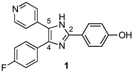
If phosphorylation of BlaR1 is important for the manifestation of resistance, could resistance to β-lactam antibiotics be attenuated (or reversed) in the presence of protein-kinase inhibitors? The strain NRS128, used above, was substituted with S. aureus MRSA252 (also known as USA200) for the following experiments. This strain exhibits high-level resistance to β-lactam antibiotics due to its expression of PBP2a. Its genome has been sequenced8 and it harbors the transposon Tn552, which encodes BlaR1. BlaR1 of MRSA252 has 99% sequence identity to that of NRS128. The minimal-inhibitory concentration (MIC) of oxacillin (a penicillin) against this strain is 256 μg/mL, consistent with high-level resistance. A protein-kinase inhibitor library of 80 known compounds was tested in a 96-well format against strain MRSA252 for this screening. We first determined MICs (broth microdilution method)9 for all the inhibitors in the library, in case some of them might have antibacterial properties of their own, which could complicate our analysis. Indeed, a few of these compounds did exhibit modest antibacterial activity and they were not studied further. Subsequently, we investigated the bacterial growth in the presence of oxacillin concentrations of 256 (MIC), 128 (½ MIC), and 64 (¼ MIC) μg/mL and one of two fixed concentrations of the protein-kinase inhibitors without antibiotic properties (0.7 μg/mL or 7 μg/mL). This rapid initial screening identified compound 1 as meeting the selection criteria of lowering the MIC for oxacillin, which was followed up by the actual evaluation of the MIC of oxacillin against S. aureus MRSA252 in the presence of the inhibitor. Inhibitor 1 at 7 μg/mL gave a reproducible four-fold decrease in the MIC of oxacillin for S. aureus MRSA252. The MIC of the kinase inhibitor alone against the same organism was ≥64 mg/mL.
Next, we tested the effect of exposure of CBAP-induced NRS128 to this kinase inhibitor. Cultures were induced with 10 μg/mL CBAP in the presence of 0, 7, or 17 μg/mL of compound 1. Whole-cell extracts of these bacteria were analyzed for BlaR1 phosphorylation by western blot using anti-Phos-Ser and anti-Phos-Tyr antibodies. Compound 1 inhibited both the phosphotyrosine and phosphoserine kinase activities by as much as 70-90% (Fig. S3). Hence, compound 1 lowered the degree of phosphorylation of BlaR1, which resulted in the lack of induction of the bla system in the presence of CBAP and at the same time the MIC for oxacillin was attenuated.
Compound 1 is a known mammalian serine/threonine-kinase inhibitor.10,11 That this compound inhibited the formation of phosphoserine and phosphotyrosine moieties in the BlaR1 protein was an important observation indicating that the kinase domain had a distinct structure that might make it a useful target for drug discovery. We undertook optimization of the structure of compound 1 for inhibition of the bacterial protein kinase(s) that phosphorylates BlaR1. A total of 70 structural variants of compound 1 were synthesized and tested.
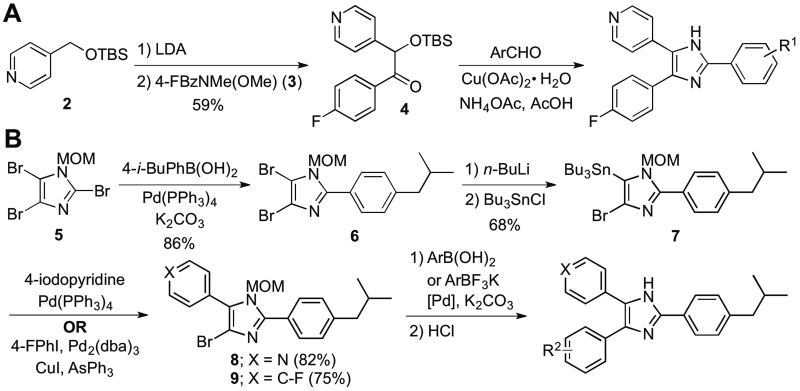
Diversification at the imidazole C2 position was achieved using the methodology of Gallagher et al. for construction of imidazoles,12 as shown in Scheme 1. Briefly, compound 2 was treated with LDA, followed by Weinreb amide 3, to give the ketone 4. This intermediate was then allowed to react with various benzaldehydes in the presence of copper(II) acetate and ammonium acetate to give a library of C2-modified imidazoles.
Scheme 1.
Synthesis of imidazole analogues with (a) C2 and (b) C4/C5 diversifications of the imidazole.
For diversification at C4 and C5, we used metal-catalyzed coupling to sequentially install the desired rings onto the imidazole (Scheme 1). Thus, tribromoimidazole derivative 513 was subjected to a Suzuki reaction with 4-isobutylphenylboronic acid to give C2-substituted imidazole 6. Suzuki reactions performed on 4,5-dibromoimidazoles such as 6 usually result in a mixture of mono- and di-coupled products.14 Therefore, 6 was converted to stannane 7 by lithiation and quenching with tributyltin chloride. Stannane 7 smoothly underwent Stille coupling15-17 with either 4-iodopyridine or 4-fluoroiodobenzene to give 8 and 9, respectively. These intermediates were then subjected to Suzuki reactions with arylboronic acids or potassium aryltrifluoroborate salts, followed by deprotection to give the desired imidazole analogues.
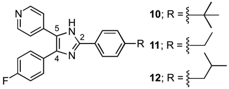
We evaluated the new imidazole analogues for their ability to lower the MIC of oxacillin against MRSA in the same manner as for the kinase-inhibitor library. In addition to MRSA252, we expanded our investigation to include strains NRS12318 and NRS70,19 both of which have 95% sequence identities between their BlaR1 proteins and that of NRS128. Interestingly, NRS123 encodes for a nonfunctional MecR1 protein and lacks the mecI gene, therefore PBP2a expression in this strain is regulated by the bla operon. The MIC values for oxacillin against the strains NRS123 and NRS70 are 16 and 32 μg/mL, respectively. Our inhibitors 10, 11, and 12 exhibited remarkable activity in lowering the MIC of oxacillin. Notably, compounds 10, 11, and 12 at 7 μg/mL are active across all three MRSA strains (Table 1). The MIC of each inhibitor by itself was >64 μg/mL across all three strains.
Table 1.
MIC values of oxacillin (μg/mL) against MRSA strains in the absence and in the presence of inhibitors 10-12 (7 μg/mL)
| Strain | No inhibitor | 10 | 11 | 12 |
|---|---|---|---|---|
| MRSA252 | 256 | 2 | 16 | 4 |
| NRS123 | 16 | 8 | 4 | 4 |
| NRS70 | 32 | 4 | 0.5 | 0.5 |
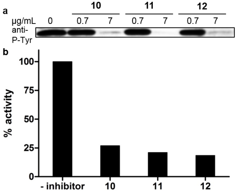
We then evaluated the ability of compounds 10, 11, or 12 to inhibit phosphorylation of BlaR1 at tyrosine residues in NRS70 extracts. The bacteria were grown with CBAP induction in the absence and presence of 0.7 μg/mL or 7 μg/mL compounds 10, 11, or 12, and analyzed by western blot using antibody against phosphotyrosine (Fig. 2A). The presence of 7 μg/mL inhibitor almost completely abolished tyrosine phosphorylation of the BlaR1 fragment in all cases. However, no inhibition of serine phosphorylation was seen with any of these synthetic compounds, even at 17 μg/mL (Fig. S4). This was in contrast to the case of the lead 1, which had inhibited both. This argues for the critical nature of tyrosine phosphorylation for regulation of BlaR1.
Figure 2.
The effect of compounds 10, 11, or 12 on BlaR1 tyrosine phosphorylation and β-lactamase activity. (a) Whole-cell extracts of NRS70 after induction with CBAP in the absence or presence of 0.7 or 7 μg/mL compounds 10, 11, or 12 were cleared of Protein A and other immunoglobin-binding proteins by incubation with IgG Sepharose and analyzed by western blot using antibodies against phosphotyrosine. (b) β-Lactamase activity of the culture media after induction with CBAP in the absence or presence of 7 μg/mL compounds 10, 11, or 12.
As we propose that phosphorylation activates the bla system, abrogation of phosphorylation should have an effect on expression of the resistance determinant(s). We have shown this to be the case in attenuation of the level of β-lactamase (product of the blaZ gene, Fig. 1). To document this, we monitored hydrolysis of the chromogenic β-lactam nitrocefin at A500 by the β-lactamase expressed in the CBAP-induced NRS70 culture media, according to the methodology reported previously.7 The initial rates of the reaction in the presence of 7 μg/mL compound 10, 11, or 12 were normalized to the activity in the absence of inhibitor (Fig. 2B). As expected, the presence of compounds 10, 11, or 12 decreased the β-lactamase activity by ~70-80%, congruent with the MIC data and the western-blot analysis. However, we cannot rule out off-target effects of these compounds, as there exist other protein targets for phosphorylation, and inhibition of the kinase(s) responsible would also impair these processes.
An observation by Tamber et al. that an stk1(pknB) gene knockout strain of the USA300 strain showed lower MIC values for β-lactam antibiotics is of interest.20 The gene stk1 (also known as pknB) encodes a highly conserved broad-specificity protein kinase in S. aureus that phosphorylates its substrates on serine, threonine, or tyrosine.21,22 To confirm Stk1 is a protein target of the inhibitors in this study, we cloned the gene and expressed and purified Stk1 from S. aureus NRS70 (Fig. S5). We found that compounds 10, 11, and 12 inhibit Stk1 autophosphorylation with IC50 values of 3.1 ± 0.8 μg/mL, 5 ± 1 μg/mL, and 6 ± 1 μg/mL, respectively. The compounds also inhibit phosphorylation of myelin-basic protein (MBP) by Stk1, with IC50 values of 2.1 ± 0.6 μg/mL, 4 ± 1 μg/mL, and 6 ± 1 μg/mL, respectively (Fig. S6).
We have shown in the present report that the BlaR1 protein of MRSA is phosphorylated in response to the challenge by β-lactam antibiotics, a step that is crucial in the signaling events leading to the induction of antibiotic resistance. The breadth of kinase/phosphatase-regulated processes in bacteria is likely vastly greater than is appreciated presently.23-25 This BlaR1 study represents the first insight as to the molecular-level regulation of a key resistance pathway in an important human pathogen. The documentation that inhibition of phosphorylation by small molecules reverses the MRSA phenotype makes available a new strategy to bring β-lactam antibiotics back from obsolescence in treatment of this insidious organism.
METHODS
General procedure for synthesis of triarylimidazole analogues with variation at C2 of the imidazole ring (Method A)
A literature procedure was followed.12 Compound 4 (1.0 equiv.), the aldehyde (1.1 equiv.), Cu(OAc)2·H2O (0.3 equiv.), and NH4OAc (10 equiv.) were dissolved in AcOH (7.5 mL/mmol 4), and the mixture was stirred at 110 °C for 1.5 h. The solution was then cooled to room temperature and was added to a mixture of conc. NH4OH (3× volume of AcOH used) and ice. After stirring for 10 minutes, the mixture was extracted with EtOAc, and the combined organic layer was washed with brine. The organic solution was dried over anhydrous Na2SO4, the suspension was filtered, and the solvent of the filtrate was removed in vacuo. Purification of the residue by flash chromatography gave the desired products in typical yields of 40-50%.
2-(4-tert-Butylphenyl)-4-(4-fluorophenyl)-5-(4-pyridyl)imidazole (10)
This product was synthesized by reacting 4 with 4-tert-butylbenzaldehyde according to Method A. Purification by flash chromatography (silica, 100% CH2Cl2 to 95:5 CH2Cl2/MeOH) gave the product as a yellow powder (49%). 1H NMR (600 MHz, CD3OD) δ 1.37 (s, 9H, C(CH3)3), 7.20 (t, 2H, J = 8.8 Hz, ArH), 7.52-7.56 (m, 6H, ArH), 7.93 (d, 2H, J = 8.8 Hz, ArH), 8.43 (br s, 2H, ArH); 13C NMR (150 MHz, CD3OD) δ 31.8, 35.8, 117.0 (d, 2JCF = 21.3 Hz), 123.3, 125.7, 127.0, 127.1, 128.2, (d, 4JCF = 2.2 Hz), 129.3, 130.3, 132.2 (d, 3JCF = 7.9 Hz), 143.5, 149.6, 150.3, 153.9, 164.4 (d, 1JCF = 246.8 Hz); HRMS (ESI): calcd for C24H23FN3 372.1871, found 372.1881 [MH]+.
2-(4-Ethylphenyl)-4-(4-fluorophenyl)-5-(4-pyridyl)imidazole (11)
This product was synthesized by reacting 4 with 4-ethylbenzaldehyde according to Method A. Purification by flash chromatography (silica, 100% CH2Cl2 to 95:5 CH2Cl2/MeOH) gave the product as a yellow powder (48%). 1H NMR (600 MHz, CD3OD) δ 1.28 (t, 3H, J = 7.6 Hz, CH3), 2.72 (q, 2H, J = 7.6 Hz, CH2), 7.21 (br s, 2H, ArH), 7.35 (d, 2H, J = 8.2 Hz, ArH), 7.53-7.55 (m, 4H, ArH), 7.91 (d, 2H, J = 8.2 Hz, ArH), 8.44 (br s, 2H, ArH); 13C NMR (150 MHz, CD3OD) δ 16.2, 29.9, 117.0 (d, 2JCF = 22.4 Hz), 123.3, 127.3, 128.5 (d, 4JCF = 4.5 Hz), 129.4, 129.6, 132.3 (d, 3JCF = 7.9 Hz), 135.1, 143.6, 147.3, 149.7, 150.3, 164.5 (d, 1JCF = 246.9 Hz); HRMS (ESI): calcd for C22H19FN3 344.1558, found 344.1572 [MH]+.
2-(4-iso-Butylphenyl)-4-(4-fluorophenyl)-5-(4-pyridyl)imidazole (12)
This product was synthesized by reacting 4 with 4-iso-butylbenzaldehyde according to Method A. Purification by flash chromatography (silica, 100% CH2Cl2 to 96:4 CH2Cl2/MeOH) gave the product as a yellow crystalline solid (49%). 1H NMR (600 MHz, CD3OD) δ 0.94 (d, 6H, J = 6.8 Hz, CH2CH(CH3)2), 1.92 (nonet, 1H, J = 6.8 Hz, CH2CH(CH3)2), 2.56 (d, 2H, J = 6.8 Hz, CH2CH(CH3)2), 7.21 (t, 2H, J = 7.8 Hz, ArH), 7.30 (d, 2H, J = 8.2 Hz, ArH), 7.53-7.55 (m, 4H, ArH), 7.91 (d, 2H, J = 8.2 Hz, ArH), 8.43 (br s, 2H, ArH); 13C NMR (150 MHz, CD3OD) δ 22.9, 31.6, 46.3, 117.0 (d, 2JCF = 22.4Hz), 123.3, 127.1, 128.6, 129.3, 129.6, 130.4, 130.9, 132.3 (d, 3JCF = 7.9 Hz), 143.6, 144.7, 149.7, 150.2, 164.4 (d, 1JCF = 248.0 Hz); HRMS (ESI): calcd for C24H23FN3 372.1871, found 372.1860 [MH]+.
Detection of BlaR1 phosphorylation in presence of antibiotic
As we had disclosed in a recent publication on fragmentation of BlaR1 during the course of induction, we detect a cleavage at position S283-F284 of BlaR1.7 This proteolytic cleavage cuts the BlaR1 protein into two fragments of roughly equal sizes (approx. 30-31 kDa). Zhang et al. identified another fragmentation site nearby, namely R293-R294.2 In order to process these protein samples for identification of the phosphorylation sites, Staphylococcus aureus NRS128 was grown in LB media to OD625 = 0.7, then was allowed to grow an additional three hours at 37 °C in the absence or presence of 10 μg/mL CBAP, a good inducer of the bla system. Extracts were prepared as previously described7 in buffer containing 50 mM Tris pH 7.5, 150 mM NaCl, 2 mM EDTA, 0.55% SDS, 2.5% Triton X-100, and Halt Protease and Phosphatase Inhibitor Cocktail (Thermo Scientific, Waltham, MA, U.S.A.) and analyzed by western blot using antibodies against phosphothreonine, phosphotyrosine, and phosphoserine. A ~30 kDa protein band was detected by the phosphotyrosine and phosphoserine specific antibodies only in cell extracts of S. aureus cells grown with CBAP (Figure S1). This band was not detected by the phosphothreonine antibody.
Initially, we had an abundance of Protein A and other immunoglobulin-binding proteins detected in the western blot, which ran in the range of 40-60 kDa and precluded visualization of the full-length BlaR1. To overcome this, we subsequently cleared all S. aureus extracts of Protein A and other immunoglobulin-binding proteins by incubation with IgG Sepharose (GE Healthcare, Little Chalfont, UK) for 1-2 h at room temperature with gentle agitation. After a brief centrifugation, the total protein in the supernatant was quantified by a BCA assay and 20 μg was loaded onto an 11% SDS-PAGE gel. Following electrophoresis, samples were transferred to a nitrocellulose membrane in a 10 mM CAPS (pH 11) buffer containing 10% methanol. Membranes were blocked in 3% BSA for phosphoserine blots or Blotto (3% BSA, 3% milk in TBS) for phosphotyrosine blots. HRP-conjugated primary antibody was applied in either 1% BSA/TBST (phosphoserine, Abcam, Cambridge, UK) or 1.1% milk/TBST (phosphotyrosine, 4G10 Platinum, EMD Millipore, Billerica, MA, U.S.A.). Phosphoserine blots were developed with Pierce ECL Substrate (Thermo Scientific) and phosphotyrosine blots were developed with SuperSignal West Dura Extended Duration Substrate (Thermo Scientific), then exposed to X-ray film for an appropriate amount of time (30-600 s).
Monitoring β-lactamase expression by nitrocefin assay
The media from CBAP-induced cultures grown in the absence or presence of 7 μg/mL compounds 10, 11, or 12 was separated from the cells by centrifugation at 3200 g, at 4 °C for 30 min. The absorbance of hydrolyzed chromogenic nitrocefin was monitored at 500 nm at room temperature for 5 min by adding 100 μM nitrocefin to 1 mL culture media. The initial rates of the reactions were determined by linear regression and the activity was normalized to the activity in the absence of inhibitor to give % activity.
Supplementary Material
ACKNOWLEDGMENTS
This work was supported by a grant from the National Institutes of Health (AI104987).
Footnotes
ASSOCIATED CONTENT
The following file is available free of charge on the ACS Publications website.
Boudreau_SI: Experimental procedures for preparation and western blot analysis of Staphylococcus aureus extracts, synthesis, and MIC determination. This information is available free of charge via the Internet at http://pubs.acs.org/.
Author contributions
MAB synthesized and evaluated the kinase inhibitors, JF characterized BlaR1 phosphorylation, LIL performed the initial experiments that led to the discovery of phosphorylation of BlaR1, QX did the work with Stk1, SM initiated the project and orchestrated its completion as the head of the lab.
Conflict of interest
The authors declare no competing financial interest.
REFERENCES
- 1.Frederick TE, Wilson BD, Cha J, Mobashery S, Peng JW. Revealing cell surface intramolecular interactions in the BlaR1 protein of methicillin-resistant Staphylococcus aureus by NMR spectroscopy. Biochemistry. 2014;53:10–12. doi: 10.1021/bi401552j. DOI: 10.1021/bi401552j. [DOI] [PMC free article] [PubMed] [Google Scholar]
- 2.Zhang HZ, Hackbarth CJ, Chansky KM, Chambers HF. A proteolytic transmembrane signaling pathway and resistance to β-lactams in Staphylococci. Science. 2001;291:1962–1965. doi: 10.1126/science.1055144. DOI: 10.1126/science.1055144. [DOI] [PubMed] [Google Scholar]
- 3.Staude MW, Frederick TE, Natarajan SV, Wilson BD, Tanner CE, Ruggiero ST, Mobashery S, Peng JW. Investigation of signal transduction routes within the sensor/transducer protein BlaR1 of Staphylococcus aureus. Biochemistry. 2015;54:1600–1610. doi: 10.1021/bi501463k. DOI: 10.1021/bi501463k. [DOI] [PMC free article] [PubMed] [Google Scholar]
- 4.Llarrull LI, Mobashery S. Dissection of events in the resistance to β-lactam antibiotics mediated by the protein BlaR1 from Staphylococcus aureus. Biochemistry. 2012;51:4642–4649. doi: 10.1021/bi300429p. DOI: 10.1021/bi300429p. [DOI] [PMC free article] [PubMed] [Google Scholar]
- 5.Arede P, Milheirico C, de Lencastre H, Oliveira DC. The anti-repressor MecR2 promotes the proteolysis of the mecA repressor and enables optimal expression of β-lactam resistance in MRSA. PLoS Pathog. 2012;8:e1002816. doi: 10.1371/journal.ppat.1002816. DOI: 10.1371/journal.ppat.1002816. [DOI] [PMC free article] [PubMed] [Google Scholar]
- 6.Blázquez B, Llarrull L, Luque-Ortega J, Alfonso C, Boggess B, Mobashery S. Regulation of the expression of the β-lactam antibiotic-resistance determinants in methicillin-resistant Staphylococcus aureus (MRSA) Biochemistry. 2014;53:1548–1550. doi: 10.1021/bi500074w. DOI: 10.1021/bi500074w. [DOI] [PMC free article] [PubMed] [Google Scholar]
- 7.Llarrull LI, Toth M, Champion MM, Mobashery S. Activation of BlaR1 protein of methicillin-resistant Staphylococcus aureus, its proteolytic processing, and recovery from induction of resistance. J. Biol. Chem. 2011;286:38148–38158. doi: 10.1074/jbc.M111.288985. DOI: 10.1074/jbc.M111.288985. [DOI] [PMC free article] [PubMed] [Google Scholar]
- 8.Holden MTG, Feil EJ, Lindsay JA, Peacock SJ, Day NPJ, Enright MC, Foster TJ, Moore CE, Hurst L, Atkin R, Barron A, Bason N, Bentley SD, Chillingworth C, Chillingworth T, Churcher C, Clark L, Corton C, Cronin A, Doggett J, Dowd L, Feltwell T, Hance Z, Harris B, Hauser H, Holroyd S, Jagels K, James KD, Lennard N, Line A, Mayes R, Moule S, Mungall K, Ormond D, Quail MA, Rabbinowitsch E, Rutherford K, Sanders M, Sharp S, Simmonds M, Stevens K, Whitehead S, Barrell BG, Spratt BG, Parkhill J. Complete genomes of two clinical Staphylococcus aureus strains: Evidence for the rapid evolution of virulence and drug resistance. Proc. Natl. Acad. Sci. U.S.A. 2004;101:9786–9791. doi: 10.1073/pnas.0402521101. DOI: 10.1073/pnas.0402521101. [DOI] [PMC free article] [PubMed] [Google Scholar]
- 9.CLSI . Performance Standards for Antimicrobial Susceptibility Testing; Twenty-Second Informational Supplement. CLSI document M100-S22. Clinical and Laboratory Standards Institute; Wayne, PA: [Google Scholar]
- 10.Lee JC, Laydon JT, McDonnell PC, Gallagher TF, Kumar S, Green D, McNulty D, Blumenthal MJ, Heys JR, Landvatter SW, Stricker JE, McLaughlin MM, Siemens IR, Fisher SM, Livi GP, White JR, Adams JL, Young PR. A protein kinase involved in the regulation of inflammatory cytokine biosynthesis. Nature. 1994;372:739–746. doi: 10.1038/372739a0. DOI: 10.1038/372739a0. [DOI] [PubMed] [Google Scholar]
- 11.Frantz B, Klatt T, Pang M, Parsons J, Rolando A, Williams H, Tocci MJ, O’Keefe SJ, O’Neill EA. The activation state of p38 mitogen-activated protein kinase determines the efficiency of ATP competition for pyridinylimidazole inhibitor binding. Biochemistry. 1998;37:13846–13853. doi: 10.1021/bi980832y. DOI: 10.1021/bi980832y. [DOI] [PubMed] [Google Scholar]
- 12.Gallagher TF, Seibel GL, Kassis S, Laydon JT, Blumenthal MJ, Lee JC, Lee D, Boehm JC, Fier-Thompson SM, Abt JW, Soreson ME, Smietana JM, Hall RF, Garigipati RS, Bender PE, Erhard KF, Krog AJ, Hofmann GA, Sheldrake PL, McDonnell PC, Kumar S, Young PR, Adams JL. Regulation of stress-induced cytokine production by pyridinylimidazoles; Inhibition of CSBP kinase. Bioorg. Med. Chem. 1997;5:49–64. doi: 10.1016/s0968-0896(96)00212-x. DOI: 10.1016/S0968-0896(96)00212-X. [DOI] [PubMed] [Google Scholar]
- 13.Niculescu-Duvaz D, Niculescu-Duvaz I, Suijkerbuijk BMJM, Ménard D, Zambon A, Nourry A, Davies L, Manne HA, Friedlos F, Ogilvie L, Hedley D, Takle AK, Wilson DM, Pons J-F, Coulter T, Kirk R, Cantarino N, Whittaker S, Marais R, Springer CJ. Novel tricyclic pyrazole BRAF inhibitors with imidazole or furan scaffolds. Bioorg. Med. Chem. 2010;18:6934–6952. doi: 10.1016/j.bmc.2010.06.031. DOI: 10.1016/j.bmc.2010.06.031. [DOI] [PMC free article] [PubMed] [Google Scholar]
- 14.Recnik L-M, Hameid MAE, Haider M, Schnürch M, Mihovilovic MD. Selective sequential cross-coupling reactions on imidazole towards neurodazine and analogues. Synthesis. 2013;45:1387–1405. DOI: 10.1055/s-0032-1316906. [Google Scholar]
- 15.Liverton NJ, Butcher JW, Claiborne CF, Claremon DA, Libby BE, Nguyen KT, Pitzenberger SM, Selnick HG, Smith GR, Tebben A, Vacca JP, Varga SL, Agarwal L, Dancheck K, Forsyth AJ, Fletcher DS, Frantz B, Hanlon WA, Harper CF, Hofsess SJ, Kostura M, Lin J, Luell S, O’Neill EA, Orevillo CJ, Pang M, Parsons J, Rolando A, Sahly Y, Visco DM, O’Keefe SJ. Design and synthesis of potent, selective, and orally bioavailable tetrasubstituted imidazole inhibitors of p38 mitogen-activated protein kinase. J. Med. Chem. 1999;42:2180–2190. doi: 10.1021/jm9805236. DOI: 10.1021/jm9805236. [DOI] [PubMed] [Google Scholar]
- 16.Liebeskind LS, Fengl RW. 3-Stannylcyclobutenediones as nucleophilic cyclobutenedione equivalents. Synthesis of substituted cyclobutenediones and cyclobutenedione monoacetals and the beneficial effect of catalytic copper iodide on the Stille reaction. J. Org. Chem. 1990;55:5359–5364. DOI: 10.1021/jo00306a012. [Google Scholar]
- 17.Farina V, Kapadia S, Krishnan B, Wang C, Liebeskind LS. On the nature of the “copper effect” in the Stille cross-coupling. J. Org. Chem. 1994;59:5905–5911. DOI: 10.1021/jo00099a018. [Google Scholar]
- 18.Baba T, Takeuchi F, Kuroda M, Yuzawa H, Aoki K, Oguchi A, Nagai Y, Iwama N, Asano K, Naimi T, Kuroda H, Cui L, Yamamoto K, Hiramatsu K. Genome and virulence determinants of high virulence community-acquired MRSA. Lancet. 2002;359:1819–1827. doi: 10.1016/s0140-6736(02)08713-5. DOI: 10.1016/S0140-6736(02)08713-5. [DOI] [PubMed] [Google Scholar]
- 19.Kuroda M, Ohta T, Uchiyama I, Baba T, Yuzawa H, Kobayashi I, Cui L, Oguchi A, Aoki K, Nagai Y, Lian J, Ito T, Kanamori M, Matsumaru H, Maruyama A, Murakami H, Hosoyama A, Mizutani-Ui Y, Takahashi NK, Sawano T, Inoue R, Kaito C, Sekimizu K, Hirakawa H, Kuhara S, Goto S, Yabuzaki J, Kanehisa M, Yamashita A, Oshima K, Furuya K, Yoshino C, Shiba T, Hattori M, Ogasawara N, Hayashi H, Hiramatsu K. Whole genome sequencing of meticillin-resistant Staphylococcus aureus. Lancet. 2001;357:1225–1240. doi: 10.1016/s0140-6736(00)04403-2. DOI: 10.1016/S0140-6736(00)04403-2. [DOI] [PubMed] [Google Scholar]
- 20.Tamber S, Schwartzman J, Cheung AL. Role of PknB kinase in antibiotic resistance and virulence in community-acquired methicillin-resistant Staphylococcus aureus strain USA300. Infect. Immun. 2010;78:3637–46. doi: 10.1128/IAI.00296-10. DOI: 10.1128/IAI.00296-10. [DOI] [PMC free article] [PubMed] [Google Scholar]
- 21.Beltramini AM, Mukhopadhyay CD, Pancholi V. Modulation of cell wall structure and antimicrobial susceptibility by a Staphylococcus aureus eukaryote-like serine/threonine kinase and phosphatase. Infect. Immun. 2009;77:1406–1416. doi: 10.1128/IAI.01499-08. DOI: 10.1128/IAI.01499-08. [DOI] [PMC free article] [PubMed] [Google Scholar]
- 22.Débarbouillé M, Dramsi S, Dussurget O, Nahori M-A, Vaganay E, Joubion G, Coxxone A, Msadek T, Duclos B. Characterization of a serine/threonine kinase involved in virulence of Staphylococcus aureus. J. Bacteriol. 2009;191:4070–4081. doi: 10.1128/JB.01813-08. DOI: 10.1128/JB.01813-08. [DOI] [PMC free article] [PubMed] [Google Scholar]
- 23.Dworkin J. Ser/Thr phosphorylation as a regulatory mechanism in bacteria. Curr. Opin. Microbiol. 2015;24C:47–52. doi: 10.1016/j.mib.2015.01.005. DOI: 10.1016/j.mib.2015.01.005. [DOI] [PMC free article] [PubMed] [Google Scholar]
- 24.Ravikumar V, Shi L, Krug K, Derouiche A, Jers C, Cousin C, Kobir A, Mijakovic I, Macek B. Quantitative phosphoproteome analysis of Bacillus subtilis reveals novel substrates of the kinase PrkC and phosphatase PrpC. Mol. Cell Proteomics. 2014;13:1965–1978. doi: 10.1074/mcp.M113.035949. DOI: 10.1074/mcp.M113.035949. [DOI] [PMC free article] [PubMed] [Google Scholar]
- 25.Cousin C, Derouiche A, Shi L, Pagot Y, Poncet S, Mijakovic I. Protein-serine/threonine/tyrosine kinases in bacterial signaling and regulation. FEMS Microbiol. Lett. 2013;346:11–19. doi: 10.1111/1574-6968.12189. DOI: 10.1111/1574-6968. [DOI] [PubMed] [Google Scholar]
Associated Data
This section collects any data citations, data availability statements, or supplementary materials included in this article.


