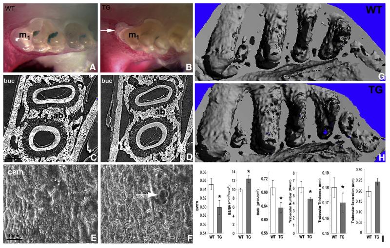
Fig. 3.
Bone loss and root resorption in Ambn over-expressor molars. (A, B) Whole-mount Alizerin red staining of mandibles from Ambn transgenic (TG) and wild-type (WT) mice at 35 days postnatal. First molars are marked M1. Note the significant bone loss at the crest of the alveolar bone (arrow) as well as the increase in periodontal ligament width in AMBN transgenic mice as compared to wild-type controls. (C, D) Micro-CT comparison of root sections of WT and TG mice. (E, F) Scanning electron microscopy images of molar teeth of 35 days old WT and Ambn transgenic mice. The arrow indicates a resorption pit on the TG molar root. (G, H) 3D μCT image reconstruction of the molar-region dentoalveolar complex of wild-type (G) and AMBN transgenic mice at age 42 days postnatal (H). Note the widened alveolar crypts, roughened alveolar bone surfaces, and reduced interradicular spaces in AMBN overexpressors (H versus G). Morphometric comparison between AMBN transgenic and WT mice. The following parameters were compared (3 samples per group, from left to right): trabecular bone volume fraction (BV/TV), surface/volume ratio (BS/BV), bone mass density (BMD), trabecular number, trabecular thickness, and trabecular separation. All but the trabecular separation were significantly different (p<0.05) between both groups. *=p<0.05.

