Abstract
Autophagy is a catabolic cellular mechanism that degrades dysfunctional proteins and organelles. Atherosclerotic plaque formation is enhanced in mice with macrophages that cannot undergo autophagy because of a deficiency of an autophagy component such as ATG5. We showed that exposure of macrophages to atherogenic lipids led to an increase in the abundance of the autophagy chaperone p62, which colocalized with polyubiquitinated proteins in cytoplasmic inclusions. p62 accumulation was increased in ATG5-null macrophages, which had large cytoplasmic ubiquitin-positive p62 inclusions. Aortas from atherosclerotic mice and plaques from human endarterectomy samples showed increased abundance of p62 and polyubiquitinated proteins that co-localized with plaque macrophages, suggesting that p62-enriched protein aggregates were characteristic of atherosclerosis. The formation of the cytoplasmic inclusions depended on p62 because lipid-loaded p62-null macrophages accumulated polyubiquitinated proteins in a diffuse cytoplasmic pattern. The failure of these aggregates to form was associated with increased secretion of IL-1β and enhanced macrophage apoptosis, which depended on the p62 ubiquitin-binding domain and at least partly involved p62-mediated clearance of NLRP3 inflammasomes. Consistent with our in vitro observations, p62-deficient mice formed greater numbers of more complex atherosclerotic plaques, and p62 deficiency further increased atherosclerotic plaque burden in mice with a macrophage-specific ablation of ATG5. Together, these data suggested that sequestration of cytotoxic ubiquitinated proteins by p62 protects against atherogenesis, a condition in which the clearance of protein aggregates is disrupted.
INTRODUCTION
Autophagy is an evolutionarily conserved cellular process with critical roles in the degradation and recycling of long-lived/damaged intracellular material including organelles (1,2). Although the regulation and function of autophagy has been studied in the context of several human diseases, its role in innate immunity and macrophage biology in particular has been a subject of intense interest. In addition to being implicated in diverse roles such as viral and microbial degradation, antibacterial reactive oxygen species (ROS) generation, antigen presentation, and modulation of proinflammatory signaling (3,4), macrophage autophagy has also been directly assessed in the context of atherosclerosis (5–7). The selective deletion of ATG5, one of the critical components of the autophagosome-forming machinery, abrogates basal autophagy as well as disrupting an ability to mount an autophagic response to atherogenic lipids (5–7). ATG5-null macrophages in turn develop several deleterious phenotypes including increased activation of the inflammasome, enhanced apoptosis/oxidative stress, and disrupted lipid efflux (5–7), mechanisms that have all been proposed to contribute to the increased atherosclerosis in mice with a deficiency in macrophage autophagy (5,6). At present, it remains unclear which of these processes or disruptions in other facets of autophagy are the predominant contributors to plaque formation. This complexity is largely due to the ability of the autophagy-lysosomal system to handle the capture and degradation of diverse cargo. Thus, determination of the relevant intracellular material in plaque macrophages might provide insight into the pathogenesis of atherosclerosis.
We have previously shown that increases in the protein abundance of p62 (also known as SQSTM1) in atherosclerotic plaques mostly localizes to plaque macrophages in a autophagy-dependent manner and directly correlates with plaque progression Given its role as a chaperone for the autophagic removal of protein and organelle cargo, increases in p62 have traditionally been used as a marker of autophagy deficiency; however, it is not unclear whether the presence of p62 is a maladaptive response or cytoprotective. If p62 serves as a marker of autophagy dysfunction and denote the accumulation of cytotoxic intracellular products, one would predict that this response is deleterious. For example, the accumulation of protein aggregates is considered a pathogenic feature of diseases of the nervous system (Alzheimer’s, Parkinson’s, and Huntington’s Diseases) (9,10), the liver (α1-antitrypsin deficiency) (11), and skeletal muscle (inclusion body myositis) (12). Thus, we sought to further define the roles of macrophage p62 and autophagy in atherosclerosis.
We showed that like plaque macrophages, cultured macrophages loaded with atherogenic lipids showed increases in the abundance of p62 and polyubiquitinated proteins, leading to the formation of p62-enriched cytoplasmic inclusion bodies. Similar observations in atherosclerotic plaques from mice and humans suggested that atherosclerosis was a disease characterized by protein aggregation and inclusion bodies. We provided evidence that p62 played a critical role in macrophage proteostasis by enabling the formation of protective inclusion bodies. In the absence of p62, protein insolubility increased because of disruptions in the formation and autophagy-dependent removal of cytoplasmic inclusions. Furthermore, these effects were associated with increased macrophage apoptosis, inflammasome activation, and IL-1β production, resulting in atherosclerotic plaque area and complexity. We concluded that the increase in p62 abundance in macrophages in the progression of atherosclerosis in the absence of autophagy was not maladaptive but rather a protective response to allow the sequestration of cytotoxic protein aggregates.
RESULTS
Atherogenic lipids result in macrophage accumulation of p62-enriched cytoplasmic inclusions
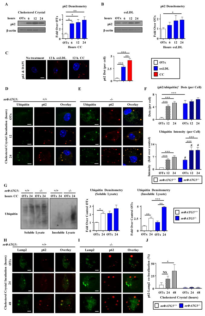
The protein abundance of p62 increases during atherosclerotic progression, an effect that is predominantly localized to plaque macrophages and appears to be related to a dysfunction in autophagic processing (5). We modeled the increase in p62 in vitro. Two frequently encountered lipid species in the atherosclerotic plaque are oxidized LDL (oxLDL) and cholesterol crystals. Although biochemically distinct, both lipid species can enter macrophages through receptor-mediated (for example, CD36) or bulk endocytic processes and traffic to the lysosomal compartment for degradation (13,14). Inefficient hydrolysis of oxidized cholesteryl esters and solubilization of cholesterol crystals leads to disruptions in lysosomal membrane integrity and lysosomal dysfunction (13,15,16), in turn disrupting cellular pathways requiring an intact lysosomal apparatus such as autophagy (5). The chaperone protein p62 shuttles intracellular polyubiquitinated proteins to autophagosomes for degradation. Thus, disruptions in autophagy induce p62 accumulation (8). Accordingly, treatment of cultured peritoneal macrophages (henceforth referred to as macrophages) with either oxLDL or cholesterol crystals led to progressive accumulation of p62 protein especially with longer incubation times (Figure 1a,b). Although both lipids transiently increased p62 transcript abundance at early time-points (Supplementary Figure 1a,b), protein abundance continued to increase indicating a sustained post-transcriptional effect. Immunofluorescence microscopy of the treated macrophages localized the p62 predominantly to distinct punctae, suggestive of aggregate formation (Figure 1c). This effect was likely mediated by disruption of the autophagy-lysosomal system by cholesterol crystals because (i) lysosomal inhibitors (chloroquine and bafilomycin) or disruptors of lysosomal membrane integrity (silica and alum crystals) also transiently increased p62 mRNA with sustained formation of p62-enriched punctae (Supplementary Figure 1c-e) and (ii) macrophages from mice with a macrophage-specific deficiency of ATG5 (mΦATG5−/−) showed increased p62 abundance and inclusion formation at baseline and with atherogenic lipid treatment (Supplementary Figure 2, Figure 1d–e).
Figure 1. Atherogenic lipid loading and autophagy deficiency lead to p62-enriched polyubiquitinated protein accumulation in macrophages.
(a to c) Western blot analysis and (c) immunofluorescence of p62 in peritoneal macrophages after cholesterol crystal (CC) or oxidized LDL (oxLDL) incubation for the indicated times. β-actin was used for loading control and densitometric quantification from three separate experiments is shown next to the representative western blot (a,b). Nuclei were counter stained with DAPI and graph represents average p62 dot numbers per cell after the indicated treatments (n≥35 cells for each group in three independent experiments; c, right). (d–e) Immunofluorescence images of control and ATG5-KO peritoneal macropahges after cholesterol crystal incubation for the indicated times using DAPI and antibodies against ubiquitinated proteins (FK-1) and p62. (f) Graphs represent average p62/ubiquitin+ dot numbers (top) and average ubiquitin intensity (bottom) per cell for immunofluorescence images in d, e (n≥30 cells for each group in three independent experiments, #P<0.05 compared to the respective time-point in Control cells). (g) Western blot analysis of polyubiquitinated proteins (FK-1 antibody) in detergent soluble and insoluble lysate fractions of cholesterol crystal-treated control and mΦATG5−/− peritoneal macrophages. Densitometric quantification from three separate experiments is shown to the right. (h–j) Immunofluorescence images of control and mΦATG5−/− peritoneal macrophages after cholesterol crystal incubation for the indicated times using antibodies against the late endosome/lysosome marker LAMP2 and p62. Graph represents percent of p62+ dots co-localizing with LAMP2 (n≥15 cells for each group in three independent experiments; j). For all panels, data are presented as mean ± SEM. (*P<0.05, **P<0.01, ***P<0.001, NS = not significant). Scale bar, 5 μm.
In addition to playing a role in protein aggregation, p62 is involved in the autophagic turnover of organelles such as peroxisomes and mitochondria (17,18). Furthermore, certain protein aggregates termed aggresomes depend on microtubules and localize to Vimentin-rich intermediate filaments (19). We used immunofluorescence microscopy to determine the localization of atherogenic lipid-induced p62 aggregates in macrophages. We did not detect co-localization of p62 with markers of early endosomes (EEA1), lysosome (LAMP2), peroxisomes (PMP70), golgi (GM130), ER (Calnexin), intermediate filaments (Vimentin), or lipid droplets (BODIPY and Nile Red) up to 24 hours of lipid incubation (Supplementary Figure 3). Finally, only a small number of p62-positive inclusions colocalized with mitochondria (MitoTracker) (Supplementary Figure 3). The absence of substantial p62 localization with any of the above subcellular organelle markers suggests that atherogenic lipid-induced p62 structures in macrophages were most likely cytoplasmic inclusion bodies akin to p62-enriched cytosolic aggregates that have been previously described (20).
Atherogenic lipid-induced inclusion bodies are enriched in p62 and polyubiquitinated proteins
The role of p62 in binding polyubiquitinated proteins led us to determine the degree of association of these components after atherogenic lipid treatment. Immunofluorescence microscopy and immunoblotting with an antibody that specifically binds polyubiquitinated proteins and not free ubiquitin (FK-1 antibody) revealed that p62 inclusions with near complete co-localization with polyubiquitinated substrates appeared as early as 6 hours after cholesterol crystal treatment (Supplementary Figure 4) and increased in abundance with longer incubation times (Figure 1d,f). In mΦATG5−/− macrophages, inclusion bodies were present independently of exposure to atherogenic lipids and the amount of polyubiquitinated proteins contained in the inclusions (as determined by fluorescence intensity) significantly increased after exposure to such lipids (Figure 1e,f). Western blot analysis with the FK-1 antibody indicated that polyubiquitinated proteins accumulated in the detergent-soluble fraction from macrophages exposed to cholesterol crystals, a response that was minimally increased in mΦATG5−/−macrophages. The detergent-insoluble fractions showed more evident increases in polyubiquitinated proteins particularly in mΦATG5−/− macrophages (Figure 1g).
To link the clearance of polyubiquitinated protein aggregates to the autophagy-lysosomal system, cholesterol crystal-treated macrophages were co-stained with p62 and the lysosomal marker LAMP2. p62-positive aggregates had little initial co-localization with LAMP2 after 24 hours of incubation (Figure 1h) and partial co-localization after 48 hours of cholesterol crystal treatment suggesting a delay but not complete cessation of lysosomal targeting (Figure 1h–j). Targeting appeared to depend on autophagic delivery of the inclusion bodies to lysosomes since mΦATG5−/− macrophages contained large p62-enriched inclusions that did not localize with lysosomes even after 48 hours (Figure 1i,j). Together, these findings suggest that p62-enriched insoluble protein aggregates form in macrophages challenged with atherogenic lipids, the removal of which is autophagy-dependent.
Polyubiquitinated protein accumulation is a hallmark of atherosclerotic plaques
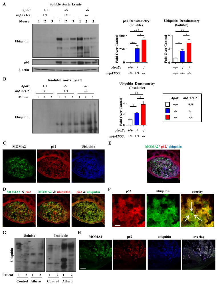
To assess whether the increase in polyubiquitinated proteins in macrophages in vitro also occurred in vivo, we compared the aortas of control, ApoE−/− (atherosclerosis-prone), and ApoE−/−/mΦATG5−/− mice fed a Western Diet. As reported previously, p62 abundance was increased in aortas in ApoE−/− mice, an effect that was increased in ApoE−/−/mΦATG5−/− mice (Figure 2a) (5). Polyubiquitinated proteins were increased in both the detergent soluble and insoluble fractions of atherosclerotic plaques from ApoE−/− mice, an effect that was increased in ApoE−/−/mΦATG5−/− mice, especially in the insoluble fraction (Figure 2a,b).
Figure 2. The accumulation of insoluble p62-enriched polyubiquitinated proteins in macrophages is characteristic of both mouse and human atherosclerosis.
(a–b) Western blot analysis of polyubiquitinated proteins and p62 in soluble (a) and insoluble (b) lysates of whole aortas from control (WT), ApoE−/−, and ApoE−/−/mΦATG5−/− mice (n=3 mice for each genotype) fed a Western Diet for 2 months. Densitometric quantification and a representative western blot are shown. (c–f) Representative immunofluorescence images of aortic roots from ApoE−/− mice co-stained with antibodies against the macrophage marker (MOMA2), ubiquitinated proteins (FK-1 antibody), and p62. Individual stains (c), overlays of 2 stains (d), overlays of all 3 stains (e), and magnified views of areas of punctate co-localization between p62 and Ubiquitin (f) are shown. Arrows indicate dots co-stained for both ubiquitin and p62. Images are representative of eight independent experiments. (g) Western blot analysis of polyubiquitinated proteins in soluble and insoluble lysates of atherosclerotic plaques and adjacent non-atherosclerotic regions extracted from post-operative human carotid endarterectomy specimens. (h) Immunofluorescence images of an atherosclerotic region from a carotid endarterectomy specimen co-stained with MOMA2, ubiquitinated proteins (FK-1 antibody), and p62. Images are representative of two carotid endarterectomy specimens. For all graphs, data are presented as mean ± SEM. *P<0.05, **P<0.01, ***P<0.001. Scale bar, 100 μm (c,h); 10 μm (f).
To determine if the increases in p62 and polyubiquitinated proteins were due to lack of degradation in macrophages, we performed immunofluorescence imaging on atherosclerotic aortic root sections stained for p62, polyubiquitinated proteins, and the macrophage marker (MOMA-2). Polyubiquitinated proteins predominantly co-localized with p62 in plaque macrophages (Figure 2c, 2d and 2e). Nonspecific auto-fluorescence of elastin fibers was seen in several fluorescence channels as verified by appropriate negative controls. High-magnification subcellular imaging of plaque macrophages revealed punctate p62- and polyubiquitin-positive staining consistent with the presence of inclusion bodies (Figure 2f).
Given the increase in insoluble polyubiquitinated proteins in lysates of mouse atherosclerotic aortas, we also performed a similar analysis in human atherosclerotic plaques obtained from specimens discarded during carotid endarterectomy. Immunoblotting revealed a significant increase in detergent insoluble polyubiquitinated protein mass in lysates from atherosclerotic carotid lesions compared to those from adjacent non-atherosclerotic tissue (Figure 2g, Supplementary Figure 5). Furthermore, immunofluorescence imaging of the carotid plaque revealed that p62 and polyubiquitinated proteins nearly completely colocalized with plaque macrophages (Figure 2h). Together with our results in the mouse aorta, these findings imply that the formation of p62-enriched insoluble protein aggregates may be a feature of atherosclerotic plaques.
p62 is necessary for atherogenic lipid-induced inclusion body formation and reducing the accumulation of insoluble polyubiquitinated proteins
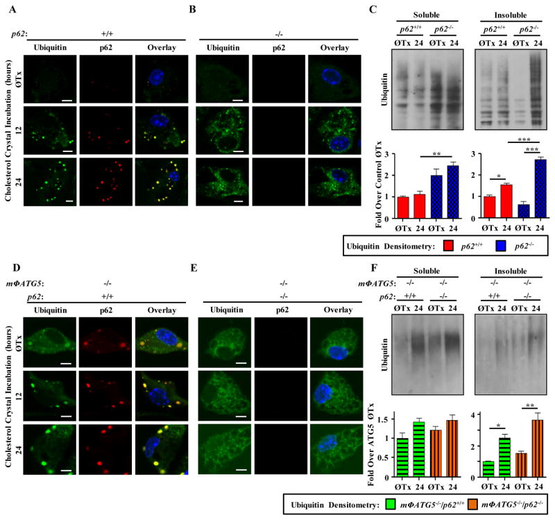
To understand the role of p62 in the inclusion body formation of atherosclerotic macrophages, we evaluated the accumulation and subcellular distribution of polyubiquitinated proteins in control and p62-deficient macrophages derived from mice with a genetically targeted p62 locus (21). Cholesterol crystal incubation over a 24 hour period resulted in the formation of p62-enriched polyubiquitinated inclusion bodies in control macrophages (Figure 3a) but not in p62−/− macrophages. In the absence of p62, polyubiquitinated proteins were present in a more diffuse and evenly distributed cytoplasmic configuration (Figure 3b). Polyubiquitinated proteins slightly increased in abundance in the detergent soluble fraction of both untreated and atherogenic lipid-treated p62−/− macrophages, and increased significantly in the insoluble fraction of p62−/− macrophages (Figure 3c).
Figure 3. p62 deficiency leads to disruption of atherogenic lipid-induced inclusion bodies and increased protein insolubility.
(a,b) Control (a) and p62−/− (b) peritoneal macrophages incubated with cholesterol crystals for the indicated times and subjected to immunofluorescence imaging using nuclear stain DAPI and antibodies against ubiquitinated protein (FK-1) and p62. (c) Detergent soluble and insoluble lysates were prepared after incubations as in (a,b) and subjected to Western blot analysis for polyubiquitinated proteins (FK-1 antibody). Densitometric quantification from three separate experiments is shown below each respective western blot. (d,e) mΦATG5−/− (d) and mΦATG5−/−/p62−/− (e) peritoneal macrophages were treated as in a,b and subjected to immunofluorescence imaging. Images are representative of five independent experiments. (f) Soluble and insoluble lysates were prepared and subjected to Western blot analysis as in (c). Densitometric quantification from three separate experiments is shown below a representative western blot. Data are presented as mean ± SEM. *P<0.05, **P<0.01, ***P<0.001. Scale bar, 5 μm.
Autophagy is the primary mechanism by which p62-enriched protein aggregates are cleared. p62-enriched polyubiquitinated protein inclusions were prevalent in mΦATG5−/− macrophages at baseline and the size of these inclusions increased upon atherogenic lipid treatment (Figure 3d). Similar to p62−/− macrophages, mΦATG5−/−/p62−/− macrophages had diffuse cytoplasmic ubiquitin staining (Figure 3e). This finding was corroborated by immunoblot analysis that demonstrated an increase in the detergent insoluble polyubiquitinated protein fraction of mΦATG5−/− macrophages that was further exacerbated in mΦATG5−/−/p62−/− macrophages (Figure 3f right). There were no appreciable baseline or compensatory changes in the proteasomal function of mΦATG5−/− or p62−/− macrophages (Supplementary Figure 6).
Together, these data suggested that (i) atherogenic lipid-mediated inclusion body formation was a p62-dependent process, (ii) p62 appeared to be required for maintaining the architecture of cytoplasmic inclusions containing polyubiquitinated proteins, and (iii) in the absence of p62, polyubiquitinated proteins accumulated as diffuse and insoluble complexes. Furthermore, although atherogenic lipids rendered the autophagy-lysosomal pathway partially dysfunctional, autophagy was required for the gradual removal of p62-enriched inclusions; the absence of both autophagy and p62 leads to further insolubility of polyubiquitinated proteins despite the overt absence of inclusion bodies.
p62 deficiency enhances atherogenic lipid-induced apoptosis and IL-1β secretion in macrophages
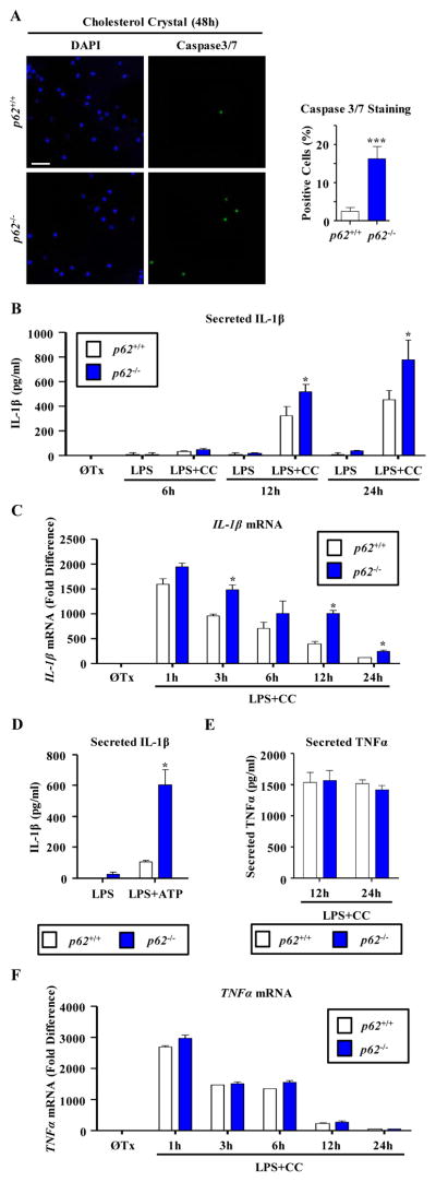
Increased inflammasome activation and IL-1β production associated with enhanced apoptosis are hallmarks of atherosclerotic lesions (22), which are also characteristic features of both autophagy-deficient macrophages (5,6) as well as neurodegenerative disorders, especially those with a substantial protein aggregation component such as Huntington’s and Parkinson’s disease (23–26). Thus, we evaluated the effect of p62 deficiency on macrophage apoptosis and IL-1β production. p62−/− macrophages were more susceptible to apoptosis after atherogenic lipid-loading (Figure 4a) and cholesterol crystals and the proinflammatory stimulus LPS synergistically increased the secretion of IL-1β from these macrophages (Figure 4b). p62−/− macrophages also showed increased transcriptional activation of IL-1β in response to LPS and cholesterol crystals (Figure 4c) and its protein product, pro-IL-1β, upon treatment with cholesterol crystals, ATP, or other inflammasome-inducing crystals such as silica and alum (Figure 4d, Supplementary Figure 7a–d). Moreover, IL-1β secretion in response to cholesterol crystal was further enhanced in p62−/−/mΦATG5−/− macrophages which are susceptible to atherogenic lipid-induced inflammasome activation (5) (Supplementary Figure 7e). The proinflammatory phenotype of p62−/− macrophages appeared to selectively increase the activation of IL-1β signaling because TNFα transcript or protein abundance was unchanged (Figure 4e,f). We concluded that p62−/− macrophages are more prone to increased apoptosis and IL-1β/inflammasome signaling, which contribute to increased atherosclerotic plaque burden and complexity.
Figure 4. p62 deficiency exacerbates atherogenic lipid-induced apoptosis and inflammatory IL-1β secretion in macrophages.
(a) Control (p62+/+) and p62−/− peritoneal macrophages were incubated with Cholesterol Crystals (CC) for 48 hours and percent of caspase 3/7-positive cells was quantified in control (n=290) and p62−/− (n=193) peritoneal macrophages in three independent experiments. Representative immunofluorescence images of Caspase 3/7 and DAPI-stained cells are shown (left) with quantification (right). (b,c) Control (p62+/+) and p62−/− peritoneal macrophages were treated with LPS +/−CC for the indicated times, cell culture media assayed for IL-1β by ELISA (n=3–4 independent wells for each treatment, b), and IL-1β transcripts were detected by quantitative PCR (c) in three independent experiments. (d) Control (p62+/+) and p62−/− peritoneal macrophages were incubated with LPS and then +/− ATP in three independent experiments. (e,f) Experiments similar to B and C were performed to detect TNFα by ELISA and quantitative PCR in three independent experiments. For all panels, data are presented as mean ± SEM. *P<0.05, ***P<0.001. Scale bar, 50 μm.
The p62-regulated pathways NRF2, ERK, p38, and NFκB do not mediate the increased IL-1β secretion from p62-deficient macrophages
The chaperone role of p62 in the selective autophagy of polyubiquitinated protein aggregates is mediated by the C-terminal ubiquitin-binding domain (27,28). However, p62 appears to also play roles in the modulation of several critical signaling pathways (29–33), thus confounding simple interpretations of the deleterious phenotypes of p62−/− macrophages. To dissect the mechanism of p62 action in macrophages, we targeted several signaling pathways affected by p62,namely nuclear factor erythroid 2-related factor 2 (NRF2), extracellular signal-regulated kinase (ERK), p38, and nuclear factor kappaB (NFκB) as the common downstream protein of atypical protein kinase Cζ (aPKCζ) and TNF receptor-associated factor 6 (TRAF-6); Supplementary Figure 8a) and analyzed the generation of IL-1β.
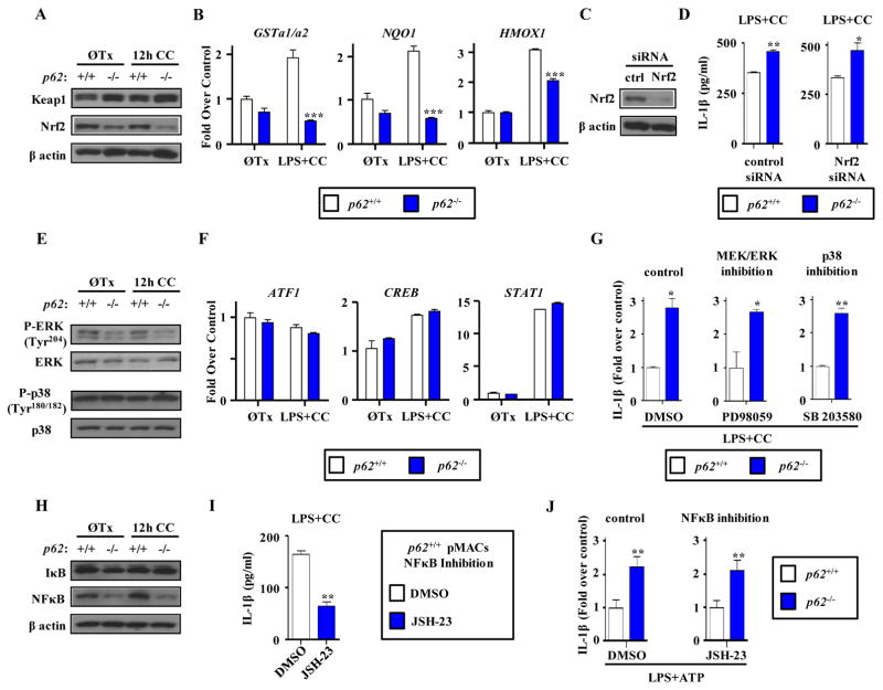
p62 activates the stress-response transcription factor NRF2 by binding to and neutralizing kelch-like ECH-associated protein 1 (KEAP1), an adaptor that facilitates NRF2 degradation through the proteasome (32). In agreement with previous reports, p62−/− macrophages had increased KEAP1 abundance, which is normally cleared by p62-dependent autophagy, and reduced NRF2 abundance (Figure 5a). Although NRF2 targets were not overtly altered in p62−/− macrophages at baseline, their transcriptional induction by co-treatment with LPS and cholesterol crystals was significantly blunted (Figure 5b). Stimulation of control and p62−/− macrophages with LPS and cholesterol crystals with NRF2 knockdown did not alter IL-1β secretion, suggesting that NRF2 was not involved (Figure 5c,d).
Figure 5. The p62-regulated pathways NRF2, ERK, p38, and NFκB are dispensible for the increased secretion of IL-1β from p62−/− macrophages.
(a,e,h) Western blot analysis of the indicated proteins in lysates from control (p62+/+) and p62−/− peritoneal macrophages following 12 hours cholesterol crystal (CC) incubation in three independent experiments. Detected residues for phospho-specific antibodies are indicated in parentheses. (b,f) Control (p62+/+) and p62−/− peritoneal macrophages were treated with LPS+CC and mRNA transcripts of NRF2 (b) or p38-dependent genes (f) were detected by quantitative PCR (n=3 independent wells for each gene). (c) Representative Western blot showing knockdown efficiency of NRF2 siRNA in three independent experiments. (d) Control (p62+/+) and p62−/− peritoneal macrophages treated with either control or NRF2 siRNA were incubated for 12 hours with LPS+CC and cell culture media assayed for IL-1β by ELISA in three independent experiments. (g,i) Control (p62+/+) and p62−/− peritoneal macrophages treated with various pharmacological pathway blockers (the MEK inhibitor PD98059, g; the p38 inhibitor SB203580, g; the NFκB inhibitor JSH-23, i) were incubated with LPS+CC and cell culture media assayed for IL-1β by ELISA in three independent experiments. (j) Control (p62+/+) and p62−/− peritoneal macrophages were incubated with LPS followed by ATP in the presence of vehicle (DMSO) or JSH-23 in three independent experiments. For ELISAs, n=3 independent wells for each condition; for all panels, data are presented as mean ± SEM. *P<0.05, **P<0.01, ***P<0.001.
Although the activation of the MEK/ERK signaling cascade has been reported to be increased in p62-deficient adipose tissue and could serve as a confounding pro-inflammatory stimulus in macrophages (29), p62−/− macrophages showed decreased activation of ERK (as gauged by phosphorylation of Tyr204) (Figure 5e) and no changes in the activity of p38 (as gauged by phosphorylation of Tyr180/182) (Figure 5e) which was also confirmed by assessing the transcript abundance of several p38-related downstream targets (Figure 5f). Furthermore, p62−/− macrophages secreted significantly higher amounts of IL-1β when we pharmacologically blocked these pathways (PD98059 to inhibit ERK and SB203580 to inhibit p38) (Figure 5g), suggesting that the ERK and p38 pathways were not involved.
NFκB is a major downstream mediator of the p62-interacting proteins aPKCζ and TRAF-6 and a key transcriptional activator of IL-1β; p62 facilitates NFκB activation (33,34). p62−/−macrophages showed reduced amounts of NFκB and no changes in IκB abundance (Figure 5h). Reduced NFκB signaling directly led to reduced IL-1β secretion (Figure 5i). To evaluate the role of NFκB more specifically in IL-1β protein processing (and not IL-1β transcription), we utilized short course LPS and ATP incubation to activate the inflammasome. Pharmacological inhibition of NFκB had no impact on the increased IL-1β secretion from p62−/− macrophages (Figure 5j).
The Ubiquitin Binding Domain of p62 is Necessary to Suppress Atherogenic Lipid-Induced IL-1β Secretion in Macrophages
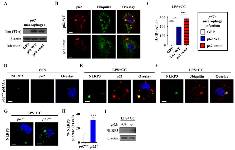
If the increase of IL-1β in p62−/− macrophages was due to an inability to sequester polyubiquitinated proteins, then selective disruption of the p62 ubiquitin-binding domain would be predicted to recapitulate the increase in IL-1β secretion in macrophages. We reconstituted p62−/− macrophages with either wild-type p62 or an I433A missense mutant of p62, which cannot bind to polyubiquitinated proteins (Figure 6a, Supplementary Figure 8b) (20). In agreement with previous reports, only the p62 mutant did not co-localize with polyubiquitinated inclusions as demonstrated by immunofluorescence analysis (Figure 6b). Moreover, reconstitution of macrophages with wild-type p62 led to significant suppression of IL-1β secretion in response to LPS and cholesterol crystal, an effect that did not occur with introduction of the p62 mutant (Figure 6c). This data demonstrates the importance of the p62 ubiquitin-binding domain in suppressing IL-1β secretion.
Figure 6. The p62 ubiquitin-binding domain is necessary to decrease secretion of IL-1β from p62−/− macrophages.
(a) Representative Western blot analysis of T2A tag after expression of constructs encoding control GFP, p62 WT or a form of p62 that cannot bind ubiquitin (umut) in p62−/− peritoneal macrophages in three independent experiments. (b) Immunofluorescence images of p62−/− peritoneal macrophages expressing either p62 WT or p62 umut constructs using antibodies against ubiquitinated proteins (FK-1) and p62. Images are representative of three independent experiments. (c) p62−/− peritoneal macrophages expressing either control GFP, p62 WT or p62 umut constructs were treated with LPS +/− CC. Cell culture media was assayed for IL-1β by ELISA (n=3 independent wells for each treatment). (d–f) Immunofluorescence images of peritoneal macrophages either without treatment (d) or after 12 hours LPS+CC treatment (e,f) using antibodies against NLRP3, p62, and ubiquitinated proteins (FK-1). Images are representative of three independent experiments. (g,h) Immunofluorescence images of control (p62+/+) and p62−/− peritoneal macrophages after LPS+CC treatment using an antibody against NLRP3 (g). Graph represents percent of cells containing NLRP3 punctae (n≥225 cells for each group; three independent experiments; h). (i) Western blot analysis of NLRP3 in LPS+CC treated cell lysates of control (p62+/+) and p62−/− peritoneal macrophages in three independent experiments. For all panels, data are presented as mean ± SEM. *P<0.05, **P<0.01, ***P<0.001. Scale bar, 5 μm.
p62 deficiency Leads to Reduced Clearance of NLRP3 Inflammasomes
Autophagy is important for clearing inflammasomes and selective delivery of ubiquitinated inflammasomes to autophagosomes involves p62 (35). We investigated whether disrupted inflammasome clearance could explain the increased IL-1β secretion in p62−/− macrophages. Immunofluorescence revealed little detectable NLRP3 or p62 in untreated macrophages (Figure 6d). Induction of the inflammasome complex with LPS and cholesterol crystals resulted in the formation of distinct NLRP3 punctae, some of which co-localized with p62 and ubiquitin (Figure 6e,f). These observations also indicate that p62-enriched polyubiquitinated aggregates have diverse cargo because only a portion are enriched in NLRP3 protein or inflammasomes in a given macrophage (Figure 6e,f).
We next assessed NLRP3 inflammasome formation in p62−/− macrophages. In contrast to polyubiquitinated proteins, NLRP3 punctae still formed in p62−/− macrophages co-incubated with LPS and cholesterol crystals (Figure 6g) and the percentage of p62−/− macrophages with readily visible NLRP3 punctae was significantly higher than control macrophages (Figure 6h). In agreement with these findings, immunoblot analysis demonstrated increased NLRP3 abundance in p62−/− macrophages (figure 6i). Taken together these data suggested that p62’s localization with and contribution in reducing NLRP3 inflammasomes was partially responsible for suppressing the inflammasome and IL-1β signaling axis in macrophages.
p62 Deficiency Leads to Increased Atherosclerotic Plaque Burden
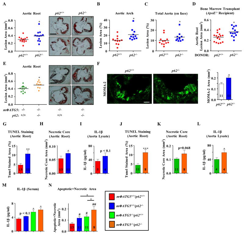
We next evaluated the impact of p62 on atherosclerosis in vivo. Western diet feeding promoted plaque formation in cohorts of p62−/− and wild-type mice, both on a proatherogenic ApoE-null background (Supplementary Figure 9). In agreement with our in vitro macrophages experiments, p62−/−/ApoE−/− mice had increased atherosclerotic plaque burden at the level of the aortic root (Figure 7a), aortic arch (Figure 7b) and whole aorta (Figure 7c). Moreover, serum metabolites (including cholesterol, triglycerides and glucose) progressively increased and were similar between both groups over the 2 month period of Western Diet feeding (Supplementary Figure 10a–c). To ascertain that the increase in atherosclerotic plaque burden was due to p62 deficiency in macrophages, we performed transplantation of bone marrow from control or p62−/− mice into ApoE−/− recipients. Those transplanted with bone marrow from p62−/− mice developed significantly increased atherosclerosis as induced through 2 months of Western diet feeding (Figure 7d).
Figure 7. p62 deficiency increases atherosclerotic plaque formation and plaque complexity.
Cohorts of control (p62+/+/mΦATG5+/+), p62−/−, mΦATG5−/− and mΦATG5−/−/p62−/− mice (all on ApoE−/− background) were fed a Western Diet for 2 months for in vivo assessment of atherosclerosis: (a–c,e) Graphs represent quantification of atherosclerotic plaque burden by computer image analysis of Oil red O-stained aortic root sections from each cohort (representative Oil red O-stained aortic roots are shown right; a,e); by en face analysis at the level of aortic arch (b) and whole aorta (c).(d) Cohorts of ApoE−/− mice transplanted with bone marrow from control (p62+/+) and p62−/− mice were fed a Western Diet for 2 months and atherosclerosis quantified as in (a). (f) Macrophage content in aortic root sections was analyzed using immunofluorescence staining using an antibody against MOMA2; representative MOMA2+ areas are shown (left). (g,h,j,k) Apoptosis and necrotic core areas of aortic root sections were determined by quantification of TUNEL immunofluorescence staining (g,j) and acellular areas respectively (h,k). (i,l) IL-1β abundance in atherosclerotic plaque lysates were determined by ELISA. (m) Graph represents IL-1β concentrations in the serum of atherosclerotic mice cohorts of indicated genotypes (n=3–4 wells pooled from at least total of 9 mice). (n) Graph represents quantification of combined apoptotic and necrotic core areas for all 4 groups. For (f–l,n), the number of mice used is shown below each figure. For all panels, data are presented as mean ± SEM. *P<0.05, **P<0.01; #P<0.05, ##P<0.01 (vs. Control). Scale bar, 0.4 mm (a,e); 100 μm (f).
Because mΦATG5−/− macrophages and aortic lysates from mΦATG5−/− mice had more insoluble p62-positive polyubiquitinated protein aggregates (Figure 1e–g, 2a,b) and that these protein aggregates were absent in mΦATG5−/−/p62−/− macrophages (Figure 3d,f), we assessed whether the absence of p62 would also have deleterious consequences in mΦATG5−/− mice. Atherosclerosis was induced by Western diet feeding in cohorts of ApoE−/−/mΦATG5−/− and ApoE−/−/mΦATG5−/−/p62−/− mice (Supplementary Figure 9). Similar to control and p62−/− mice, serum metabolic parameters including total cholesterol, triglycerides, and glucose were unchanged between the two groups (Supplementary Figure 10d–f). Compared to ApoE−/−/mΦATG5−/− mice, ApoE−/−/mΦATG5−/−/p62−/− mice had significantly increased atherosclerotic lesion area (Figure 7e). Thus, atherosclerotic plaque burden in ApoE−/−/mΦATG5−/− mice was (i) significantly higher than controls in agreement with previous reports, (ii) similar to that in p62−/− mice suggesting that p62 deficiency was as atherogenic as macrophage autophagy deficiency, and (iii) increased when combined with p62 deficiency. Together, these data suggest that when the autophagic processing of polyubiquitinated cargo is disrupted, the ability of p62 to form cellular inclusion bodies is atheroprotective.
p62 Deficiency Increases Atherosclerotic Plaque Complexity
Because the absence of p62 in atherogenic lipid-treated macrophages led to insoluble polyubiquitinated protein accumulation that was associated with increased IL-1β secretion and enhanced apoptosis (Figure 3,4), we assessed whether the pro-inflammatory and pro-apoptotic phenotype of p62−/− macrophages was recapitulated in vivo. p62−/− mice had significantly higher macrophage content than controls (Figure 7f). We next quantified several hallmarks of plaque complexity in vivo by evaluating the degree of plaque apoptosis and necrotic core formation in aortic root sections and IL-1β in whole aorta lysates. Plaques in p62−/− mice showed significantly higher amounts of apoptosis (Figure 7g, Supplementary Figure 11) and increased necrotic core area (Figure 7h) than those in control mice. Wild-type and p62−/− atherosclerotic aortas and serum did not show statistically significant changes in IL-1β and/or TNFα concentrations (Fig. 7i, Supplementary Figure 12). Moreover, mΦATG5−/−/p62−/− mice had markedly increased plaque apoptotic markers, a trend toward larger necrotic cores, and significant increases in aortic and serum IL-1β were observed (Figure 7j–m), suggesting a worsening of the pathologic features characteristic of mΦATG5-KO plaques (5,6). Quantification of the combined apoptotic and necrotic plaque regions showed that lesions in mΦATG5−/−/p62−/− mice had greater amounts of cell death than those in mΦATG5−/− or p62−/− mice (Figure 7n). This increase in plaque complexity mirrored the effects seen with lesion size (Figure 7a,e). Thus, we concluded that the main factor contributing to the increased plaque burden and complexity in p62−/− mice were increased numbers of macrophages prone to increased apoptosis and IL-1β signaling. Together, these data support a role for p62 and the formation of p62-enriched polyubiquitinated protein-containing inclusion bodies in suppressing atherogenesis.
DISCUSSION
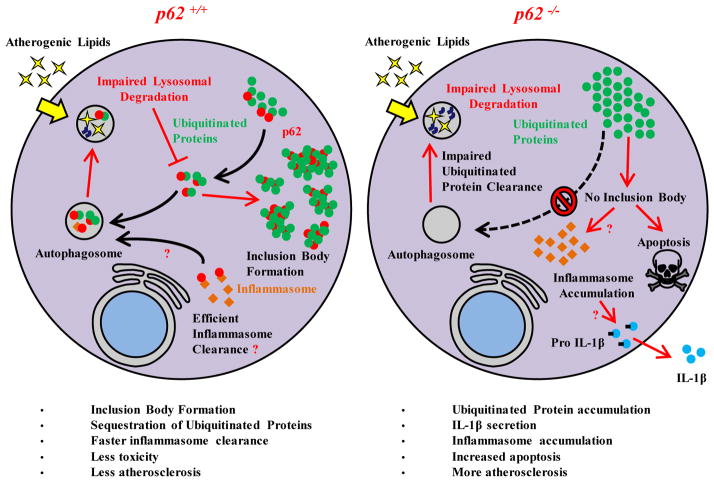
In this study, we provided evidence supporting a protective role for p62-enriched inclusion bodies in atherosclerotic macrophages and the pathogenesis of atherosclerosis. Our data suggested that the previously reported increase in p62 in atherosclerotic plaques was due to the accumulation of p62-enriched detergent insoluble polyubiquitinated protein aggregates. Atherogenic lipids induced the formation of p62-enriched protein inclusions in cultured macrophages in vitro and these inclusions were also detected in the macrophages of mouse and human atherosclerotic plaques, suggesting that protein aggregation is a previously unrecognized feature of atherosclerosis. The removal of these p62-enriched inclusions was autophagy-dependent with p62 functioning as a chaperone; the absence of p62 led to disruption in the formation of inclusion bodies and overall reduced protein solubility. p62 deficiency has functional consequences leading to increased macrophage apoptosis, inflammatory signaling (IL-1β), and the development of complex atherosclerotic plaques. We confirmed in cultured macrophages that it was p62’s ability to bind and clear ubiquitinated aggregates, including inflammasomes (and not its effect on several other signaling pathways), that likely mediated these functional effects. The critical role for p62 in autophagic removal of inclusion bodies was further supported by the observation that p62 deficiency exacerbated the proatherogenic phenotype of mice with macrophage-specific autophagy deficiency. The role of p62 and polyubiquitinated protein aggregates in atherosclerotic macrophages is summarized in Figure 8.
Figure 8. Schematic depiction of the links between macrophage p62, polyubiquitinated protein inclusion body formation, and atherosclerosis.
p62 is essential for the clearance of polyubiquitinated proteins by autophagy. Although atherogenic lipids impair this degradative process by affecting lysosomal degradation, the subsequent formation of p62-positive inclusion bodies are less cytotoxic, which is at least partly due to more efficient p62-dependent clearance of inflammasomes (left). In the absence of p62, inclusion bodies cannot form leading to the accumulation of poorly formed polyubiquitinated protein aggregates and increased numbers of inflammasomes. These defects in turn initiate deleterious cellular pathways such as IL-1β secretion and apoptosis which exacerbate atherosclerotic progression (right).
The development of p62-enriched protein aggregates in atherosclerosis is reminiscent of those described in neurodegenerative disorders. Tau-enriched Neurofibrillary tangles in Alzheimer’s Disease, α-synuclein-enriched Lewy Bodies in Parkinson’s Disease, and inclusion bodies composed of mutant polynucleotide repeat proteins in Huntington’s Disease and Amyotrophic Lateral Sclerosis (9,10,36–38) appear to require autophagy-mediated degradation (20,39–42). The progressive accumulation and defective clearance of these protein aggregates are associated with observed increases in cytotoxicity, inflammatory signaling, and neurodegeneration (23,43). Although the formation of inclusion bodies is regarded as indispensable for neurodegeneration, the notion that aggregate formation is a direct contributor to cytotoxicity has been challenged. For example, work in Huntington’s Disease demonstrates that p62 promotes the formation of protein aggregates composed of mutant Huntingtin; in its absence, cytotoxicity and cell death occurs (20,44). Studies of amyloid proteins of various types suggest that pre-fibrillar aggregates rather than mature amyloid fibrils contribute most heavily to cytotoxicity. Our observation that p62−/− macrophages in atherosclerotic mice did not form inclusion bodies and showed increased protein insolubility, concomitant with increases in cell death and inflammation, support the concept that inclusion bodies are more likely a protective cellular response.
Although the identity of the pathogenic protein in protein aggregation disorders is often known (for example Tau, α-synuclein, Huntingtin, α1-antitrypsin), the identity of the proteins in the inclusion bodies of the atherosclerotic plaque are not known. Insoluble lipid oxidation products called ceroid have been described in atherosclerotic macrophages (45), but our observation of poor co-localization between lipid markers and p62-enriched inclusion bodies preclude this possibility. One interesting lead is the inflammasome protein complex itself. We found that some of the inflammasome complexes (detected as NLRP3+ punctae) that were induced and activated by cholesterol crystals in macrophages co-localized with both p62 and ubiquitin. This appeared to be critical for the eventual clearance of these complexes (and dampening of IL-1β secretion) because the absence of p62 resulted in significantly increased NLRP3+ punctae and IL-1β. The ubiquitination and p62- and autophagy-mediated clearance of inflammasomes has been described previously by another group and is a component of the regulation of the IL-1β response (35). Aside from ubiquitin-mediated clearance, there is also evidence that formation of the inflammasome complex itself undergoes prion-like polymerization (46). It would be interesting to see whether the p62-dependent sequestration of protein aggregates into large inclusions could also serve to modulate this polymerization process. Going beyond the candidate approach, systematic identification of p62-bound polyubiquitinated proteins and protein aggregates could provide critical insights into the pathogenesis of atherosclerosis. For example, our immunoblot analysis revealed that certain insoluble polyubiquitinated proteins were prominent in p62-bound aggregates and potentially amenable to mass spectrometric identification.
The role of p62 in mammalian pathophysiology appears to be context-dependent, especially if one considers the phenotypes of autophagy and p62 deficiency in the liver (21). Disruption of hepatocyte autophagy results in formation of cytoplasmic protein inclusions and liver injury, which is surprisingly rescued by concomitant genetic ablation of p62. In this regard, the p62-dependent induction of detoxifying enzymes mediated by activation of NRF2 has been implicated in the liver phenotype rather than the link between p62, protein aggregation, and liver degeneration. As discussed above, in addition to serving as a chaperone for the transport of polyubiquitinated proteins to autophagosomes, p62 activates several other pathways including NRF2 signaling by disrupting the interaction of NRF2 with KEAP1, an adaptor that facilitates the proteasomal degradation of NRF2 (21,47). Indeed, we confirmed that a similar relationship between p62, KEAP1, and NRF2 existed in macrophages (Figure 5a). However, it was unlikely that the proatherogenic phenotype of p62−/− mice is a result of relative NRF2 deficiency since NRF2−/− mice have reduced atherosclerosis (48,49) and targeting the NRF2 pathway in cultured macrophages either had no effect (Figure 5d) or blunted cholesterol crystal-mediated inflammasome activation (50). Furthermore, our assessment of the other prominent p62-modulated pathways demonstrated unchanged (p38) or blunted (ERK and NFκB) signaling (Figure 5e–j). Thus, the ubiquitin-independent functions of p62 are likely to play a minor role in its atheroprotective effect.
In atherosclerosis and macrophages from atherosclerotic plaques, protein aggregation is a marker of disease progression but this appears to be a protective response in minimizing cytotoxicity and inflammation. Our data support the notion that methods of enhancing the removal of aggregates are likely to be a more fruitful atheroprotective measure than reducing their formation.
MATERIALS AND METHODS
Animals
Protocols were approved by the Washington University Animal Studies Committee. All mice used in this study were in C57BL/6 background (>N7). Mice with the floxed ATG5 locus (51) were crossed with Cre-recombinase transgenic mice under the control of the Lysosomal-M promoter to generate macrophage specific ATG5 knock-out (mΦATG5−/−) mice (5). p62−/− mice were as described previously (21). Crosses between mΦATG5−/−, p62−/−, and ApoE−/− mice generated mice with p62−/−,, p62−/−/mΦATG5−/− or littermate controls on a ApoE−/− background. p62, ATG5, Cre, and ApoE genotyping was performed as described previously (21,51,52). Mice housed in a specific pathogen-free barrier facility were weaned at 3 weeks of age to a standard mouse chow providing 6% calories as fat, then started on a Western-type diet containing 0.15% cholesterol providing 42% calories as fat (TD 88137, Harlan, Madison, WI) at ~2 months of age.
Human Carotid Samples
The material removed at the time of carotid endarterectomy was de-identified and collected as part of a Washington University Institutional Review Board approved protocol. Samples were immediately placed in cold saline and kept refrigerated on ice for transport to the laboratory. Samples were then rapidly dissected into atherosclerotic regions and adjacent regions with minimal atherosclerotic plaque and snap frozen in liquid nitrogen or placed in tissue freezing medium for Western blotting or sectioning respectively.
Macrophage Culture and Treatment
Macrophages were isolated as previously described (53). Briefly, mice were injected intraperitoneally with 4% thioglycollate media (Sigma, T9032) and 4 days later, peritoneal macrophages were isolated, washed, counted, and plated in DMEM supplemented with 10% FBS. Macrophages treated with LPS (100 ng/ml, Invivogen), cholesterol crystals (500 μg/ml), oxidized-LDL (50 μg/ml), silica crystals (200 μg/ml, Invivogen), alum (100 μg/ml, Invivogen), bafilomycin (100 nM, Sigma B1793), chloroquine (10 μM, Sigma C6628), PD98059 (30 μM, EMD Millipore 513000), SB 203580 (10 μM, EMD Millipore 559389), and/or JSH-23 (25 μM, EMD Millipore 481408) were harvested at various times for mRNA or protein using standard techniques or were fixed with 4% paraformaldehyde for immunofluorescence microscopy. Cholesterol crystals were generated by subjecting cholesterol powder (Sigma, C8667) to an ethanol precipitation technique as described (54,55). Oxidized LDL was generated by Cu2+-oxidation of LDL (Sigma, L7914) as described (56).
Lentiviral Transduction and siRNA Knock-down
Wild-type or mutant p62 (C-terminally tagged with the T2A sequence) was overexpressed in cultured macrophages using the lentiviral expression system pCDH-MCS-T2A-Puro-MSCV (system Biosciences, CD522A-1) essentially as described (57). The p62 ubiquitin-binding mutant (p62 umut) was generated using standard polymerase chain reaction (PCR)-based methods to introduce an I433A point mutation that was confirmed by sequencing. Lentivirus was generated in 293T cells by co-transfection of the p62-containing transfer vector with packaging (psPAX2) and envelope (pMD2.G) vectors using standard calcium phosphate precipitation method. Harvested lentivirus was first concentrated (Clontech Takara Bio, 631231), added to cultured macrophages twice (6 hours and 24 hours respectively), and production confirmed by Western blot of the T2A tag (using an antibody against 2A peptide).
To knock-down NRF2, SMARTpool ON-TARGETplus Mouse NRF2 and control (non-targeting) siRNA was used (Dharmacon Inc, L-040766-00-0005 and D-001810-10-05 respectively). siRNAs were transfected into macrophages using DeliverX Plus (Affymetrix Inc, DX0052) per manufacturer’s protocols and knock-down confirmed by Western blotting of NRF2.
Preparation of Soluble and Insoluble Fractions and Western Blotting
Cells were lysed in a standard lysis buffer containing 1% Triton X-100 (Cell Signaling, #9803), sonicated and centrifuged at 17000 × g for 15 min. Supernatant was defined as the soluble fraction while the pellet (insoluble fraction) was washed with lysis buffer, suspended in boiling SDS-PAGE loading buffer, and sonicated for protein solubilization. For mouse aorta or human carotid tissues, a Glas-Col homogenizer was first used to disrupt cells followed by a similar lysis protocol. Protein quantification, separation, transfer, and blotting was performed as described (58) using the following antibodies: p62 (abcam, ab56416), β-actin (Sigma-Aldrich, A2066), polyubiquitinated proteins (FK2, Millipore, 04-263), 2A peptide (Millipore, ABS31), NRF2 (Santa Cruz Biotechnology, sc-13032), KEAP1 (Proteintech, 10503-2-AP), ERK1 (Santa Cruz Biotechnology, sc-94), p-ERK (Santa Cruz Biotechnology, sc-7383), p38α/β (Santa Cruz Biotechnology, sc-7972), p-p38 (Santa Cruz Biotechnology, sc-17852-R), IκB-α (Santa Cruz Biotechnology, sc-371), NLRP3 (R&D Systems, MAB7578), Caspase-1 (Santa Cruz Biotechnology, M-20), IL-1β (R&D Systems, AF-401-NA).
Immunofluorescence Microscopy
Immunofluorescence imaging of frozen-tissue sections and peritoneal macrophages was performed as previously described (58). For all imaging of cultured macrophages, at least two to three mice of each genotype was used in each experiment and experiments were performed at least three times. Where images needed quantification, the number of cells quantified is included in the figure legend. For non-quantified images, a representative image was chosen after analysis of n>10 cells. For all imaging of frozen-tissue sections, a representative image was chosen after analysis of three sections per aortic root from at least six mice. Specificity of observed staining was tested in parallel experiments omitting primary or secondary antibodies or using samples from mice with genetic deficiencies where available. The following primary antibodies were used: p62 (Progen Biotechnik, GP62-C), polyubiquitinated proteins (FK1, Enzo Life Sciences, BML-PW8805), MOMA-2 (AbD Serotec, MCA519C), LAMP2 (Abcam, GL2A7), NLRP3 (R&D Systems, MAB7578), EEA1 (BD Biosciences, 610457), Calnexin (Santa Cruz Biotech, SC6465), GM130 (BD Biosciences, 610822), PMP70 (Sigma, P0090), Vimentin (Abcam, ab45939). Species-specific fluorescent secondary antibodies were obtained from Invitrogen/Life technologies. MitoTracker Red CMXRos (Life Technologies, M7512) was used per manufacturer’s protocol. CellEvent Caspase-3/7 Green Detection Reagent (Life Technologies, C10423) and DeadEnd Fluorometric TUNEL System (Promega, G3250) were used per manufacturer’s protocol. Imaging was performed using a Zeiss LSM-700 Confocal microscope; regions of interest and thresholds were determined and signal over the threshold was quantified using ZEN microscope software (Carl Zeiss Microscopy).
Analytical Procedures and Lesion Quantification
Secreted IL-1β and TNF-α from peritoneal macrophage culture media were measured using ELISA assays from R&D Systems (MLB00B) and BD Biosciences (560478). Cholesterol, triglycerides, and glucose were assayed in serum following a 6-hour fast per manufacturer’s protocols (Thermo Scientific TR13421, TR22421, and TR15408). Proteasome activity was assayed in macrophages using the Proteasome-Glo luminescent kit per manufacturer’s protocol (Promega G8660). For assessment of transcripts, extraction of total RNA, reverse transcription, and quantitative RT-PCR was performed as described (59). Sequences for the oligonucleotide primers used were designed using the qPrimerDepot database- http://primerdepot.nci.nih.gov. Assays were performed in duplicate and normalized to amplified mRNA for the ribosomal protein 36B4.
The en face and aortic root cross-section techniques previously described (60) were used to quantify atherosclerosis throughout the aorta as well as at the vessel origin. En face regions analyzed included the aortic arch, thoracic aorta, and abdominal aorta (spanning from the aortic valve to the bifurcation of the iliac arteries).
Bone Marrow Transplantation
Transplantation and assessment of atherosclerosis was essentially as described (61). Briefly, marrow was isolated from the femurs/tibias of 2 month old wild-type and p62−/− mice (both on an ApoE−/− background), subjected to erythrocyte lysis, and unfractionated bone marrow cells were counted to obtain a defined concentration. A cohort of ApoE−/− recipient mice were lethally irradiated with 10 Gy from a cesium-137 γ-irradiator and were reconstituted with retro-orbital injection of ~2 × 10⁶ donor marrow within 4 hours. Donor marrow was allowed to repopulate for 3 weeks before recipient mice were placed on a Western-type diet for 8 weeks followed by quantification of aortic root atherosclerosis.
Statistical Analyses
Statistical significance of differences was calculated using the Student unpaired t test or ANOVA (for multiple groups) followed by Tukey’s multiple comparison test for parametric data (serum metabolites and in vitro macrophage experiments except qPCR) or the Mann-Whitney U test for non-parametric data (qPCR and atherosclerosis quantitation). Graphs containing error bars show the mean +/− standard error of the mean (SEM). Statistical significance is represented as follows: *P < 0.05, **P < 0.01, ***P < 0.001.
Supplementary Material
Supplementary Figure 1. Effect of atherogenic lipids and other lysosomal inhibitors on p62 transcription and the formation of p62-enriched inclusion bodies.
Supplementary Figure 2. Autophagy deficiency exacerbates atherogenic lipid-induced p62 accumulation.
Supplementary Figure 3. p62 aggregates induced by atherogenic lipids do not colocalize with organelle or lipid markers.
Supplementary Figure 4. Atherogenic lipid loading leads to p62-enriched polyubiquitinated protein accumulation as early as 6 hours in macrophages.
Supplementary Figure 5. Densitometric quantification of ubiquitin immunoblots of human carotid endarterectomy specimens.
Supplementary Figure 6. Proteasome activity is not induced in mΦATG5−/− and p62−/− peritoneal macrophages.
Supplementary Figure 7. The effect of p62 deficiency on the IL-1β signaling axis.
Supplementary Figure 8. Cartoons summarizing p62-regulated pathways and p62-reconstituting constructs.
Supplementary Figure 9. Chart summarizing experimental protocol and mice cohorts used for in vivo assessment of atherosclerosis.
Supplementary Figure 10. Serum metabolic parameters of control, p62−/−, mΦATG5−/−, and mΦATG5−/−/p62−/− mice are similar after 2 months of Western diet feeding.
Supplementary Figure 11. p62 deficiency increases plaque apoptosis.
Supplementary Figure 12. Serum TNFα concentrations are not increased in p62−/− mice fed a high-fat diet.
Supplementary Figure 13. Representative Ponceau S stains of ubiquitin immunoblots in the manuscript.
Acknowledgments
We thank Dr. Herbert Virgin for providing the p62−/− mice, Drs. Katsu Funai, Irfan Lodhi, and Rithwick Rajagopal for providing antibody reagents to stain intracellular organelles, Dr. Joel Schilling for providing reagents to assess the macrophage inflammasome, and the technical assistance provided by Ms. Joan Avery (confocal microscopy), Ms. Karyn Holloway (mouse husbandry), Dr. Trey Coleman (frozen tissue sectioning), and Dr. Xiaochao Wei and Ms. Li Yin (bone marrow transplantation).
Funding: This work was supported by 5K08HL098559, 1R01HL125838, 1R01AG037120, the Foundation for Barnes-Jewish Hospital, and the Washington University Diabetic Cardiovascular Disease Center.
Footnotes
Author contributions: I.S. and B.R. designed the research. I.S. performed and analyzed the experiments and prepared the figures. S.B., R.E., E.E., C.J.S., and T.D.E. assisted in collection and analysis of mouse samples. B.A. and J.A.C. collected human carotid specimens and assisted in their analysis. I.S. and B.R. wrote the manuscript.
Competing interests: The authors declare that they have no competing interests.
REFERENCES AND NOTES
- 1.Levine B, Kroemer G. Autophagy in the pathogenesis of disease. Cell. 2008;132:27–42. doi: 10.1016/j.cell.2007.12.018. [DOI] [PMC free article] [PubMed] [Google Scholar]
- 2.Singh R, Kaushik S, Wang Y, Xiang Y, Novak I, Komatsu M, Tanaka K, Cuervo AM, Czaja MJ. Autophagy regulates lipid metabolism. Nature. 2009;458:1131–1135. doi: 10.1038/nature07976. [DOI] [PMC free article] [PubMed] [Google Scholar]
- 3.Sumpter R, Jr, Levine B. Autophagy and innate immunity: triggering, targeting and tuning. Semin Cell Dev Biol. 2010;21:699–711. doi: 10.1016/j.semcdb.2010.04.003. [DOI] [PMC free article] [PubMed] [Google Scholar]
- 4.Levine B, Mizushima N, Virgin HW. Autophagy in immunity and inflammation. Nature. 2011;469:323–335. doi: 10.1038/nature09782. [DOI] [PMC free article] [PubMed] [Google Scholar]
- 5.Razani B, Feng C, Coleman T, Emanuel R, Wen H, Hwang S, Ting JP, Virgin HW, Kastan MB, Semenkovich CF. Autophagy links inflammasomes to atherosclerotic progression. Cell Metab. 2012;15:534–544. doi: 10.1016/j.cmet.2012.02.011. [DOI] [PMC free article] [PubMed] [Google Scholar]
- 6.Liao X, Sluimer JC, Wang Y, Subramanian M, Brown K, Pattison JS, Robbins J, Martinez J, Tabas I. Macrophage autophagy plays a protective role in advanced atherosclerosis. Cell Metab. 2012;15:545–553. doi: 10.1016/j.cmet.2012.01.022. [DOI] [PMC free article] [PubMed] [Google Scholar]
- 7.Ouimet M, Franklin V, Mak E, Liao X, Tabas I, Marcel YL. Autophagy regulates cholesterol efflux from macrophage foam cells via lysosomal acid lipase. Cell Metab. 2011;13:655–667. doi: 10.1016/j.cmet.2011.03.023. [DOI] [PMC free article] [PubMed] [Google Scholar]
- 8.Sergin I, Razani B. Self-eating in the plaque: what macrophage autophagy reveals about atherosclerosis. Trends Endocrinol Metab. 2014;25:225–234. doi: 10.1016/j.tem.2014.03.010. [DOI] [PMC free article] [PubMed] [Google Scholar]
- 9.Ross CA, Poirier MA. Opinion: What is the role of protein aggregation in neurodegeneration? Nat Rev Mol Cell Biol. 2005;6:891–898. doi: 10.1038/nrm1742. [DOI] [PubMed] [Google Scholar]
- 10.Eisenberg D, Jucker M. The amyloid state of proteins in human diseases. Cell. 2012;148:1188–1203. doi: 10.1016/j.cell.2012.02.022. [DOI] [PMC free article] [PubMed] [Google Scholar]
- 11.Perlmutter DH. Alpha-1-antitrypsin deficiency: importance of proteasomal and autophagic degradative pathways in disposal of liver disease-associated protein aggregates. Annu Rev Med. 2011;62:333–345. doi: 10.1146/annurev-med-042409-151920. [DOI] [PubMed] [Google Scholar]
- 12.Askanas V, Engel WK, Nogalska A. Inclusion body myositis: a degenerative muscle disease associated with intra-muscle fiber multi-protein aggregates, proteasome inhibition, endoplasmic reticulum stress and decreased lysosomal degradation. Brain Pathol. 2009;19:493–506. doi: 10.1111/j.1750-3639.2009.00290.x. [DOI] [PMC free article] [PubMed] [Google Scholar]
- 13.Jerome WG. Lysosomes, cholesterol and atherosclerosis. Clinical lipidology. 2010;5:853–865. doi: 10.2217/clp.10.70. [DOI] [PMC free article] [PubMed] [Google Scholar]
- 14.Moore KJ, Sheedy FJ, Fisher EA. Macrophages in atherosclerosis: a dynamic balance. Nat Rev Immunol. 2013;13:709–721. doi: 10.1038/nri3520. [DOI] [PMC free article] [PubMed] [Google Scholar]
- 15.Duewell P, Kono H, Rayner KJ, Sirois CM, Vladimer G, Bauernfeind FG, Abela GS, Franchi L, Nunez G, Schnurr M, Espevik T, Lien E, Fitzgerald KA, Rock KL, Moore KJ, Wright SD, Hornung V, Latz E. NLRP3 inflammasomes are required for atherogenesis and activated by cholesterol crystals. Nature. 2010;464:1357. doi: 10.1038/nature08938. [DOI] [PMC free article] [PubMed] [Google Scholar]
- 16.Emanuel R, Sergin I, Bhattacharya S, Turner JN, Epelman S, Settembre C, Diwan A, Ballabio A, Razani B. Induction of lysosomal biogenesis in atherosclerotic macrophages can rescue lipid-induced lysosomal dysfunction and downstream sequelae. Arterioscler Thromb Vasc Biol. 2014;34:1942–1952. doi: 10.1161/ATVBAHA.114.303342. [DOI] [PMC free article] [PubMed] [Google Scholar]
- 17.Kim PK, Hailey DW, Mullen RT, Lippincott-Schwartz J. Ubiquitin signals autophagic degradation of cytosolic proteins and peroxisomes. Proc Natl Acad Sci U S A. 2008;105:20567–20574. doi: 10.1073/pnas.0810611105. [DOI] [PMC free article] [PubMed] [Google Scholar]
- 18.Kubli DA, Gustafsson AB. Mitochondria and mitophagy: the yin and yang of cell death control. Circ Res. 2012;111:1208–1221. doi: 10.1161/CIRCRESAHA.112.265819. [DOI] [PMC free article] [PubMed] [Google Scholar]
- 19.Kopito RR. Aggresomes, inclusion bodies and protein aggregation. Trends Cell Biol. 2000;10:524–530. doi: 10.1016/s0962-8924(00)01852-3. [DOI] [PubMed] [Google Scholar]
- 20.Bjorkoy G, Lamark T, Brech A, Outzen H, Perander M, Overvatn A, Stenmark H, Johansen T. p62/SQSTM1 forms protein aggregates degraded by autophagy and has a protective effect on huntingtin-induced cell death. J Cell Biol. 2005;171:603–614. doi: 10.1083/jcb.200507002. [DOI] [PMC free article] [PubMed] [Google Scholar]
- 21.Komatsu M, Waguri S, Koike M, Sou YS, Ueno T, Hara T, Mizushima N, Iwata J, Ezaki J, Murata S, Hamazaki J, Nishito Y, Iemura S, Natsume T, Yanagawa T, Uwayama J, Warabi E, Yoshida H, Ishii T, Kobayashi A, Yamamoto M, Yue Z, Uchiyama Y, Kominami E, Tanaka K. Homeostatic levels of p62 control cytoplasmic inclusion body formation in autophagy-deficient mice. Cell. 2007;131:1149–1163. doi: 10.1016/j.cell.2007.10.035. [DOI] [PubMed] [Google Scholar]
- 22.Tabas I. Macrophage death and defective inflammation resolution in atherosclerosis. Nat Rev Immunol. 2010;10:36–46. doi: 10.1038/nri2675. [DOI] [PMC free article] [PubMed] [Google Scholar]
- 23.Masters SL, O’Neill LA. Disease-associated amyloid and misfolded protein aggregates activate the inflammasome. Trends Mol Med. 2011;17:276–282. doi: 10.1016/j.molmed.2011.01.005. [DOI] [PubMed] [Google Scholar]
- 24.Zuccato C, Valenza M, Cattaneo E. Molecular mechanisms and potential therapeutical targets in Huntington’s disease. Physiol Rev. 2010;90:905–981. doi: 10.1152/physrev.00041.2009. [DOI] [PubMed] [Google Scholar]
- 25.Venderova K, Park DS. Programmed cell death in Parkinson’s disease. Cold Spring Harb Perspect Med. 2012;2 doi: 10.1101/cshperspect.a009365. [DOI] [PMC free article] [PubMed] [Google Scholar]
- 26.Nixon RA, Yang DS. Autophagy and neuronal cell death in neurological disorders. Cold Spring Harb Perspect Biol. 2012;4 doi: 10.1101/cshperspect.a008839. [DOI] [PMC free article] [PubMed] [Google Scholar]
- 27.Komatsu M, Ichimura Y. Physiological significance of selective degradation of p62 by autophagy. FEBS Lett. 2010;584:1374–1378. doi: 10.1016/j.febslet.2010.02.017. [DOI] [PubMed] [Google Scholar]
- 28.Lin X, Li S, Zhao Y, Ma X, Zhang K, He X, Wang Z. Interaction domains of p62: a bridge between p62 and selective autophagy. DNA Cell Biol. 2013;32:220–227. doi: 10.1089/dna.2012.1915. [DOI] [PubMed] [Google Scholar]
- 29.Rodriguez A, Duran A, Selloum M, Champy MF, Diez-Guerra FJ, Flores JM, Serrano M, Auwerx J, Diaz-Meco MT, Moscat J. Mature-onset obesity and insulin resistance in mice deficient in the signaling adapter p62. Cell Metab. 2006;3:211–222. doi: 10.1016/j.cmet.2006.01.011. [DOI] [PubMed] [Google Scholar]
- 30.Muller TD, Lee SJ, Jastroch M, Kabra D, Stemmer K, Aichler M, Abplanalp B, Ananthakrishnan G, Bhardwaj N, Collins S, Divanovic S, Endele M, Finan B, Gao Y, Habegger KM, Hembree J, Heppner KM, Hofmann S, Holland J, Kuchler D, Kutschke M, Krishna R, Lehti M, Oelkrug R, Ottaway N, Perez-Tilve D, Raver C, Walch AK, Schriever SC, Speakman J, Tseng YH, Diaz-Meco M, Pfluger PT, Moscat J, Tschop MH. p62 links beta-adrenergic input to mitochondrial function and thermogenesis. J Clin Invest. 2013;123:469–478. doi: 10.1172/JCI64209. [DOI] [PMC free article] [PubMed] [Google Scholar]
- 31.Duran A, Linares JF, Galvez AS, Wikenheiser K, Flores JM, Diaz-Meco MT, Moscat J. The signaling adaptor p62 is an important NF-kappaB mediator in tumorigenesis. Cancer Cell. 2008;13:343–354. doi: 10.1016/j.ccr.2008.02.001. [DOI] [PubMed] [Google Scholar]
- 32.Komatsu M, Kurokawa H, Waguri S, Taguchi K, Kobayashi A, Ichimura Y, Sou YS, Ueno I, Sakamoto A, Tong KI, Kim M, Nishito Y, Iemura S, Natsume T, Ueno T, Kominami E, Motohashi H, Tanaka K, Yamamoto M. The selective autophagy substrate p62 activates the stress responsive transcription factor Nrf2 through inactivation of Keap1. Nat Cell Biol. 2010;12:213–223. doi: 10.1038/ncb2021. [DOI] [PubMed] [Google Scholar]
- 33.Sanz L, Diaz-Meco MT, Nakano H, Moscat J. The atypical PKC-interacting protein p62 channels NF-kappaB activation by the IL-1-TRAF6 pathway. Embo J. 2000;19:1576–1586. doi: 10.1093/emboj/19.7.1576. [DOI] [PMC free article] [PubMed] [Google Scholar]
- 34.Wooten MW, Seibenhener ML, Mamidipudi V, Diaz-Meco MT, Barker PA, Moscat J. The atypical protein kinase C-interacting protein p62 is a scaffold for NF-kappaB activation by nerve growth factor. J Biol Chem. 2001;276:7709–7712. doi: 10.1074/jbc.C000869200. [DOI] [PubMed] [Google Scholar]
- 35.Shi CS, Shenderov K, Huang NN, Kabat J, Abu-Asab M, Fitzgerald KA, Sher A, Kehrl JH. Activation of autophagy by inflammatory signals limits IL-1beta production by targeting ubiquitinated inflammasomes for destruction. Nat Immunol. 2012;13:255–263. doi: 10.1038/ni.2215. [DOI] [PMC free article] [PubMed] [Google Scholar]
- 36.Irvine GB, El-Agnaf OM, Shankar GM, Walsh DM. Protein aggregation in the brain: the molecular basis for Alzheimer’s and Parkinson’s diseases. Mol Med. 2008;14:451–464. doi: 10.2119/2007-00100.Irvine. [DOI] [PMC free article] [PubMed] [Google Scholar]
- 37.Arrasate M, Finkbeiner S. Protein aggregates in Huntington’s disease. Exp Neurol. 2012;238:1–11. doi: 10.1016/j.expneurol.2011.12.013. [DOI] [PMC free article] [PubMed] [Google Scholar]
- 38.Mori K, Weng SM, Arzberger T, May S, Rentzsch K, Kremmer E, Schmid B, Kretzschmar HA, Cruts M, Van Broeckhoven C, Haass C, Edbauer D. The C9orf72 GGGGCC repeat is translated into aggregating dipeptide-repeat proteins in FTLD/ALS. Science. 2013;339:1335–1338. doi: 10.1126/science.1232927. [DOI] [PubMed] [Google Scholar]
- 39.Wooten MW, Hu X, Babu JR, Seibenhener ML, Geetha T, Paine MG, Wooten MC. Signaling, polyubiquitination, trafficking, and inclusions: sequestosome 1/p62’s role in neurodegenerative disease. J Biomed Biotechnol. 2006;2006:62079. doi: 10.1155/JBB/2006/62079. [DOI] [PMC free article] [PubMed] [Google Scholar]
- 40.Gal J, Strom AL, Kilty R, Zhang F, Zhu H. p62 accumulates and enhances aggregate formation in model systems of familial amyotrophic lateral sclerosis. J Biol Chem. 2007;282:11068–11077. doi: 10.1074/jbc.M608787200. [DOI] [PubMed] [Google Scholar]
- 41.Ramesh Babu J, Lamar Seibenhener M, Peng J, Strom AL, Kemppainen R, Cox N, Zhu H, Wooten MC, Diaz-Meco MT, Moscat J, Wooten MW. Genetic inactivation of p62 leads to accumulation of hyperphosphorylated tau and neurodegeneration. J Neurochem. 2008;106:107–120. doi: 10.1111/j.1471-4159.2008.05340.x. [DOI] [PubMed] [Google Scholar]
- 42.Watanabe Y, Tatebe H, Taguchi K, Endo Y, Tokuda T, Mizuno T, Nakagawa M, Tanaka M. p62/SQSTM1-dependent autophagy of Lewy body-like alpha-synuclein inclusions. PLoS One. 2012;7:e52868. doi: 10.1371/journal.pone.0052868. [DOI] [PMC free article] [PubMed] [Google Scholar]
- 43.Stefani M, Dobson CM. Protein aggregation and aggregate toxicity: new insights into protein folding, misfolding diseases and biological evolution. J Mol Med (Berl) 2003;81:678–699. doi: 10.1007/s00109-003-0464-5. [DOI] [PubMed] [Google Scholar]
- 44.Arrasate M, Mitra S, Schweitzer ES, Segal MR, Finkbeiner S. Inclusion body formation reduces levels of mutant huntingtin and the risk of neuronal death. Nature. 2004;431:805–810. doi: 10.1038/nature02998. [DOI] [PubMed] [Google Scholar]
- 45.Mitchinson MJ, Ball RY, Carpenter KL, Enright JH. Macrophages and ceroid in human atherosclerosis. Eur Heart J. 1990 Nov;(Suppl E):116–121. doi: 10.1093/eurheartj/11.suppl_e.116. [DOI] [PubMed] [Google Scholar]
- 46.Cai X, Chen J, Xu H, Liu S, Jiang QX, Halfmann R, Chen ZJ. Prion-like polymerization underlies signal transduction in antiviral immune defense and inflammasome activation. Cell. 2014;156:1207–1222. doi: 10.1016/j.cell.2014.01.063. [DOI] [PMC free article] [PubMed] [Google Scholar]
- 47.Lau A, Wang XJ, Zhao F, Villeneuve NF, Wu T, Jiang T, Sun Z, White E, Zhang DD. A noncanonical mechanism of Nrf2 activation by autophagy deficiency: direct interaction between Keap1 and p62. Mol Cell Biol. 2010;30:3275–3285. doi: 10.1128/MCB.00248-10. [DOI] [PMC free article] [PubMed] [Google Scholar]
- 48.Sussan TE, Jun J, Thimmulappa R, Bedja D, Antero M, Gabrielson KL, Polotsky VY, Biswal S. Disruption of Nrf2, a key inducer of antioxidant defenses, attenuates ApoE-mediated atherosclerosis in mice. PLoS One. 2008;3:e3791. doi: 10.1371/journal.pone.0003791. [DOI] [PMC free article] [PubMed] [Google Scholar]
- 49.Barajas B, Che N, Yin F, Rowshanrad A, Orozco LD, Gong KW, Wang X, Castellani LW, Reue K, Lusis AJ, Araujo JA. NF-E2-related factor 2 promotes atherosclerosis by effects on plasma lipoproteins and cholesterol transport that overshadow antioxidant protection. Arterioscler Thromb Vasc Biol. 2011;31:58–66. doi: 10.1161/ATVBAHA.110.210906. [DOI] [PMC free article] [PubMed] [Google Scholar]
- 50.Freigang S, Ampenberger F, Spohn G, Heer S, Shamshiev AT, Kisielow J, Hersberger M, Yamamoto M, Bachmann MF, Kopf M. Nrf2 is essential for cholesterol crystal-induced inflammasome activation and exacerbation of atherosclerosis. Eur J Immunol. 2011;41:2040–2051. doi: 10.1002/eji.201041316. [DOI] [PubMed] [Google Scholar]
- 51.Hara T, Nakamura K, Matsui M, Yamamoto A, Nakahara Y, Suzuki-Migishima R, Yokoyama M, Mishima K, Saito I, Okano H, Mizushima N. Suppression of basal autophagy in neural cells causes neurodegenerative disease in mice. Nature. 2006;441:885–889. doi: 10.1038/nature04724. [DOI] [PubMed] [Google Scholar]
- 52.Chakravarthy MV, Pan Z, Zhu Y, Tordjman K, Schneider JG, Coleman T, Turk J, Semenkovich CF. “New” hepatic fat activates PPARalpha to maintain glucose, lipid, and cholesterol homeostasis. Cell Metab. 2005;1:309–322. doi: 10.1016/j.cmet.2005.04.002. [DOI] [PubMed] [Google Scholar]
- 53.Febbraio M, Podrez EA, Smith JD, Hajjar DP, Hazen SL, Hoff HF, Sharma K, Silverstein RL. Targeted disruption of the class B scavenger receptor CD36 protects against atherosclerotic lesion development in mice. J Clin Invest. 2000;105:1049–1056. doi: 10.1172/JCI9259. [DOI] [PMC free article] [PubMed] [Google Scholar]
- 54.Rajamaki K, Lappalainen J, Oorni K, Valimaki E, Matikainen S, Kovanen PT, Eklund KK. Cholesterol crystals activate the NLRP3 inflammasome in human macrophages: a novel link between cholesterol metabolism and inflammation. PLoS One. 2010;5:e11765. doi: 10.1371/journal.pone.0011765. [DOI] [PMC free article] [PubMed] [Google Scholar]
- 55.Whiting MJ, Watts JM. Cholesterol crystal formation and growth in model bile solutions. J Lipid Res. 1983;24:861–868. [PubMed] [Google Scholar]
- 56.Steinbrecher UP, Witztum JL, Parthasarathy S, Steinberg D. Decrease in reactive amino groups during oxidation or endothelial cell modification of LDL. Correlation with changes in receptor-mediated catabolism. Arteriosclerosis. 1987;7:135–143. doi: 10.1161/01.atv.7.2.135. [DOI] [PubMed] [Google Scholar]
- 57.Hwang S, Maloney NS, Bruinsma MW, Goel G, Duan E, Zhang L, Shrestha B, Diamond MS, Dani A, Sosnovtsev SV, Green KY, Lopez-Otin C, Xavier RJ, Thackray LB, Virgin HW. Nondegradative role of Atg5-Atg12/Atg16L1 autophagy protein complex in antiviral activity of interferon gamma. Cell Host Microbe. 2012;11:397–409. doi: 10.1016/j.chom.2012.03.002. [DOI] [PMC free article] [PubMed] [Google Scholar]
- 58.Razani B, Engelman JA, Wang XB, Schubert W, Zhang XL, Marks CB, Macaluso F, Russell RG, Li M, Pestell RG, Di Vizio D, Hou H, Jr, Kneitz B, Lagaud G, Christ GJ, Edelmann W, Lisanti MP. Caveolin-1 null mice are viable but show evidence of hyperproliferative and vascular abnormalities. J Biol Chem. 2001;276:38121–38138. doi: 10.1074/jbc.M105408200. [DOI] [PubMed] [Google Scholar]
- 59.Razani B, Zhang H, Schulze PC, Schilling JD, Verbsky J, Lodhi IJ, Topkara VK, Feng C, Coleman T, Kovacs A, Kelly DP, Saffitz JE, Dorn GW, 2nd, Nichols CG, Semenkovich CF. Fatty acid synthase modulates homeostatic responses to myocardial stress. J Biol Chem. 2011;286:30949–30961. doi: 10.1074/jbc.M111.230508. [DOI] [PMC free article] [PubMed] [Google Scholar]
- 60.Semenkovich CF, Coleman T, Daugherty A. Effects of heterozygous lipoprotein lipase deficiency on diet-induced atherosclerosis in mice. J Lipid Res. 1998;39:1141–1151. [PubMed] [Google Scholar]
- 61.Schneider JG, Finck BN, Ren J, Standley KN, Takagi M, Maclean KH, Bernal-Mizrachi C, Muslin AJ, Kastan MB, Semenkovich CF. ATM-dependent suppression of stress signaling reduces vascular disease in metabolic syndrome. Cell Metab. 2006;4:377–389. doi: 10.1016/j.cmet.2006.10.002. [DOI] [PubMed] [Google Scholar]
Associated Data
This section collects any data citations, data availability statements, or supplementary materials included in this article.
Supplementary Materials
Supplementary Figure 1. Effect of atherogenic lipids and other lysosomal inhibitors on p62 transcription and the formation of p62-enriched inclusion bodies.
Supplementary Figure 2. Autophagy deficiency exacerbates atherogenic lipid-induced p62 accumulation.
Supplementary Figure 3. p62 aggregates induced by atherogenic lipids do not colocalize with organelle or lipid markers.
Supplementary Figure 4. Atherogenic lipid loading leads to p62-enriched polyubiquitinated protein accumulation as early as 6 hours in macrophages.
Supplementary Figure 5. Densitometric quantification of ubiquitin immunoblots of human carotid endarterectomy specimens.
Supplementary Figure 6. Proteasome activity is not induced in mΦATG5−/− and p62−/− peritoneal macrophages.
Supplementary Figure 7. The effect of p62 deficiency on the IL-1β signaling axis.
Supplementary Figure 8. Cartoons summarizing p62-regulated pathways and p62-reconstituting constructs.
Supplementary Figure 9. Chart summarizing experimental protocol and mice cohorts used for in vivo assessment of atherosclerosis.
Supplementary Figure 10. Serum metabolic parameters of control, p62−/−, mΦATG5−/−, and mΦATG5−/−/p62−/− mice are similar after 2 months of Western diet feeding.
Supplementary Figure 11. p62 deficiency increases plaque apoptosis.
Supplementary Figure 12. Serum TNFα concentrations are not increased in p62−/− mice fed a high-fat diet.
Supplementary Figure 13. Representative Ponceau S stains of ubiquitin immunoblots in the manuscript.


