Figure 3.
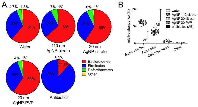
Relative abundance of bacterial phyla from mouse cecal contents after 28 days of oral gavage with 10 mg/kg bw/day of 110 nm, 20 nm citrate-coated, or 20 nm PVP-coated AgNP in comparison to 28 days oral gavage with water or 12 days of antibiotic (cefoperazone) administration in the drinking water. A. Mean % relative abundance of predominant phyla for mice from each group. All AgNP and water- dosed groups had a preponderance of Bacteroidetes and Firmicutes, with lesser numbers of Deferribacteres. Other phyla (combined) were present at <2% in each group and consisted of unclassified phyla, Actinobacteria, Proteobacteria, Tenericutes, Verrucomicrobia, and TM7. B. Median (horizontal bar), interquartile range (box), and minimum/maximum for major phyla identified in all groups. Only the antibiotics-treated group had significant differences from other groups, consisting of increased Firmicutes and decreased Bacteroidetes (ANOVA, Tukey’s multiple comparisons test, p<0.0001). N=5–6 for water or AgNP groups (see text). Only two points are shown for the antibiotics group since only two animals had sufficient recovery of gut bacterial DNA for sequencing remaining after antibiotic dosing, despite the 24 hour washout period.

