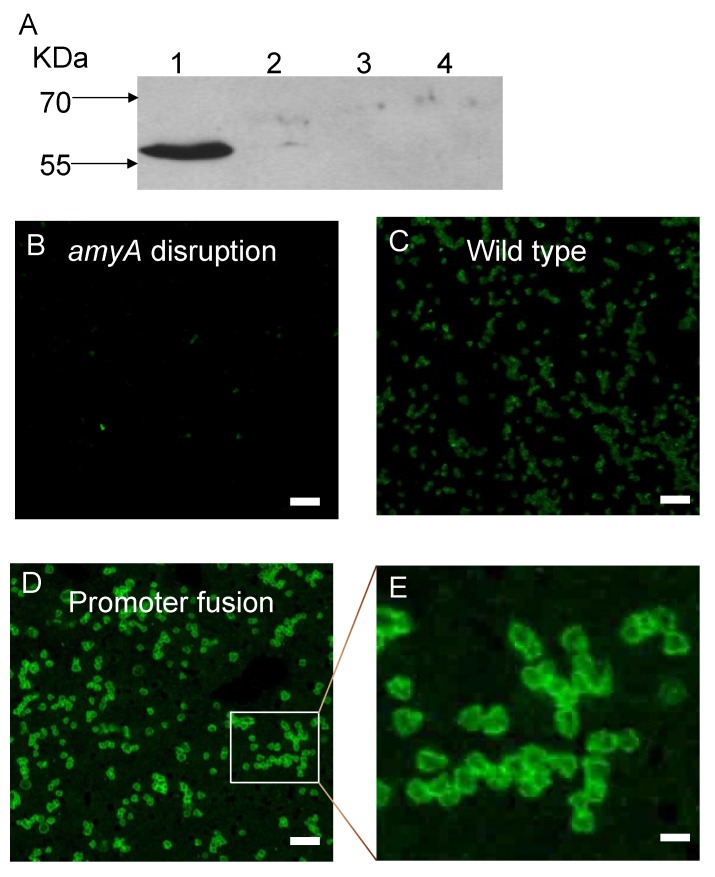
Figure 7.
Panel A, Western blot analysis of β-glycosidase (LacS) from S. solfataricus wild-type strain, 98/2. Samples were: cytoplasmic fractions (Lane 1), membrane fractions (Lane 2), S-layer fractions (Lane 3); and supernatant (Lane 4). Samples were loaded in an amount equivalent to 1.0 OD540 of cells. The western blot was probed with a 1:5000 dilution of the anti-LacS antibody, followed by a 1:5000 dilution of HRP goat anti-rabbit IgG secondary antibody. Immunofluorescence analysis of cell-associated α-Amylase. Fixed cells were treated with anti-α-Amylase antibodies conjugated to Alexafluor dye. Panel B. Cells lacking the α-amylase gene. Panel C. Wild type cells. Panel D. Cells encoding the α-amylase promoter fusion. Panel E. Inset of panel C. Bars in Panels A–C 5 microns, bar in panel D 0.5 microns.

