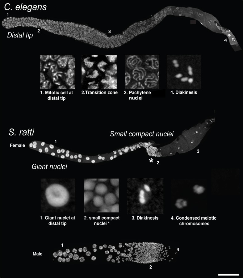
Fig. 2.
Comparisons between the C. elegans and Strongyloides gonads. Top: a DAPI-stained dissected C. elegans hermaphroditic (adult) gonad, showing progression of germ cells in the germline (distal tip is to the left). The numbers 1–4 indicate the immediate insets below, with each inset showing the characteristic morphology (mitotically dividing cells at distal tip, crescent-shaped nuclei at transition zone, “bowl of spaghetti” in the pachytene zone and condensed chromosomes at diakinesis, respectively) of germ nuclei for those regions. Bottom: DAPI-stained dissected gonads from S. ratti adult females (top) and males (bottom) showing the completely different gonadal organization in comparison to C. elegans. Note the shorter but broad nature of the S. ratti male gonad in comparison to the female (adult males are smaller in size to adult females; adults are approximately 28–30 h post-culturing). Here, the entire distal arm is occupied by intensely staining giant nuclei, followed by a band of small compact nuclei at the gonadal loop (asterisk). Except for even more strongly condensed small nuclei in males, the organization is identical in both sexes. Insets 1 and 2 are derived from female gonads. The band of small nuclei is followed proximally in females by nuclei, which might be in diakinesis (shown in inset 3 taken from a female germline) and in males with condensed presumably meiotic chromosomes (inset 4, taken from a male germline). Scale bar 50 μm

