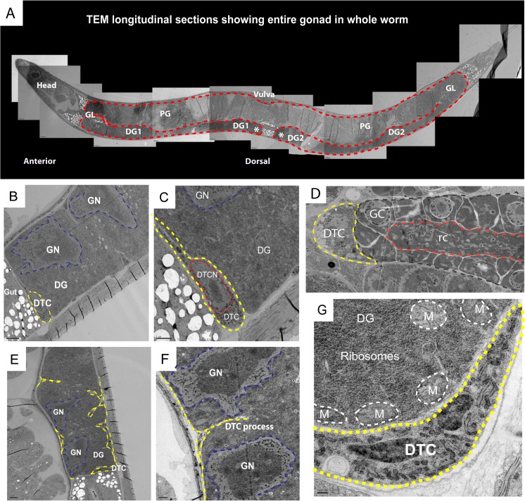
Fig. 3.
Transmission electron microscopy (TEM) of the S. papillosus free-living female gonad. a Semithin longitudinal TEM sections of a S. papillosus female showing the entire gonad (outlined in red) in the body of the adult worm, with the vulva (top, central). The two distal gonad arms are marked as DG1 and DG2 (distal tips are marked by asterisks); the gonadal loop is labeled as GL and the proximal gonad as PG. Female adults that were approximately 28–30 h post-fecal culturing were used for this analysis. b TEM section showing the distal tip of the gonad, DG, with giant nuclei, GN (outlined in blue), and the distal tip cell, DTC (in yellow). c Zoom in of the DTC (yellow) showing its nucleus, DTCN (in red) in addition to the giant nuclei, GN (in blue) in the distal gonad, DG. d TEM section from Pristionchus pacificus showing the DTC (outlined in yellow) sitting as a cap on the distal tip (outlined in gray), with germ cells GC, around a central rachis “rc” (in red). This organization is similar to what is found in C. elegans (image courtesy of Metta Riebesell). e The distal tip cell and its processes (outlined in yellow) making contact with the distal gonad at regular intervals. f Magnified view showing a DTC process (in yellow) contacting the distal gonad close to two giant nuclei GN (in blue). g Zoom in at the distal tip (with DTC outlined in yellow) showing the high density of ribosomes (electron dense regions) and mitochondria, M (outlined in white) in the distal gonad, DG

