Abstract
David Kasner, MD (1927–2001), used his extensive dissections of eye bank eyes and experiences in teaching cataract surgery to resident physicians to realize that excision of vitreous when present in the anterior chamber of eyes undergoing cataract surgery was preferable to prior intraoperative procedures. Noting that eyes tolerated his maneuvers, he then performed planned subtotal open-sky vitrectomies; first on a traumatized eye in 1961, then on two eyes of patients with amyloidosis (1966–1967). The success of these operations was noted by others, most particularly Robert Machemer, MD. Kasner’s work directly led to further surgical developments, including closed pars plana vitrectomy.
Keywords: history of ophthalmology, vitrectomy, retinal surgery, retina
The Early Years
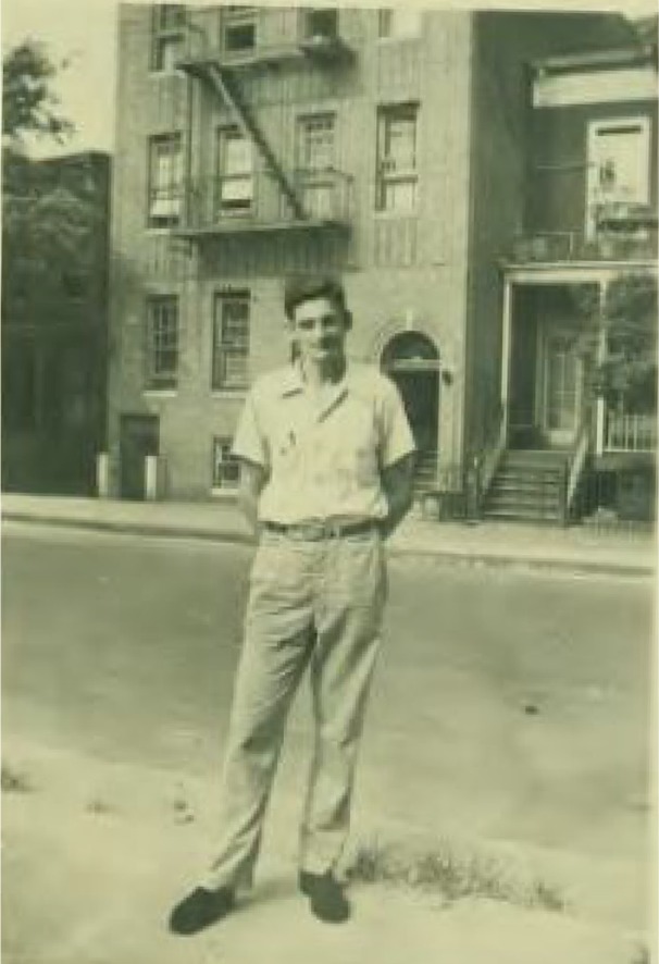
David Kasner was born in New York City (NYC) on August 11, 1927, to Russian Jewish immigrant parents (Fig. 1). His father was a tailor who worked relentlessly to support his family. Kasner spoke Yiddish as a first language, grew to an imposing 6′8″ and graduated from Morris High School in the Bronx’s rough-and-tumble streets. After one year of city college, Kasner was drafted into the U.S. Army (February 1946). His nonconformist nature clashed with Army routine and southern culture during his 18-month stint in the Army Air Corps in Biloxi, MS (Louis Kasner, MD, written communication, April 2013).
Figure 1.

Kasner as a teen in New York City (provided with permission, courtesy of Joan Kasner).
After serving his country, Kasner returned to college in New York for more studies. Nonconformity may have intervened again when he chose to transfer to Tulane University in New Orleans, LA, where he earned his BS degree in 1950. He immediately enrolled at Tulane University Medical School and received his MD degree in 1954.
It is unclear as to why Kasner decided to become an ophthalmologist, but he may have been markedly affected by the traumatic injury that blinded his brother Phil’s left eye sometime in their teen years. After an internship at Maimonides Hospital in NYC, Kasner spent a year at the University of Pennsylvania (UPA), where he enrolled in a basic science course in ophthalmology.
That year at UPA must have counted for one of the three years’ residency training required by the American Board of Ophthalmology for certification at that time (John Clarkson, MD, written communication, February 2014). Kasner then found his way to Chicago where he served as a resident at the University of Illinois Eye and Ear Infirmary 1956–1958.
Landing in Miami: Early Years in Practice
Kasner sought out private practice opportunities in far-flung sites. After considering Los Angeles, Seattle, and New York, he finally chose Miami, joining Harry Horwich, MD, in private practice in the adjacent suburb of Coral Gables.
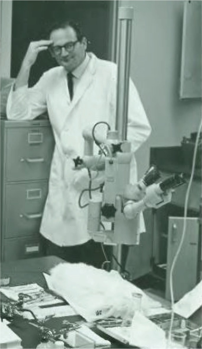
When Edward Norton, MD, began the Ophthalmology Department (Bascom Palmer Eye Institute) at the University of Miami in 1962, Kasner joined the voluntary faculty, helping teach cataract surgery to resident physicians at the Miami Veterans Administration Hospital (Fig. 2). While at Bascom Palmer, Kasner began to avail himself of its eye bank eyes to perform autopsy dissections (Fig. 2). Norton allowed and encouraged Kasner’s research curiosity (Jerome Fisher, MD, written communication, September 2013).
Figure 2.

Kasner in the Bascom Palmer Eye Institute pathology laboratory (provided with permission, courtesy of Joan Kasner).
Garage Research
Interestingly, Kasner performed most of his dissections in his garage at his Coral Gables home. (Many years later, the city rebuked him for always leaving his car in the driveway, as his laboratory consumed all the parking space inside.) (Jerome Fisher, MD, oral communication, September 2013).
Kasner’s private garage laboratory had air conditioning, microscopes, cameras, and dissection tables to help him tease apart the mysteries of limbus anatomy and other anterior segment structures relevant to cataract surgery–in the hopes of finding safer techniques and better outcomes for such surgeries. At that time, almost all such procedures were done with a large superior entry wound at the limbus to allow for removal of the lens and its capsule en toto (intracapsular cataract extraction).
A major intraoperative complication of intracapsular extraction was vitreous “loss.” Vitreous gel would move forward into the anterior chamber, the incision, or even out of the eye after the crystalline lens had been removed.1 Cystoid macular edema (CME) would develop frequently; that often meant substantial and permanent central visual loss. Vitreous loss also raised the rate of subsequent retinal detachment. Three strategies were used to deal with this event.2 Often the wound was closed, and the surgeon would “hope for the best.” Castroviejo introduced an additional step of sweeping vitreous out of the closed wound. Maumanee recommended closing the wound and aspirating liquid vitreous from the mid-vitreous cavity in hopes of encouraging the displaced gel to fall back and away from the wound.
An apparent barrier to Kasner’s exploratory work ultimately provided the impetus for a new technique and a solution to this age-old problem. The laboratory eyes from the eye banks were embedded in a clear acrylic resin that turned the vitreous cloudy, spoiling its appearance and hindering a clear view of the anterior segment. Kasner learned that he could remove the vitreous using scissors and blunt forceps. Later, he utilized wadded-up paper to engage the vitreous (Jerome Fisher, MD, written communication, September 2013). This technique not only solved Kasner’s immediate visibility problem for his research but also opened the door for a solution to a persistent surgical problem.
Research Becomes Real Life
Kasner realized that his extramural laboratory studies gave him a different answer to the surgical problem of vitreous loss. During cataract extraction, he started using cellulose sponges to engage a portion of the displaced vitreous gel, cutting it free, and removing it from the surgical field. He would repeat this over and over until the remaining vitreous was at or behind the plane of the pupil. Then he would close the cataract incision in the usual manner. He called this technique radical anterior vitrectomy.3
Kasner became profoundly convinced that vitreous could be removed safely from the eye. He tested his radical theory in 1961 on an eight-year-old boy whose eye was injured traumatically by a tent stake. The eye suffered a horizontal corneal laceration with loss of intraocular contents and retained lens material. Kasner operated on the child on July 28, 1961, an unspecified time after the initial surgical repair of the laceration. Kasner opened the eye with a very large limbal incision of more than 300°. This allowed the cornea to be folded back and gave access to the pupil and vitreous cavity. Kasner used his cellulose sponge/scissors technique for subtotal removal of the vitreous (and retained lens material), then closed the corneal incision. Even decades later, the patient retained 20/50 corrected vision (Jerome Fisher, MD, oral and written communications, September 2014).
Kasner performed two more cases of planned subtotal vitrectomy in 1966 and 1967, both on patients who had developed opacified vitreous secondary to amyloidosis. Those patients were operated on at Jackson Memorial Hospital in Miami. Gordon Miller, MD, chief resident at Bascom Palmer Eye Institute at the time, assisted on each case (Gordon Miller, MD, oral communication, October 2013). The operations consisted of the 300° corneal incision, intracapsular extraction of the crystalline lens, and forceps/cellulose sponge scissors subtotal vitrectomy, followed by limbal wound closure. The operating microscope (not typically used by eye surgeons at the time) and general anesthesia were utilized for the operations, which lasted about 1.5 hours. Both patients experienced marked vision improvement: one from hand motions to 20/30 and the other from 20/100 to 20/40. This technique was termed “open-sky vitrectomy.” The results were presented at the annual meeting of the American Academy of Ophthalmology and Otolaryngology in 1967 and published in 1968.4
Kasner used his “open-sky” technique on his own patients, as well as the patients he operated on at Miami Veterans Administration Medical Center, where Kasner supervised cataract surgery performed by resident physicians from the Bascom Palmer Eye Institute/University of Miami (Howard Lieberman, MD, oral communication, September 2013). Postoperative follow-up of those patients seemed to show less CME than those treated with prior techniques. Additionally, there did not appear to be any substantial untoward effects such as an increased rate of subsequent retinal detachment.
Radical Dedication
Kasner’s technique led to many eye surgeons calling for advice. Often Kasner would help other surgeons in the community repair complications that occurred at the initial procedure.
Throughout his career, Kasner videotaped many of his surgical procedures. This allowed him to see how complications developed. His tapes also augmented his lectures and were a great teaching tool for residents to view.
Kasner’s nonconformist attitude persisted throughout his life. For example, when a surgical technologist handed him the wrong kind of scissors, he did not ask for the right kind. Instead, he turned to the resident he was assisting and said something like, “Let’s do the surgery with this instead and pretend that the surg tech had dropped the right scissors–because you never know when that may happen.” (Christopher Blodi, MD, firsthand observation, January 1982)
Publications (or their lack)
Kasner’s published case series of 21 patients5 in 1971, which was a long-delayed written report of the technique that he had started performing around 1960. During intervening years, Kasner frequently gave presentations of his technique for “vitreous loss” at Bascom Palmer Eye Institute’s grand rounds and annual meetings of the prestigious Welsh Cataract Congress.
Regrettably, Kasner didn’t publish most of his findings. Perhaps his busy private practice responsibilities prohibited it. Some speculate that Kasner may have been dyslexic (Jerome Fisher, MD, oral communication, September 2013). Still, a few presentations found their way into print.3,6 However, the clear evidence that an eye could tolerate removal of virtually all vitreous piqued the interest of a young retinal surgeon on Bascom Palmer’s full-time faculty: Robert Machemer, MD.
Springboard for Further Development
More than intrigued by Kasner’s grand rounds discussions on vitrectomy surgery, Machemer and his fellow at the time (Helmut Buettner, MD) visited Kasner at his garage laboratory. They watched and photographed Kasner perform open-sky vitrectomy on an eye bank eye (Helmut Buettner, MD, oral communication, December 2013).
Machemer tried Kasner’s open-sky technique on rabbits; and then, in 1970, on a patient with diabetic vitreous hemorrhage. Instead of using cellulose sponges and scissors, Machemer removed the vitreous with a battery-powered, hollowed-out drill with a side opening, which allowed the drill bit’s rotation to cut small pieces of vitreous that could then be manually aspirated through the drill. He added an infusion tube attached to the side of the cutting device to allow the eye to stay formed as the vitreous was removed.7,8
Additionally, Machemer decided that a less stressful approach to the vitreous cavity would be through a pars plana incision. Machemer performed his first pars plana vitrectomy on a patient with a diabetic vitreous hemorrhage on April 20, 1970. After suffering for five years with 2/200 vision, the patient regained 20/50 vision.9
In those days, leading-edge medical devices were largely one of a kind: custom made, not mass produced. Such was the case with Machemer’s “vitrectomy machine” [or “vitreous nibbler,” later officially termed vitreous infusion suction cutter (VISC)]. Jean-Marie Parel, the bioengineer who built all of Machemer’s equipment, worked full-time at Bascom Palmer, as did Machemer. Although Kasner only had a clinical appointment at Bascom and did not operate there except to help teach cataract surgery to residents, Kasner frequented Parel’s laboratory to talk and to teach–both eye anatomy and eye surgery ideas–using his garage-laboratory cadaver eyes as illustrations. Parel deeply appreciated this, so he would give Kasner each prior-generation VISC every time he built a slightly more advanced one for Machemer (Jean-Marie Parel, PhD, oral communication, October 2013). And so Kasner started performing pars plana vitrectomies as well.
Later Life
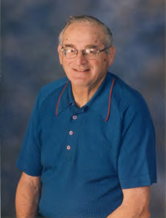
Kasner continued his professional life as he always had: in private practice in Coral Gables and teaching residents at the Miami Veterans Administration how to do initial cataract operations (Fig. 3). Later in life, Kasner’s son, Louis, joined him in practice; and the two occasionally operated together. Kasner continued to experiment in his home laboratory, trying to find a way to chemically dissolve vitreous, but never finding the answer (Louis Kasner, written communication, April 2013).
Figure 3.

Kasner in later years (provided with permission, courtesy of Joan Kasner).
Kasner had few hobbies outside of ophthalmology. He met his wife, Joan, through ophthalmology. She was the sister of a Bascom Palmer resident who introduced the two. Kasner enjoyed fishing in later years, often with his brother Sam who had moved down to south Florida upon retirement (Louis Kasner, written communication, April 2013).
In 1975, Kasner was diagnosed with chronic lymphocytic leukemia. He went through years of chemotherapy and a splenectomy but continued to work in spite of developing chronic anemia. He worked until the last week of his life. Kasner died of a ruptured aortic aneurysm on January 6, 2001 (Louis Kasner, written communication, April 2013).
Conclusion
David Kasner’s approaches to vitreous removal, initially as a technique to deal with cataract surgery complications, and later to perform intentional, subtotal removal, proved safe and effective. His work led others to more fully advance vitrectomy surgery.
Was Kasner really the first?
Kasner historically has been credited as the originator of open-sky vitrectomy. However, in the murky past, a similar operation had been performed years earlier in Hiroshima, Japan. Tsugio Dodo, MD, used a technique he termed “diapupillary resection” for subtotal removal of a vitreous hemorrhage from a patient. His results were published in 1955 in a Japanese journal.** However, it strains credulity to think that Kasner knew of this work. The article was published in Japanese and was not referenced by any [Western] retinal surgery article or textbook until many decades later. In fact, it is unclear whether Dodo performed the operation on any other patients. It is also unclear as to why the technique did not gain popularity in Japan in subsequent years (Kirk Packo, MD; written communication February 2014).
Dodo T. Diapupillary Resection of Vitreous Opacity. Nippon Ganka Gakkai Zasshi. 1955;59: 1737–45.
Acknowledgments
Many thanks to Joan Kasner for sharing the photographs of her husband and her account of Kasner’s personal life. Also, thanks to Louis Kasner (David Kasner’s son) for his contributions to the details of Kasner’s personal life. Credit goes to Kirk Packo, MD, for the sidebar content. Packo learned about Dodo from Dr. Yasuo Tano (a famous, now-deceased Japanese retinal surgeon). Finally, thanks go to Lana Christian of CreateWrite Inc. for lending her editing skills to this paper.
Footnotes
ACADEMIC EDITOR: Joshua Cameron, Editor in Chief
PEER REVIEW: Three peer reviewers contributed to the peer review report. Reviewers’ reports totaled 574 words, excluding any confidential comments to the academic editor.
FUNDING: Author discloses no external funding sources.
COMPETING INTERESTS: Author discloses no potential conflicts of interest.
Paper subject to independent expert blind peer review. All editorial decisions made by independent academic editor. Upon submission manuscript was subject to anti-plagiarism scanning. Prior to publication all authors have given signed confirmation of agreement to article publication and compliance with all applicable ethical and legal requirements, including the accuracy of author and contributor information, disclosure of competing interests and funding sources, compliance with ethical requirements relating to human and animal study participants, and compliance with any copyright requirements of third parties. This journal is a member of the Committee on Publication Ethics (COPE).
Author Contributions
Conceived and designed the experiments: CFB. Analyzed the data: CFB. Wrote the first draft of the manuscript: CFB. Contributed to the writing of the manuscript: CFB. Agree with manuscript results and conclusions: CFB. Jointly developed the structure and arguments for the paper: CFB. Made critical revisions and approved final version: CFB. The author reviewed and approved of the final manuscript.
REFERENCES
- 1.Vail D. After results of vitreous loss. In: Theodore FH, editor. Complications After Cataract Surgery. Boston, MA: Little, Brown; 1964. pp. 9–33. [DOI] [PubMed] [Google Scholar]
- 2.Jaffe NS. Cataract Surgery and its Complications. St. Louis, MO: CV Mosby; 1981. pp. 266–7. [Google Scholar]
- 3.Boyd B, Kasner D. Vitrectomy: a new approach to the management of vitreous [personal interview] Highl Ophthalmol. 1969;11:304–9. [Google Scholar]
- 4.Kasner D, Miller GR, Taylor WH, Sever RJ, Norton EW. Surgical treatment of amyloidosis of the vitreous. Trans Am Acad Ophthalmol Otolaryngol. 1968;72:410–8. [PubMed] [Google Scholar]
- 5.Cerasoli JR, Kasner D. A follow-up study of vitreous loss during cataract surgery managed by anterior vitrectomy. Am J Ophthalmol. 1971;71:1040–3. doi: 10.1016/0002-9394(71)90572-1. [DOI] [PubMed] [Google Scholar]
- 6.Kasner D. Vitreous loss during cataract surgery. Trans Am Acad Ophthamol Otolaryngol. 1973;77(2):O184–5. [PubMed] [Google Scholar]
- 7.Machemer R. The development of pars plana vitrectomy: a personal account. Graefe’s Arch Clin Exp Ophthalmol. 1995;233:453–468. doi: 10.1007/BF00183425. [DOI] [PubMed] [Google Scholar]
- 8.Anderson WB., Jr Excerpt: Oral history of Robert Machemer, MD Foundation of the American Academy of Ophthalmology Museum of Vision & Ophthalmic Heritage. 2009. [Accessed May 17, 2016]. Available at: http://museumofvision.org/dynamic/files/uploaded_files_filename_109.pdf.
- 9.Machemer R, Buettner H, Norton EWD, Parel JM. Vitrectomy: a pars plana approach. Trans Am Acad Ophthalmol Otolaryngol. 1973;75:813–20. [PubMed] [Google Scholar]


