

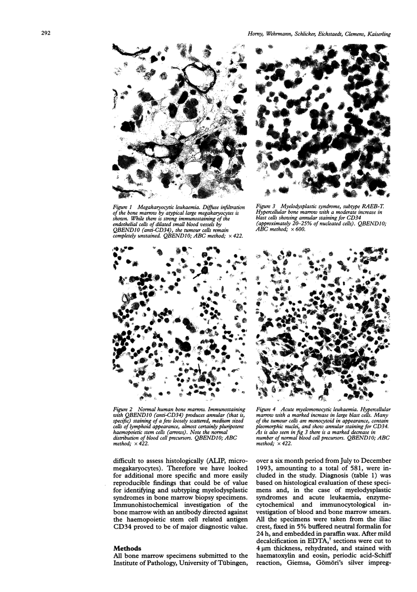
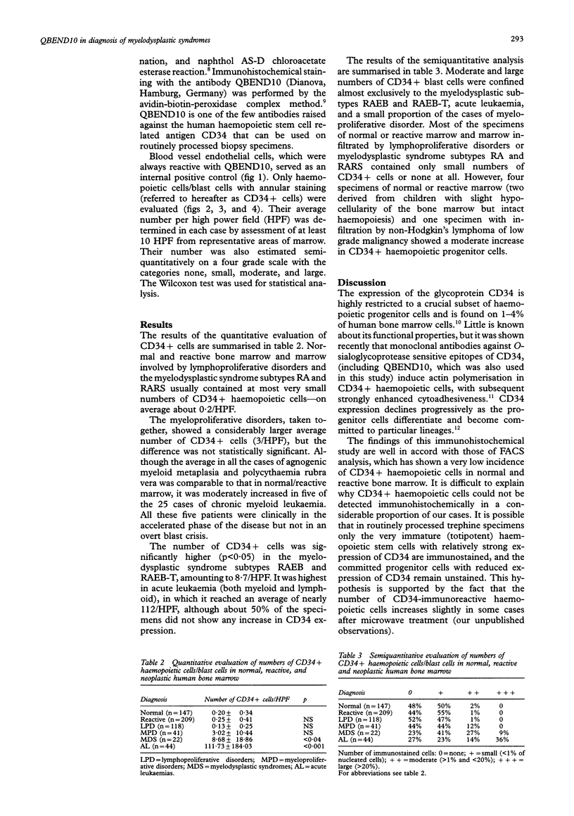
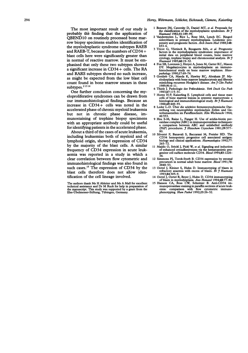
Abstract
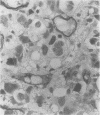
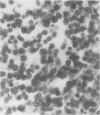
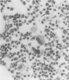
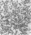
AIM--The assessment of the value of the antibody QBEND10, which is directed against the haemopoietic stem cell related antigen CD34, in the immunohistochemical diagnosis of myelodysplastic syndrome in routinely processed bone marrow biopsy specimens. METHODS--581 formalin fixed, paraffin embedded trephine biopsy specimens of the iliac crest were immunostained with QBEND10 (avidin-biotin complex/ABC method). The number of CD34+ haemopoietic stem cells/blast cells (referred to hereafter as CD34+ cells) was determined in each case. The Wilcoxon test was used for statistical analysis. RESULTS--The following diagnostic categories were defined: (1) normal or reactive bone marrow (n = 356), (2) lymphoproliferative disorders, usually non-Hodgkin's lymphoma of low grade malignancy or multiple myeloma (n = 118), (3) myelodysplastic syndrome (n = 22), (4) acute leukaemia (n = 44), and (5) myeloproliferative diseases (n = 41). The average number of CD34+ cells was very low (0.2/HPF) in normal and reactive bone marrow, in lymphoproliferative disorders and in the myelodysplastic syndrome subtypes RA and RARS. Myeloproliferative diseases showed an average of three CD34+ cells/HPF. However, the average number of CD34+ cells was significantly higher (p < 0.05) in the myelodysplastic syndrome subtypes RAEB and RAEB-T (8.7/HPF) and in acute leukaemia (including both myeloid and lymphoblastic leukaemia; 111.7/HPF). CONCLUSIONS--QBEND10 is of value for the identification of RAEB and RAEB-T in routinely processed bone marrow biopsy specimens because it enables the detection of even small increases in the number of CD34+ cells.
Full text
PDF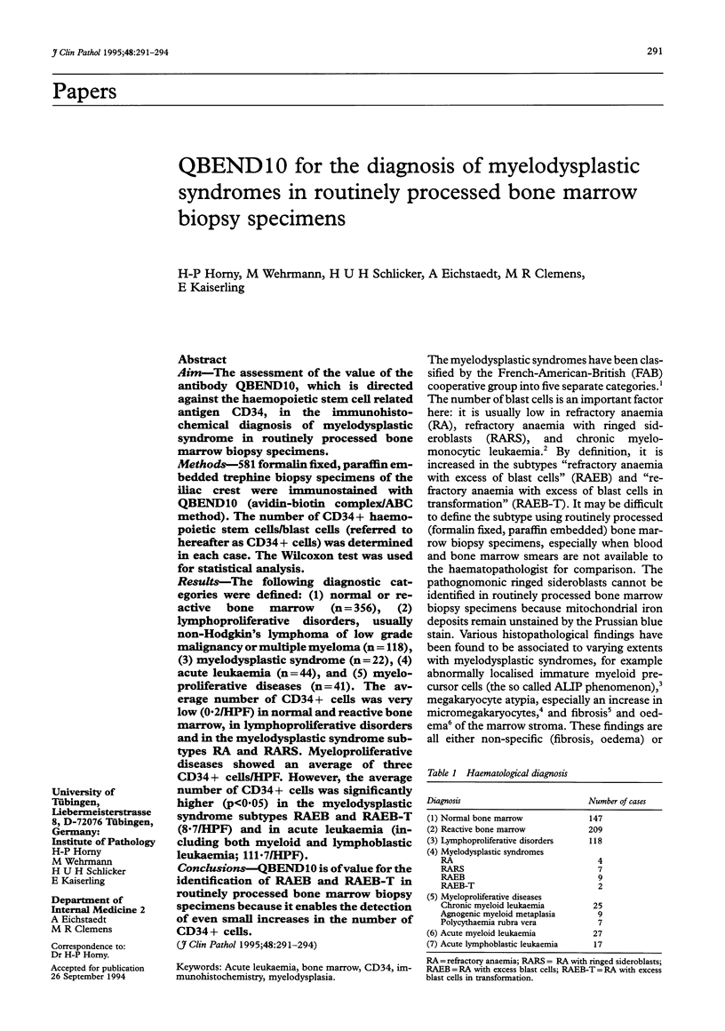
Images in this article
Selected References
These references are in PubMed. This may not be the complete list of references from this article.
- Bennett J. M., Catovsky D., Daniel M. T., Flandrin G., Galton D. A., Gralnick H. R., Sultan C. Proposals for the classification of the myelodysplastic syndromes. Br J Haematol. 1982 Jun;51(2):189–199. [PubMed] [Google Scholar]
- Fox S. B., Lorenzen J., Heryet A., Jones M., Gatter K. C., Mason D. Y. Megakaryocytes in myelodysplasia: an immunohistochemical study on bone marrow trephines. Histopathology. 1990 Jul;17(1):69–74. doi: 10.1111/j.1365-2559.1990.tb00665.x. [DOI] [PubMed] [Google Scholar]
- Gottlieb C. A., Maeda K., Hawley R. C., Abraham J. P. Myelodysplasia with bone marrow lymphocytosis and fibrosis mimicking recurrent Hodgkin's disease. Am J Clin Pathol. 1989 Jan;91(1):6–11. doi: 10.1093/ajcp/91.1.6. [DOI] [PubMed] [Google Scholar]
- Hanson C. A., Ross C. W., Schnitzer B. Anti-CD34 immunoperoxidase staining in paraffin sections of acute leukemia: comparison with flow cytometric immunophenotyping. Hum Pathol. 1992 Jan;23(1):26–32. doi: 10.1016/0046-8177(92)90006-o. [DOI] [PubMed] [Google Scholar]
- Horny H. P., Kaiserling E. Lymphoid cells and tissue mast cells of bone marrow lesions in systemic mastocytosis: a histological and immunohistological study. Br J Haematol. 1988 Aug;69(4):449–455. doi: 10.1111/j.1365-2141.1988.tb02397.x. [DOI] [PubMed] [Google Scholar]
- Hsu S. M., Raine L., Fanger H. Use of avidin-biotin-peroxidase complex (ABC) in immunoperoxidase techniques: a comparison between ABC and unlabeled antibody (PAP) procedures. J Histochem Cytochem. 1981 Apr;29(4):577–580. doi: 10.1177/29.4.6166661. [DOI] [PubMed] [Google Scholar]
- LEDER L. D. UBER DIE SELEKTIVE FERMENTCYTOCHEMISCHE DARSTELLUNG VON NEUTROPHILEN MYELOISCHEN ZELLEN UND GEWEBSMASTZELLEN IM PARAFFINSCHNITT. Klin Wochenschr. 1964 Jun 1;42:553–553. doi: 10.1007/BF01486688. [DOI] [PubMed] [Google Scholar]
- Majdic O., Stöckl J., Pickl W. F., Bohuslav J., Strobl H., Scheinecker C., Stockinger H., Knapp W. Signaling and induction of enhanced cytoadhesiveness via the hematopoietic progenitor cell surface molecule CD34. Blood. 1994 Mar 1;83(5):1226–1234. [PubMed] [Google Scholar]
- Oertel J., Kleiner S., Huhn D. Immunotyping of blasts in refractory anaemia with excess of blasts. Br J Haematol. 1993 Jun;84(2):305–309. doi: 10.1111/j.1365-2141.1993.tb03069.x. [DOI] [PubMed] [Google Scholar]
- Oertel J., Oertel B., Beyer J., Huhn D. CD 34 immunotyping of blasts in myelodysplasia. Ann Hematol. 1994 Feb;68(2):77–80. doi: 10.1007/BF01715135. [DOI] [PubMed] [Google Scholar]
- Silvestri F., Banavali S., Baccarani M., Preisler H. D. The CD34 hemopoietic progenitor cell associated antigen: biology and clinical applications. Haematologica. 1992 May-Jun;77(3):265–273. [PubMed] [Google Scholar]
- Simmons P. J., Torok-Storb B. CD34 expression by stromal precursors in normal human adult bone marrow. Blood. 1991 Dec 1;78(11):2848–2853. [PubMed] [Google Scholar]
- Thiele J. Pathologie der Präleukämie. Verh Dtsch Ges Pathol. 1983;67:115–131. [PubMed] [Google Scholar]
- Tricot G., Vlietinck R., Boogaerts M. A., Hendrickx B., De Wolf-Peeters C., Van den Berghe H., Verwilghen R. L. Prognostic factors in the myelodysplastic syndromes: importance of initial data on peripheral blood counts, bone marrow cytology, trephine biopsy and chromosomal analysis. Br J Haematol. 1985 May;60(1):19–32. doi: 10.1111/j.1365-2141.1985.tb07381.x. [DOI] [PubMed] [Google Scholar]
- Vandermolen L., Rice L., Rose M. A., Lynch E. C. Ringed sideroblasts in primary myelodysplasia. Leukemic propensity and prognostic factors. Arch Intern Med. 1988 Mar;148(3):653–656. [PubMed] [Google Scholar]