Abstract
Introduction
Chronic myofascial temporomandibular disorders (TMD) may have multiple etiological and maintenance factors. One potential factor, central pain sensitization, was quantified here as the response to the temporal summation (TS) paradigm, and that response was compared between case and control groups.
Objectives
As previous research has shown that fibromyalgia (FM) is diagnosed iñ20% of TMD patients, Aim 1 determined whether central sensitization is found preferentially in myofascial TMD cases that have orofacial pain as a regional manifestation of FM. Aim 2 determined if the report of after-sensations (AS) following TS varied depending on whether repeated stimuli were rated as increasingly painful.
Methods
One hundred sixty-eight women, 43 controls, 100 myofascial TMD-only cases, and 25 myofascial TMD + FM cases, were compared on thermal warmth and pain thresholds, thermal TS, and decay of thermal AS. All cases met Research Diagnostic Criteria for TMD; comorbid cases also met the 1990 American College of Rheumatology criteria for FM.
Results
Pain thresholds and TS were similar in all groups. When TS was achieved (~60%), significantly higher levels of AS were reported in the first poststimulus interval, and AS decayed more slowly over time, in myofascial TMD cases than controls. By contrast, groups showed similar AS decay patterns following steady state or decreasing responses to repetitive stimulation.
Conclusion
In this case–control study, all myofascial TMD cases were characterized by a similar delay in the decay of AS. Thus, this indicator of central sensitization failed to suggest different pain maintenance factors in myofascial TMD cases with and without FM.
Keywords: temporomandibular joint dysfunction syndrome, temporal summation of pain, women, central sensitization, QST
Introduction
The cause(s) of pain complaints in myofascial pain syndrome, the most common type of temporomandibular disorders (TMD), are not known. One theory holds that pain results from a dysregulation of endogenous pain mechanisms, and this theory is partially supported by quantitative sensory testing studies showing that myofascial TMD patients have lower thresholds to noxious thermal and pressure stimuli than controls (hyperalgesia), as well as more painful responses to innocuous stimuli (allodynia),1–7 higher levels of temporal summation (TS, participant reports increased painfulness of repeated stimuli, despite constant stimulus intensity)6,8–10 and greater persistence of after-sensations (AS, sensations that remain after active stimulation ceases).11 Prospective data12 have shown that elevated thresholds and heightened levels of thermal TS of the hand precede the diagnosis of myofascial TMD,13 suggesting that TS responses are a marker of vulnerability, if not part of a causal chain. However, increased sensitivity is not present in all myofascial TMD patients, suggesting that there may be hypersensitive subgroups.14,15
Fibromyalgia (FM), a widespread pain syndrome, is comorbid in ~20% of myofascial TMD cases.16,17 (Myofascial TMD has also been reported to be comorbid with other chronic pain states, including migraine and chronic fatigue syndrome,18 irritable bowel syndrome,19 and multiple comorbid pain conditions.20) Hypersensitivity to somatic stimulation is a widely accepted sign in FM.21,22 Psychophysical studies in FM patients generally show increased sensitivity to a multitude of laboratory pain stimuli,23 suggesting a higher “gain” when processing afferent nociceptive signals and a delayed resolution of AS.24–26 A parsimonious inference is that facial pain in comorbid patients is a sign of undiagnosed FM.27,28 Indeed, the Research Diagnostic Criteria (RDC) for TMD do not assess pain in areas other than the head,29 and so a diagnosis of FM could be missed in someone whose primary complaint was facial pain. Similarly, the 1990 American College of Rheumatology (ACR) criteria for FM do not assess pain in the head.30 Whether pain dysregulation in myofascial TMD cases without FM is also attributable to central factors has not been widely studied. Pfau et al.28 compared TS between myofascial TMD cases with localized (facial) or widespread pain, but did not specifically diagnose FM, and did not study AS. Thus, one innovative goal of this report is to test the hypothesis that central sensitization, measured as both TS and AS, is limited to the subset of myofascial TMD cases with comorbid FM.
A second goal of this report is to estimate the efficiency with which the thermal TS protocol provokes TS, evaluate differences in AS depending on whether or not TS was provoked, and compare both of these outcomes between both case groups and controls. Previous research has shown that even when individuals are presented with a train of identical thermal stimuli at an ideal temperature and rate (>45°C and ~0.3 Hz), there is only sometimes an increase in the apparent strength of stimuli appearing later in the train.21,31–34 One question that arises from this inconsistency is whether AS, which provide the particularly interesting view of central nervous system processing that is uncoupled from ongoing stimulation, depend on the development of TS. We hypothesize increased persistence of AS in the subset of TS trials that provoke TS.
Methods
Participants
Participants, all women, all fluent in English, were enrolled solely on the basis of the presence (cases) or absence (controls) of a myofascial TMD. Each gave written informed consent and all examinations were conducted at the Bluestone Center for Clinical Research at the NYU College of Dentistry (NYUCD). The study was approved by the New York University (NYU) School of Medicine Institutional Review Board. Data were collected between January and November 2011.
Myofascial TMD patients
Cases were recruited primarily from those seeking care at the Facial Pain Clinic at the NYUCD, but also from public postings at the NYU College of Dentistry. Participating myofascial TMD patients met RDC/TMD for TMD Group I29 that is, pain of muscle origin, including a complaint of pain and pain associated with localized areas of tenderness to palpation in muscle. They may have also met criteria for Groups 2 and 3, but this was not exclusionary. All patients reported facial pain of at least 1 year duration, and a minimum intensity of four of ten. The study coordinator (an MD) participated in biannual calibration training sessions to ensure consistency in this tender point examination as well as that for FM.
Controls
Controls were recruited from other NYUCD dental clinics and acquaintances of participating cases, constituting a sample that was a demographic match to cases on age, socioeconomic status, race, and Hispanic ethnicity. Controls could not have reported 1+ weeks of facial pain in the last 2 years or more than one painful site upon masticatory muscle palpation.
Exclusion criteria
Potential cases or controls indicating that they had a motor vehicle accident or another major and identifiable physical trauma involving the face were excluded, as were those with dental treatment within 48 hours of the RDC/TMD eligibility examination.
Measures
Pain history and intensity
The clinical research coordinator gathered RDC/TMD standardized pain history data,29 including questions about pain onset, pain severity, and pain-related disability. Characteristic pain intensity was computed as the average of present/current, worst, and average pain in the past 6 months, each rated on a 0–10 scale (0, no pain. 10, pain as bad as could be).
FM examination
All participants were evaluated according to the 1990 ACR criteria for the diagnosis of FM syndrome.30 Participants were enrolled on the basis of their TMD status, and their FM status was determined later.
Quantitative sensory testing
Heat stimuli were delivered by the Pathway Stimulator (Medoc Ltd., Ramat Yishai, Israel) through a contact heat-evoked potential stimulator thermode (27 mm diameter). Warm and pain thresholds were determined at three sites: the thenar eminence (the muscular area that flexes the thumb) of the nondominant hand and the skin on the right and left face overlying the belly of the masseter muscle. We sought to avoid sensitization by allowing several minutes of recovery between stimulation trials.
Determination of warm and pain thresholds by double random staircase
Procedure. The thermode resting temperature was 32°C for the determination of warm thresholds and 38°C for the determination of pain thresholds. On any particular trial, stimulus intensity was selected at random from one of two lists, or staircases. The beginning of a trial was signaled to the participant by a “beep”. The temperature then increased to the target temperature at a rate of 1°C/s for warmth (2°C/s for pain) and was held for 0.5 seconds for warmth (1.0 seconds for pain) before returning to the resting temperature at a rate of 40°C/s. If the participant failed to indicate that the stimulus increment in that interval was detected (or painful), by pushing “N” on the mouse, the temperature of the next stimulus in that staircase was adjusted upward by either 1°C or 3°C, respectively, for warmth and pain staircases. Stimulus temperatures continued increasing until the participant indicated that the stimulus increment was either detected or labeled painful, by pushing “Y” on the mouse, at which point the temperature of the next stimulus in that staircase was adjusted downward by 0.5°C (for warmth) or 1.0°C (for pain). Stimulus temperatures continued decreasing until the participant again pressed “N”, at which point the temperature was increased in 0.5° steps until the participant pressed “Y”. Then, the stimulus decreased and increased in 0.25° steps. In this way, the procedure continued to the threshold temperature, the point at which the participant was as likely to say “yes” as “no”.
Participant instructions: For this test, hold the surface against the muscular part of your thumb (or substitute face, as appropriate). At the beginning of each cycle you will hear two beeps separated by 1 or 2 seconds. Your job is to press the ‘Y’ button on the mouse if you felt an increase in warmth (substitute ‘pain’ for pain threshold) during that time and the ‘N’ button if you did not. After a brief pause, the cycle will repeat. If you are unsure how to respond, please take your best guess. Any questions?
Evaluation of TS of second pain and AS
These procedures quantify the development of TS and the resolution of AS. The challenges are twofold: first, to teach the participant to focus on the late sensations that reflect the activity of the C-nociceptive fibers that are necessary for the development of TS and second, to find a stimulus intensity that becomes more painful with repeated stimulation.
Training phase: qualitative observation of peak pain, late sensations, and AS. Stimulus intensities of 45°C, 47°C, 49°C, and 51°C. were presented in a train of 15 pulses (700 ms duration), an interpulse interval of 2 seconds, producing stimulation rates of ~0.3 Hz (higher temperatures take a little longer to reach peak temperature and return to baseline). Training occurred on the thenar eminence of the nondominant hand.
Procedure. The participant was instructed to look for three characteristics of the stimulation that suggest C-nociceptor activation: 1) sensations described as burning, prickling, or sizzling; 2) late sensations, those that first appear after stimulus offset; and 3) AS, feelings that linger in the stimulated area well after the last stimulus was presented. The stimulus intensity was increased only if the participant did NOT detect at least two of these signs of C-nociceptive activity given the current intensity. If these signs were detected, that temperature was recorded and training was terminated. Participants experienced as many as four training trials, one for each stimulus intensity. They did not practice using verbal rating scale during training, and training data were not analyzed.
-
Participant instructions. The reason for this test is to see how the very smallest nerves in your body are working. Because they are small, they send information slowly. We are going to start out with some practice, so you know what I want you to pay attention to.
As before, I want you to hold the surface against your thumb. The machine will present you with a series of 15 stimuli, each lasting ~1 second, and spaced ~2 seconds apart. Unless you absolutely cannot stand it, please hold the surface in place until the last temperature is delivered (~30 seconds), and then remove it from your skin.
I want you to pay attention to three things that you might feel. First, I want you to focus on each temperature in the series, and notice when the feeling of heat or pain becomes strongest. You may notice that feelings continue after a temperature has returned to its beginning level or first appear after a temperature has returned to its beginning level. We call these late sensations, and you might not feel this until after the temperature presented to your hand is over. Second, I want you to tell me whether you recognize several feelings that might have occurred, what some people call “prickling, sizzling, or burning”. And third, I want you to let me know if you continue to feel sensations after you remove the stimulator from your skin. Any questions?
Testing phase: quantitative rating of late sensations and/or peak pain
These tests quantify the intensity of late thermal sensations during stimulation and any AS that may linger. Stimulation parameters were identical to those described for training. All ratings employed a 100 point numerical rating scale (NRS), with several categorical reference points (see Participant instructions below).
-
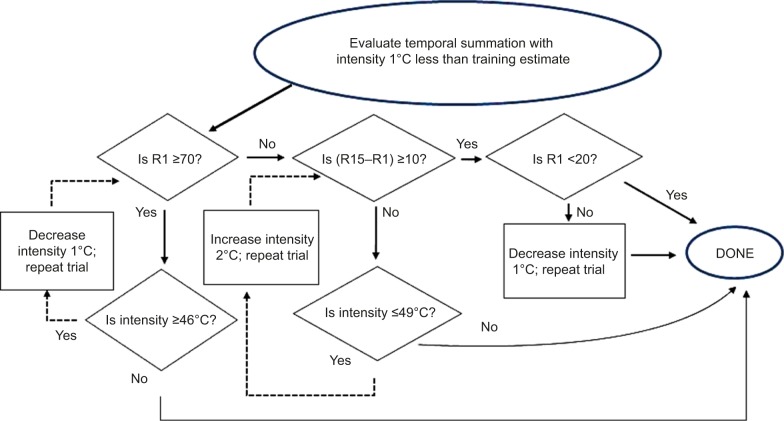
Procedure. The starting temperature for this procedure was 1°C less than that shown to produce late sensations, C-nociceptive qualities, or AS during training. If the training procedure failed to locate an appropriate temperature, 45°C was used. Figure 1 summarizes the decision points that were used to tailor the stimulation temperature to each participant’s needs.
Participants were cued to rate intensity after presentation of the 1st, 5th, 10th, and 15th stimulus in the train, and were also cued to rate the intensity of residual AS at 15 seconds intervals for 2 minutes afterward. When AS were reported at the “0” level for two successive periods, the trial was terminated before 2 minutes and “0” was imputed to each remaining time point. After each attempt to achieve TS, there was a break of several minutes before retesting.
Participant instructions. This test uses a series of temperatures like those you just experienced, except now I want you to use a number to rate the intensity of the late sensations, or peak pain/maximum pain, if there are no late sensations. Please make your ratings using numbers from this scale (present chart), with these anchors: 10, warm; 20, a barely painful sensation; 30, very weak pain; 40, weak pain; 50, moderate pain; 60, slightly strong pain; 70, strong pain; 80, very strong pain; 90, nearly intolerable pain; and 100, intolerable pain. Do you understand how to use the scale?
Figure 1.
Flow chart shows decision points in selecting stimulus intensities for, and ending, the temporal summation procedure.
Note: R1 and R15 are responses to the first and last stimuli in the train, respectively.
As before, hold the surface against your thumb. The machine will present you with a series of 15 temperatures, each lastinĝ1 second and followed within 2 seconds by another temperature. Now and then you’ll hear a beep. When you hear the beep, make your intensity ratings for the temperature which immediately follows. Your first judgment is usually best, so answer as quickly as possible. Unless you absolutely cannot stand it, please hold the surface in place until the last temperature is delivered (~30 seconds), and then remove it from your skin. For up to 2 minutes after that, I’ll again ask you to rate the intensity. Any questions?
Data analysis
Group differences in mean thresholds were analyzed with one-way analysis of variance. Responses to TS used a linear mixed models procedure (Statistical Package for the Social Sciences, v. 22, IBM Corporation, Armonk, NY, USA) to evaluate fixed effects of group and time while adjusting the analysis for covariates of temperature, and random effects of repeated observations over time within a trial, and multiple trials in some persons. Primary analysis of AS used general estimating equations to model responses in a binomial distribution with a survival link and assuming an autoregressive one covariance relationship among the time points. This model estimated the probability of reporting little-to-no sensation (NRS <5) at each poststimulation interval, while censoring observations that already reached this end point, and adjusting the analysis for multiple trials and varying stimulus temperatures or final pain rating during stimulation. A significance level of 5% was used. The study planned to recruit more cases than controls (2:1 ratio), while maintaining 80% power to detect moderately sized effects (0.5 standard deviation [SD]) between controls and equal sized subgroups of cases with and without FM.
Results
Participants
Among likely eligible patients who were approached in the clinic, 19 did not provide consent to an RDC/TMD screening examination. Of the 145 patients who agreed, 138 met RDC criteria. The reasons for subsequent exclusion were a history of trauma to the face (n=6), practical exclusions related to other study aims (n=5) (for details, see Raphael et al35). Among 63 potential controls, six were excluded with more than one tender point on RDC/TMD examination. Of the 54 meeting eligibility criteria, eight canceled or failed to appear. In all, 126 women with a diagnosis of myofascial TMD and 48 controls participated. Of these, six cases and one control did not provide complete quantitative sensory testing data due to equipment failures. Twenty-six cases also met 1990 ACR criteria for FM, as did one control; that control was excluded from quantitative sensory testing analyses. For simplicity, the two case groups are referred to as TMD-only and TMD + FM (or comorbid) groups in the following: “case”, without modification, refers to both case groups.
Both case groups and controls showed similar demographic characteristics: most indicated that their race was white (62.6%) or black (14.4%). Hispanic ethnicity was indicated by 22.5%. Mean ± SD age was 39.2±14.6 years (range 19–78), and mean ± SD education was 15.0±2.2 years (range 11–20). Among concomitant medications used by at least 5% of the sample, there was greater use of nonsteroidal anti-inflammatory drugs (NSAIDs), muscle relaxants, and opioids in cases than controls (P<0.05), and similar use of selective serotonin reuptake inhibitors, anticonvulsants, contraceptives, bronchodilators, and allergy medications. Only NSAID use was higher in comorbid than other cases. No one was asked to change usual medications for study purposes. As shown in Table 1, cases with comorbid FM were older and reported longer pain duration than TMD-only. Cases with comorbid FM reported somewhat greater intensities of characteristic facial pain than others, and significantly greater pain when the facial TPs were palpated. By contrast, counts of painful facial TPs were statistically similar in each case group. In addition, TMD-only patients were more likely than controls to report widespread pain during the FM diagnostic evaluation.
Table 1.
Mean ± SD age and clinical pain characteristics in each group
| Group (N) | Age (years) | Facial TPs severity (0–10) | Facial TPs (count) | Facial pain duration (months) | Characteristic pain intensity (0–10) | Widespread pain (%) |
|---|---|---|---|---|---|---|
| Controls (48) | 36.7±14.2a | 3±6.4a | ||||
| TMD-only (99) | 36.3±17.3a | 2.0±1.1 | 13.0±4.1 | 86.5±78.9 | 4.9±1.9 | 23±24.0b |
| TMD + FM (26) | 43.4±20.4b | 2.6±1.1 | 14.2±4.0 | 183.5±153.8 | 6.0±1.3 | 26±100c |
| Omnibus P-value | 0.16 | 0.02 | 0.19 | 0.008 | 0.07 | 0.01 |
Note: Different superscript letters (a–c) indicate a statistical difference, P<0.05.
Abbreviations: FM, fibromyalgia; SD, standard deviation; TMD, temporomandibular disorders.
Thermal thresholds
Table 2 shows mean warm thresholds of ~34°C on both sides of the face of control and TMD-only participants. By contrast, cases with FM averaged about a degree higher than other participants. While pain thresholds were also about a degree higher in cases than controls, there was no statistical difference. There was little difference among groups in warm or pain thresholds sampled from the hand. These data fail to support a hypothesis of increased sensitivity in either case group.
Table 2.
Mean ± SD warm and pain thresholds °C determined by staircase procedure in each group
| Group (N) | Warm L face | Warm R face | Warm hand | Pain L face | Pain R face | Pain hand |
|---|---|---|---|---|---|---|
| Controls (47) | 33.8±1.2a | 33.7±1.5a | 32.7±0.6a | 41.4±2.5a | 41.5±2.4a | 42.0±2.4a |
| TMD-only (98) | 33.7±1.4a | 33.9±1.5a | 32.9±1.0a | 42.7±2.3b | 42.0±2.3a | 42.3±2.2a |
| TMD + FM (26) | 35.0±2.7b | 34.8±2.0b | 33.1±1.0a | 42.6±2.1b | 42.6±2.4a | 41.7±2.4a |
| Omnibus P-value | 0.002 | 0.01 | 0.30 | 0.08 | 0.15 | 0.45 |
Note: Different superscript letters (a and b) indicate a statistical difference, P<0.05.
Abbreviations: FM, fibromyalgia; L, left; R, right; SD, standard deviation; TMD, temporomandibular disorders.
Temporal summation
Participants were exposed to multiple stimuli, starting at 45°C or as determined in the training phase, and continuing until summation was shown or the 51°C stimulus was presented (Figure 1). Six participants did not tolerate repetition of the lowest intensity stimulus and were excluded from analysis. There remained a total of 621 trials from 161 participants (Median =3 trials/participant). Stimulus intensities of 45°C, 46°C, 47°C, 48°C, 49°C, 50°C, and 51°C were judged, respectively, by 105, 141, 145, 104, 71, 37, and 18 participants.
TS response patterns
Based on the ratings given to each stimulus intensity that attempted to elicit TS, each trial was categorized as one of three types: those that produced an increase of at least 10 points between the response to the 1st and 15th stimulus (summation); those that produced a change between −10 and +10 (steady state); and those that produced a decrease of at least ten points on the NRS (decreasing). Figure 2 shows mean ± SE ratings during and after stimulation for each response pattern. Summation trials produced mean increases of almost 20 points, and steady state responses were, by definition, flat over the stimulation interval. Surprisingly, trials that elicited a declining response started from a high initial rating, an NRS of above 40, despite the fact that the stimulus intensity on those trials was similar to that presented on steady state and summation trials. Ratings to the final stimulus in the train were similar in declining and steady state trials.
Figure 2.
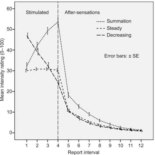
Mean ± SE numerical rating scale reports during the temporal summation and AS phases of response.
Notes: Different lines show responses for the three response patterns, decreasing, steady, and summating trials, averaged over all participants. The vertical line separates the four reports given during the stimulation period from the eight AS. AS appear elevated when the stimulation produced a summating pattern (n=167).
Abbreviations: AS, after-sensations; SE, standard error.
Summation was seen on at least one trial in 94 (58.4%) participants. Of the 67 participants who did not summate, 14 (8.7%) were exposed to the complete range of stimuli (and so the absence of a summation responses is determinate). The procedure was terminated early (ie, without summation and before the maximum intensity was reached, called “indeterminate trials”) in 53 participants (32.9%). While two participants terminated after one trial and two after two trials, participants typically stopped after they failed to show summation after three or four trials. Thus, while all stimulus temperatures were sufficiently intense to activate C-nociceptor and support summation, testing provided a definitive result in only ~2/3 of the sample.
Effects of stimulus temperature
Some of the variability in achieving TS may be attributed to stimulus temperature. Figure 3 shows that stimulus temperature <48°C did not, on average, elicit summating responses. Table 3 shows that the probability of a summating response increased with stimulus intensity, while steady state responses were largely independent of intensity and decreasing responses were inversely proportional to intensity. Linear mixed model analysis showed that the summation pattern was associated with the presence of higher stimulation temperatures (mean [M] =47.8°C) than the steady state or decreasing response patterns (M =46.8°C and 46.7°C, respectively; P<0.001). TMD cases and controls summated at statistically similar temperatures (interaction P=0.30), failing to support a “hyperalgesia-like” response to the TS paradigm among cases. Thus, while summation responses were more likely as stimulus temperature increased, temperature accounted for <10% of pattern variability. Higher stimulus temperatures did not guarantee summation.
Figure 3.
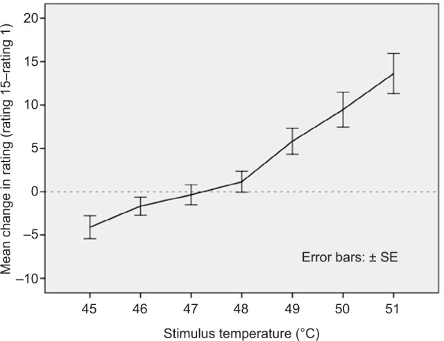
Mean ± SE change in numerical rating scale reports between the 1st and 15th stimulus in the train, as a function of stimulus temperature.
Note: Temporal summation was unlikely to occur at temperatures <48°C.
Abbreviation: SE, standard error.
Table 3.
Distribution of TS patterns by stimulation temperature, for the total sample (responses to adjacent temperatures were averaged and the range is shown)
| TS pattern
|
|||
|---|---|---|---|
| Temperature range (°C) | Decreasing (139 trials in 75 participants) | Steady state (312 trials in 130 participants) | Increasing (summation) (170 trials in 99 participants) |
| 45–46 | 31.7% | 49.6% | 18.7% |
| 47–48 | 22.1 | 52.2 | 25.7 |
| 49–51 | 4.8 | 47.6 | 47.6 |
Abbreviation: TS, temporal summation.
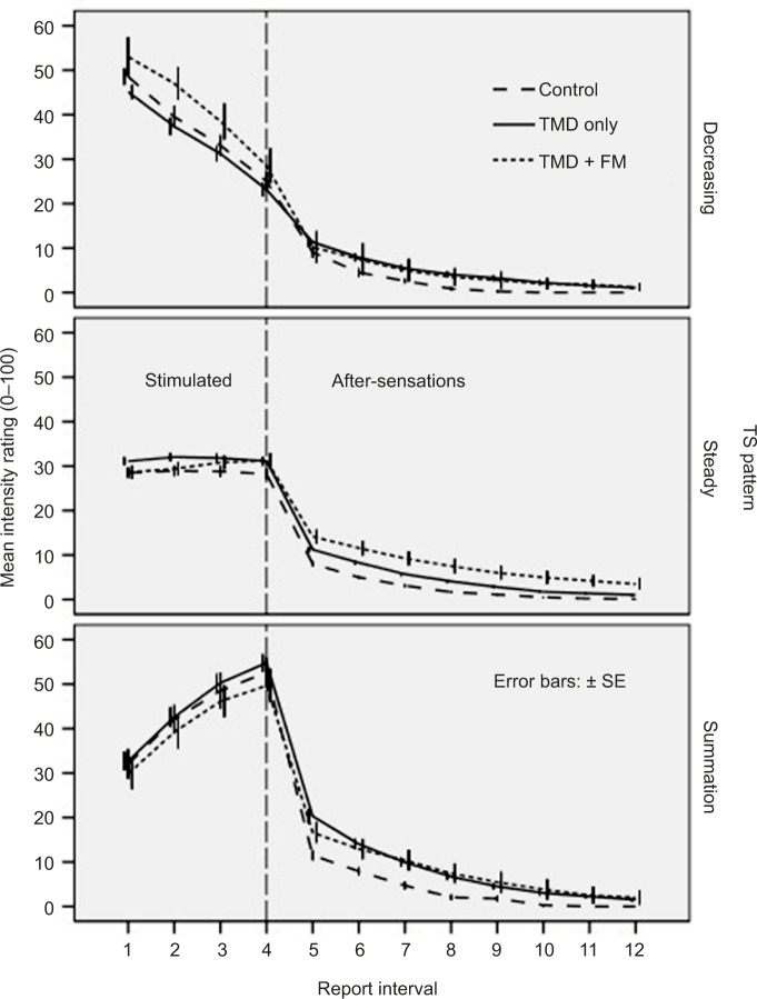
Figure 4 shows mean ratings of stimulus intensity over the 12 report intervals for each clinical group. Few differences in TS are apparent between clinical groups. Intensity reports drop dramatically upon stimulus termination, and this drop appears greater after summation trials, and among case groups. Ratings then continue to decline during the remaining 105 seconds of recovery, where the lowest ratings are seen in controls.
Figure 4.
Mean ± SE numerical rating scale reports during the TS and AS phases of response.
Notes: Different lines show responses in each clinical group for each of the three response patterns, decreasing, steady, and summating trials. The vertical line separates the four reports given during the stimulation period (TS) from the eight AS. AS appear elevated in both case groups when stimulation produced steady state or summating patterns.
Abbreviations: AS, after-sensations; FM, fibromyalgia; SE, standard error; TMD, temporomandibular disorders; TS, temporal summation.
In the analyses below, we compare the clinical groups on three aspects of response: the change in ratings over the course of repeated stimulation; the first rating after stimulus termination; and the slope during the remainder of recovery.
Responses during repetitive stimulation
To evaluate the development of TS as a function of group and time, a linear mixed model tested main and interaction effects in a three groups × four periods analysis of each TS response pattern, while adjusting for the stimulus temperature on that trial. For decreasing pattern trials, analysis showed an effect of temperature (P<0.001), indicating an increase of 3.9 rating points for every 1°C and period (P<0.001). The latter effect predicted mean ratings of 44.0, 37.8, 32.2, and 25.7 to stimuli 1, 5, 10, and 15 (P<0.05 between each successive level) at the average covariate temperature of 46.4°C. Thus, ratings decreased significantly with repeated stimulation, similarly in cases and controls, after adjusting for differences in stimulus temperature. For steady state trials, analysis showed only an effect of temperature (P<0.001), indicating a 4.5 point higher rating for a unit increase in temperature. For summation trials, analysis showed an effect of temperature (P<0.001), indicating an increase of 7.3 points for every 1°C and period (P<0.001). The latter effect predicted mean ratings of 30.5, 38.0, 43.4, and 46.3 to stimuli 1, 5, 10, and 15 (P<0.05 between each successive level save the last) at the average covariate temperature of 47.8°C. The increase with repeated stimulation was similar in each group (interaction P=0.77). Thus, after adjusting for temperature, TS to repetitive stimulation proceeded to a similar endpoint and at a similar rate in the two case groups and controls.
After-sensations
Do the different response patterns during repetitive stimulation affect the report of AS? Inspection of Figure 2 suggests that the first report of AS (interval 5, 15 seconds after delivery of the last stimulus) following a summating response is approximately twice that seen following either decreasing or steady-state response patterns, but those means were not controlled for the higher temperatures judged during summation trials. The analyses include that control to address three hypotheses: AS are more intense when stimulus repetition leads to TS; AS are more intense in cases than controls; and AS are more intense in TMD cases with FM than without FM.
First report of AS
During the first poststimulus period, AS ratings increased ~1.9 points for each additional 1°C of stimulus temperature used during stimulation (P<0.001). After adjusting for stimulus temperature (or the final rating, which produced similar effects), summation trials still produced higher mean estimated ratings than steady state or decreasing patterns (M ± SE =14.7±1.0 vs 11.5±0.9 and 12.9±1.2, respectively; P<0.001). Thus, while AS reports increased in proportion to stimulating temperatures for all patterns, they were maximized following summating trials.
First reports of AS were also higher in TMD-only and TMD + FM groups than controls (M ± SE =13.7±0.9 and 16.2±1.8 vs 9.2±1.3, respectively; P=0.005). Analysis failed to show a difference between TMD cases with and without FM (P=0.23). Because the TMD + FM group was relatively small, there is some risk of a type 2 error. In fact, however, the standardized difference (Cohen’s d) between the TMD-only and TMD + FM groups was small, <0.25 SD, relative to a difference of 0.5 SD between the case and control groups. There was no evidence that AS varied with the interaction between group and TS pattern (P=0.25). Thus, initial AS were reported as more intense by cases than controls, but there was no difference in AS report between cases with and without FM.
Together, these analyses suggest that first AS reports increase with the temperature of the repetitive stimuli, were higher following trials that produced TS and were higher in cases than controls. Data failed to support the hypothesis that AS are more intense in the TMD + FM group.
First to last AS
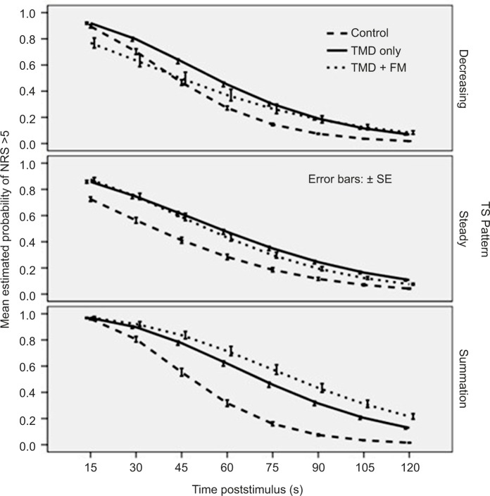
Figure 5 shows mean estimated probabilities of a positive AS report (NRS> 5) during the first poststimulus interval to be near 1.0 for summation trials, and at somewhat lower levels for trials showing steady state or declining patterns. Also apparent is a higher probability of AS reports in cases than controls, particularly following summation trials. Report levels dropped to virtually zero after 2 minutes in all participants following decreasing or steady state patterns, and controls also dropped to zero following summation trials. By contrast, ~20% of case responses remain elevated following summation trials. For each summation pattern, a general estimating model evaluated the probability of NRS> 5 as a function of case status and time while adjusting for the final stimulated report level, which produced a more precise model than achieved by adjusting stimulus temperature. For the decreasing and steady state response patterns, AS reports decreased over time (both P<0.001), and were somewhat more likely to be reported by cases than controls (P=0.06 and 0.02, respectively), but the rate of decline was similar in all groups (interaction P=0.17 and 0.52). By contrast, AS following summating trials decayed more slowly in cases than controls (interaction P=0.01), although there was a similar rate of decay in cases with and without FM (P=0.32). These data support the hypothesized slower decay of AS in cases specifically during trials that produced TS.
Figure 5.
Mean ± SE estimated probabilities of a sensation report following stimulus termination in each clinical group and for each TS pattern.
Notes: Probabilities are estimated from a general estimating equation with a survival link that modeled responses as a function of clinical group, summation pattern, and time, while adjusting for the final stimulated intensity ratings. AS reports appear to decay more slowly in the case groups, particularly during trials that elicited TS.
Abbreviations: AS, after-sensations; FM, fibromyalgia; NRS, numerical rating scale; SE, standard error; TMD, temporomandibular disorders; TS, temporal summation.
Discussion
This study sought to address two sets of questions, one substantive and one methodological. The substantive question asked whether TMD cases evidence increased sensitivity to noxious heat, and whether those cases who also met 1990 ACR criteria for FM were more sensitive than those who did not. To summarize, warm thresholds were significantly higher in the FM than TMD-only or control groups; this effect was unrelated to older age or higher pain ratings in the FM group, and remains puzzling. While pain thresholds trended higher in the cases, that effect was only about half as strong as for warm thresholds. Higher heat pain thresholds have been inconsistently reported among TMD cases.1,4,36,37 There was no statistical difference between cases and controls in the development of TS during repetitive thermal simulation, consistent with other reports stating that increased levels of TS are only sometimes reported to characterize chronic pain patients.31,33,34 Also consistent with prior research,3,11,31,38,39 AS were reported at low levels and decayed rapidly in controls. By contrast, initial reports of AS in cases tended to increase with final stimulated report levels, and then decayed more slowly than controls. In addition, ~20% of the cases, but no controls, continued to report sensations 2 minutes after stimulation ended. Taken together, these data suggest increased persistence of AS following noxious thermal stimulation in women with myofascial TMD.
We had hypothesized more TS and increased persistence of AS in the subset of TMD cases with comorbid FM. Instead, data showed that TMD-only and TMD + FM cases showed similar rates of acceleration of TS and decay of AS. While results showed more intense AS ratings in the comorbid group, that effect was less than half the size of the difference between cases and controls (0.2 SD vs 0.5 SD). Consistent with other reports,28,40 21% of this sample selected for TMD also met criteria for FM, suggesting that it is representative. Pfau et al.,28 who addressed a similar question, showed increasing sensitivity to a wide variety of sensory stimuli as one moves from controls to those with regional TMD and then to those with TMD and widespread pain. By contrast, our data do not confirm increased vulnerability in those with both signs of FM. Nevertheless, we would agree with their conclusion that TMD patients, with or without FM, evidence a “disturbance of central pain processing”.
Our first methodological question asked how often repetitive thermal stimulation resulted in increased report levels, defined as an increase of at least ten points. TS was seen in 60% of the participants. Most attempts to elicit summation showed a steady sate response, and a small number showed a decreasing pattern. In general, summation success increased with stimulus temperature, and TS was less likely to be seen when judging stimuli below 48°C. Ironically, some of these summation failures may be attributed to a tactic designed to foster success in summation, tailoring stimulus temperatures to potentially hyperalgesic individuals. (For example, FM patients have been reported to show TS at temperatures of 45°C.41) Thus, TS was indeterminate in the 30% who experienced one or more lower stimulus temperatures and then terminated the procedure. Because they may have shown TS to higher stimulus intensities, the true rate of summation success could approach 90%. (Similar results, not shown, were obtained when gauging the difference between the first response and the maximal change.) Thus, while we anticipated summation to stimuli at low as 45°C, TMD cases were no more likely than controls to show TS at those lower stimulus levels. This suggests that the efficiency of TS paradigm may be increased by using stimuli of at least 48°C, if tolerated, when participants fail to summate at lower temperatures.
Nevertheless, even if there was a definitive result in 90% of individuals, this would still be problematic – a good test produces a definitive result in all takers. For example, it is rarely not possible to define a pain threshold for an individual. If the TS paradigm does not reliably produce a summating pattern, despite extensive training in the recognition of second pain, its utility is diminished. Using the current definition of summation, a ten point increase, does not seem to be the problem, as others have defined TS as any positive change and still find a substantial number of summation failures.34 Because the procedure allowed each participant to hold the thermode, it is possible that some failures could be attributed to insufficient contact. There have also been recent reports that stimulus characteristics, sex, and stimulus location33,34 influence the success of the TS paradigm. Two studies report that dextromethorphan reverses TS in some chronic pain patients,42,43 suggesting that individual differences in TS may be explained by differences in N-methyl-d-aspartate receptor activity. In addition, all of our participants continued to take their regular medications, and NSAID, muscle relaxant and opioid use, which were increased in cases, may have influenced TS.
Our second methodological question asked whether summation affected AS. AS ratings decayed more slowly following trials that produced summating responses than the decreasing or steady state TS pattern, and more slowly in cases than controls. This suggests that summation is a necessary indicator of windup of wide dynamic range and nociceptive specific neurons within the dorsal horn of the spinal cord.21,24,25,44,45 Consistent with others,39 however, AS were also apparent following nonsummating responses and may indicate another mechanism, perhaps peripheral sensitization.46 Thus, while summation may produce a special “kind” of AS, it is not necessary to produce “all” AS. If these response patterns do indicate activation of different circuits, they could be separately analyzed. Both kinds of AS decayed more slowly in cases than controls, and at different rates in summating and nonsummating trials. Together, these data suggest that TS does affect the intensity and quality of AS.
On the other hand, steady state and decreasing response patterns differed primarily in the response to the first stimulus in the train. Responses to the last stimulus, as well as the magnitude and rate of AS decay, were otherwise similar for those patterns. If the first stimulus sometimes elicited a reflexive startle, there may be no difference in processing of the stimuli related to the development of TS. The analysis of AS comes to similar conclusion when these two patterns are treated as a single set of nonsummating responses. Thus, steady state and decreasing response patterns may activate similar neural circuits related to TS and the decay of AS, and they need not be treated as distinct response patterns.
Strengths of this study include the use of a very large sample of patients and controls in a study specifically designed to compare TS and AS in TMD cases with and without FM. Limitations might include the use of a participant-held thermode, which does not guarantee a consistent contact pressure, and may have been responsible for some instances of summation failure. As well, TS efficiency may have been increased if all participants had been encouraged to experience temperatures of at least 48°C, if they failed to summate at lower temperatures.
Conclusion
Slow decay of thermal AS appears to characterize this treatment-seeking sample of TMD cases regardless of whether they meet criteria for FM. Attempts to elicit thermal TS did not always evoke a summating response pattern, and this failure was not fully explained by case status or stimulation temperatures. Cases reported more AS than controls, regardless of TS pattern. However, only AS following summating TS patterns displayed the sustained response anticipated from a centrally mediated mechanism, and cases showed significantly more of this activation than controls. Given that AS occur after stimulation had been terminated, these data are consistent with a pain maintenance mechanism in TMD based on a potentiated central response to thermal stimulation.
Acknowledgments
The study was approved by the Institutional Review Board at the New York University (NYU) School of Medicine. This study was supported by the National Institute of Dental and Craniofacial Research, DE 18569.
Footnotes
Disclosure
The authors report no conflicts of interest in this work.
References
- 1.Maixner W, Fillingim R, Booker D, Sigurdsson A. Sensitivity of patients with painful temporomandibular disorders to experimentally evoked pain. Pain. 1995;63(3):341–351. doi: 10.1016/0304-3959(95)00068-2. [DOI] [PubMed] [Google Scholar]
- 2.Malow RM, Grimm L, Olson RE. Differences in pain perception between myofascial pain dysfunction patients and normal subjects: a signal detection analysis. J Psychosom Res. 1980;24(6):303–309. doi: 10.1016/0022-3999(80)90021-5. [DOI] [PubMed] [Google Scholar]
- 3.Greenspan JD, Slade GD, Bair E, et al. Pain sensitivity risk factors for chronic TMD: descriptive data and empirically identified domains from the OPPERA case control study. J Pain. 2011;12(11 Suppl):T61–T74. doi: 10.1016/j.jpain.2011.08.006. [DOI] [PMC free article] [PubMed] [Google Scholar]
- 4.Sarlani E, Greenspan JD. Evidence for generalized hyperalgesia in temporomandibular disorders patients. Pain. 2003;102(3):221–226. doi: 10.1016/S0304-3959(03)00095-2. [DOI] [PubMed] [Google Scholar]
- 5.Svensson P, List T, Hector G. Analysis of stimulus-evoked pain in patients with myofascial temporomandibular pain disorders. Pain. 2001;92(3):399–409. doi: 10.1016/S0304-3959(01)00284-6. [DOI] [PubMed] [Google Scholar]
- 6.Fernández-de-las-Peñas C, Galán-del-Río F, Fernández-Carnero J, Pesquera J, Arendt-Nielsen L, Svensson P. Bilateral widespread mechanical pain sensitivity in women with myofascial temporomandibular disorder: evidence of impairment in central nociceptive processing. J Pain. 2009;10(11):1170–1178. doi: 10.1016/j.jpain.2009.04.017. [DOI] [PubMed] [Google Scholar]
- 7.Classification of chronic pain. Descriptions of chronic pain syndromes and definitions of pain terms. Prepared by the International Association for the Study of Pain, Subcommittee on Taxonomy. Pain Suppl. 1986;3:S1–S226. [PubMed] [Google Scholar]
- 8.Maixner W, Fillingim R, Sigurdsson A, Kincaid S, Silva S. Sensitivity of patients with painful temporomandibular disorders to experimentally evoked pain: evidence for altered temporal summation of pain. Pain. 1998;76(1–2):71–81. doi: 10.1016/s0304-3959(98)00028-1. [DOI] [PubMed] [Google Scholar]
- 9.Sarlani E, Grace EG, Reynolds MA, Greenspan JD. Evidence for up-regulated central nociceptive processing in patients with masticatory myofascial pain. J Orofac Pain. 2004;18(1):41–55. [PubMed] [Google Scholar]
- 10.Vierck CJ, Jr, Cannon RL, Fry G, Maixner W, Whitsel BL. Characteristics of temporal summation of second pain sensations elicited by brief contact of glabrous skin by a preheated thermode. J Neurophysiol. 1997;78(2):992–1002. doi: 10.1152/jn.1997.78.2.992. [DOI] [PubMed] [Google Scholar]
- 11.Raphael KG, Janal MN, Anathan S, Cook DB, Staud R. Temporal summation of heat pain in temporomandibular disorder patients. J Orofac Pain. 2009;23(1):54–64. [PMC free article] [PubMed] [Google Scholar]
- 12.Fillingim RB, Slade GD, Diatchenko L, et al. Summary of findings from the OPPERA baseline case–control study: implications and future directions. J Pain. 2011;12(11 Suppl):T102–T107. doi: 10.1016/j.jpain.2011.08.009. [DOI] [PMC free article] [PubMed] [Google Scholar]
- 13.Maixner W. Is pain sensitivity a predictive factor for the development of painful temporomandibular disorders?; Paper presented at: American Pain Society Annual Meeting 2003; Chicago, IL: [Google Scholar]
- 14.Slade GD, Diatchenko L, Bhalang K, et al. Influence of psychological factors on risk of temporomandibular disorders. J Dent Res. 2007;86(11):1120–1125. doi: 10.1177/154405910708601119. [DOI] [PubMed] [Google Scholar]
- 15.Slade GD, Diatchenko L, Ohrbach R, Maixner W. Orthodontic treatment, genetic factors and risk of temporomandibular disorder. Semin Orthod. 2008;14(2):146–156. doi: 10.1053/j.sodo.2008.02.005. [DOI] [PMC free article] [PubMed] [Google Scholar]
- 16.Janal MN, Raphael KG, Nayak S, Klausner J. Prevalence of myofascial temporomandibular disorder in US community women. J Oral Rehabil. 2008;35(11):801–809. doi: 10.1111/j.1365-2842.2008.01854.x. [DOI] [PubMed] [Google Scholar]
- 17.Plesh O, Wolfe F, Lane N. The relationship between fibromyalgia and temporomandibular disorders: prevalence and symptom severity. J Rheumatol. 1996;23(11):1948–1952. [PubMed] [Google Scholar]
- 18.Dahan H, Shir Y, Nicolau B, Keith D, Allison P. Self-reported migraine and chronic fatigue syndrome are more prevalent in people with myofascial vs nonmyofascial temporomandibular disorders. J Oral Facial Pain Headache. 2016;30(1):7–13. doi: 10.11607/ofph.1550. [DOI] [PubMed] [Google Scholar]
- 19.Aaron LA, Burke MM, Buchwald D. Overlapping conditions among patients with chronic fatigue syndrome, fibromyalgia, and temporomandibular disorder. Arch Intern Med. 2000;160(2):221–227. doi: 10.1001/archinte.160.2.221. [DOI] [PubMed] [Google Scholar]
- 20.Bair E, Gaynor S, Slade GD, et al. Identification of clusters of individuals relevant to temporomandibular disorders and other chronic pain conditions: the OPPERA study. Pain. 2016;157(6):1266–1278. doi: 10.1097/j.pain.0000000000000518. [DOI] [PMC free article] [PubMed] [Google Scholar]
- 21.Price DD, Hu JW, Dubner R, Gracely RH. Peripheral suppression of first pain and central summation of second pain evoked by noxious heat pulses. Pain. 1977;3(1):57–68. doi: 10.1016/0304-3959(77)90035-5. [DOI] [PubMed] [Google Scholar]
- 22.Pud D, Granovsky Y, Yarnitsky D. The methodology of experimentally induced diffuse noxious inhibitory control (DNIC)-like effect in humans. Pain. 2009;144(1–2):16–19. doi: 10.1016/j.pain.2009.02.015. [DOI] [PubMed] [Google Scholar]
- 23.Petzke F, Clauw DJ, Ambrose K, Khine A, Gracely RH. Increased pain sensitivity in fibromyalgia: effects of stimulus type and mode of presentation. Pain. 2003;105(3):403–413. doi: 10.1016/S0304-3959(03)00204-5. [DOI] [PubMed] [Google Scholar]
- 24.Staud R, Vierck CJ, Cannon RL, Mauderli AP, Price DD. Abnormal sensitization and temporal summation of second pain (wind-up) in patients with fibromyalgia syndrome. Pain. 2001;91(1–2):165–175. doi: 10.1016/s0304-3959(00)00432-2. [DOI] [PubMed] [Google Scholar]
- 25.Price DD, Staud R, Robinson ME, Mauderli AP, Cannon R, Vierck CJ. Enhanced temporal summation of second pain and its central modulation in fibromyalgia patients. Pain. 2002;99(1–2):49–59. doi: 10.1016/s0304-3959(02)00053-2. [DOI] [PubMed] [Google Scholar]
- 26.Staud R, Cannon RC, Mauderli AP, Robinson ME, Price DD, Vierck CJ., Jr Temporal summation of pain from mechanical stimulation of muscle tissue in normal controls and participants with fibromyalgia syndrome. Pain. 2003;102(1–2):87–95. doi: 10.1016/s0304-3959(02)00344-5. [DOI] [PubMed] [Google Scholar]
- 27.Marbach JJ. Is myofascial face pain a regional expression of fibromyalgia? J Musculoskel Pain. 1995;3(2):93–97. [Google Scholar]
- 28.Pfau DB, Rolke R, Nickel R, Treede R-D, Daublaender M. Somatosensory profiles in subgroups of patients with myogenic temporomandibular disorders and fibromyalgia syndrome. Pain. 2009;147(1–3):72–83. doi: 10.1016/j.pain.2009.08.010. [DOI] [PubMed] [Google Scholar]
- 29.Dworkin SF, LeResche L. Research diagnostic criteria for temporomandibular disorders: review, criteria, examinations and specifications, critique. J Craniomandib Disord. 1992;6(4):301–355. [PubMed] [Google Scholar]
- 30.Wolfe F, Smythe HA, Yunus MB, et al. The American College of Rheumatology 1990 criteria for the classification of fibromyalgia. Report of the Multicenter Criteria Committee. Arthritis Rheum. 1990;33(2):160–172. doi: 10.1002/art.1780330203. [DOI] [PubMed] [Google Scholar]
- 31.Staud R, Robinson ME, Price DD. Temporal summation of second pain and its maintenance are useful for characterizing widespread central sensitization of fibromyalgia patients. J Pain. 2007;8(11):893–901. doi: 10.1016/j.jpain.2007.06.006. [DOI] [PMC free article] [PubMed] [Google Scholar]
- 32.Price DD. Characteristics of second pain and flexion reflexes indicative of prolonged central summation. Exp Neurol. 1972;37(2):371–387. doi: 10.1016/0014-4886(72)90081-7. [DOI] [PubMed] [Google Scholar]
- 33.Kong J-T, Johnson K, Balise R, Mackey S. Test–retest reliability of thermal temporal summation using an individualized protocol. J Pain. 2013;14(1):79–88. doi: 10.1016/j.jpain.2012.10.010. [DOI] [PMC free article] [PubMed] [Google Scholar]
- 34.Anderson RJ, Craggs JG, Bialosky JE, et al. Temporal summation of second pain: variability in responses to a fixed protocol. Eur J Pain. 2013;17(1):67–74. doi: 10.1002/j.1532-2149.2012.00190.x. [DOI] [PMC free article] [PubMed] [Google Scholar]
- 35.Raphael KG, Sirois DA, Janal MN, et al. Sleep bruxism and myofascial temporomandibular disorders: a laboratory-based polysomnographic investigation. J Am Dent Assoc. 2012;143(11):1223–1231. doi: 10.14219/jada.archive.2012.0068. [DOI] [PMC free article] [PubMed] [Google Scholar]
- 36.Park JW, Clark GT, Kim YK, Chung JW. Analysis of thermal pain sensitivity and psychological profiles in different subgroups of TMD patients. Int J Oral Maxillofac Surg. 2010;39(10):968–974. doi: 10.1016/j.ijom.2010.06.003. [DOI] [PubMed] [Google Scholar]
- 37.Bragdon EE, Light KC, Costello NL, et al. Group differences in pain modulation: pain-free women compared to pain-free men and to women with TMD. Pain. 2002;96(3):227–237. doi: 10.1016/S0304-3959(01)00451-1. [DOI] [PubMed] [Google Scholar]
- 38.Staud R, Price DD, Robinson ME, Mauderli AP, Vierck CJ. Maintenance of windup of second pain requires less frequent stimulation in fibromyalgia patients compared to normal controls. Pain. 2004;110(3):689–696. doi: 10.1016/j.pain.2004.05.009. [DOI] [PubMed] [Google Scholar]
- 39.Sato H, Saisu H, Muraoka W, Nakagawa T, Svensson P, Wajima K. Lack of temporal summation but distinct after sensations to thermal stimulation in patients with combined tension-type headache and myofascial temporomandibular disorder. J Orofac Pain. 2012;26(4):288–295. [PubMed] [Google Scholar]
- 40.Raphael KG, Marbach JJ, Klausner J. Myofascial face pain. Clinical characteristics of those with regional vs. widespread pain. J Am Dent Assoc. 2000;131(2):161–171. doi: 10.14219/jada.archive.2000.0143. [DOI] [PubMed] [Google Scholar]
- 41.Potvin S, Paul-Savoie E, Morin M, Bourgault P, Marchand S. Temporal summation of pain is not amplified in a large proportion of fibromyalgia patients. Pain Res Treat. 2012;2012:938595. doi: 10.1155/2012/938595. [DOI] [PMC free article] [PubMed] [Google Scholar]
- 42.Staud R, Vierck CJ, Robinson ME, Price DD. Effects of the N-methyl-D-aspartate receptor antagonist dextromethorphan on temporal summation of pain are similar in fibromyalgia patients and normal control participants. J Pain. 2005;6(5):323–332. doi: 10.1016/j.jpain.2005.01.357. [DOI] [PubMed] [Google Scholar]
- 43.Zhou Q, Price DD, Callam CS, Woodruff MA, Verne GN. Effects of the N-methyl-d-aspartate receptor on temporal summation of second pain (wind-up) in irritable bowel syndrome. J Pain. 2011;12(2):297–303. doi: 10.1016/j.jpain.2010.09.002. [DOI] [PMC free article] [PubMed] [Google Scholar]
- 44.Staud R, Weyl EE, Price DD, Robinson ME. Mechanical and heat hyperalgesia highly predict clinical pain intensity in patients with chronic musculoskeletal pain syndromes. J Pain. 2012;13(8):725–735. doi: 10.1016/j.jpain.2012.04.006. [DOI] [PMC free article] [PubMed] [Google Scholar]
- 45.Craggs JG, Staud R, Robinson ME, Perlstein WM, Price DD. Effective connectivity among brain regions associated with slow temporal summation of C-fiber-evoked pain in fibromyalgia patients and healthy controls. J Pain. 2012;13(4):390–400. doi: 10.1016/j.jpain.2012.01.002. [DOI] [PMC free article] [PubMed] [Google Scholar]
- 46.Graven-Nielsen T, Arendt-Nielsen L. Peripheral and central sensitization in musculoskeletal pain disorders: an experimental approach. Curr Rheumatol Rep. 2002;4(4):313–321. doi: 10.1007/s11926-002-0040-y. [DOI] [PubMed] [Google Scholar]


