Abstract
Chronic kidney disease (CKD) is becoming a worldwide problem. However, current treatment options are limited. In the current study we showed that QiShenYiQi (QSYQ), a water-ethanol extract from several Chinese medicines, is a potent inhibitor of renal interstitial fibrosis. QSYQ inhibited transforming growth factor-β1 (TGF-β1)-responsive α-smooth muscle actin (α-SMA), collagen I, and fibronectin up-regulation in obstructive nephropathy and cultured cells. Administration of QSYQ also inhibited the established renal interstitial fibrosis in obstructive nephropathy. Interestingly, QSYQ selectively inhibited TGF-β1-induced β-catenin up-regulation and downstream gene transcription. Taken together, our study suggests that QSYQ selectively inhibits TGF-β1-induced β-catenin up-regulation and might have significant therapeutic potential for the treatment of renal fibrosis.
Introduction
Chronic kidney disease (CKD) has a high prevalence and mortality rate, and is thus becoming a worldwide problem. [1–3] However, there are still few clinical treatment options which can block the progression of CKD. Renal fibrosis is recognized as a final common pathway of progressive CKD [4–6]. Inhibition of renal fibrosis may be a key factor to develop new clinical treatment options.
Transforming growth factor-β1 (TGF-β1), via downstream signaling molecules, such as Smad2/3, p38, PI3K, and ERK, plays a critical role in the pathogenesis of renal fibrosis [7–12]. However, β-catenin, a key protein in Wnt signaling, also plays a great role in renal interstitial fibrosis [13,14]. It is now clear that all these pathways play a critical role in a wide variety of fibrotic CKDs, such as obstructive nephropathy [15], diabetic nephropathy [16], and drug toxicity-induced nephropathy[17]. Thus, these molecules might be a potential target for therapeutic intervention of fibrotic CKD.
QiShenYiQi (QSYQ) is a water-ethanol extract from Radix astragali, Salvia miltiorrhiza, Panaxnotoginseng, and rosewood. Qishenyiqi dropping pills, produced by Tasly Pharmaceutical Co. Ltd., was approved for clinical use for coronary heart diseases and myocardial infarction rehabilitation by the State Food and Drug Administration of China in 2003. Recently, QSYQ has been reported to be an inhibitor of hepatic fibrosis and myocardial inflammation and fibrosis [18–21]. Thus, we determined whether or not this compound derived from Chinese medicines can inhibit renal interstitial fibrosis, and if so, the underlying mechanisms by which QSYQ exert a renal reparative effect.
Materials and Methods
QSYQ Preparation
QSYQ (Batch number 20090122) was a gift from Tasly Pharmaceutical Co., Ltd. (Tianjin, P.R. China). QSYQ was prepared from water-ethanol extracts of R. astragali, S. miltiorrhiza, P. notoginseng, and rosewood according to the guidelines of Good Manufacturing and Laboratory Practices, and verified by a Chinese government agency.
Animal Model and Treatment
Experimental animals
Male Sprague-Dawley rats with a body weight of 200–250 g were purchased from the Animal Experiment Committee of Southern Medical University (Guangzhou, China). Rats were maintained under standard room temperature (22 ± 2°C) and relative humidity (60% ± 10%) with a 12 h light/dark cycle. Rats were allowed free access to food and water throughout the acclimatization and experimental period.
Ethical statement
This study was carried out in strict accordance with the recommendations in the Guide for the Care and Use of Laboratory Animals of the National Institutes of Health. The protocol was approved by the Committee on the Ethics of Animal Experiments of the Nanfang Hospital (Permit number NFYY-2013-046).
All efforts were made to minimize suffering as follows. Rats were anaesthetized with an intra-peritoneal injection of sodium pentobarbital at a concentration of 3%. Operations involving the unilateral ureteral obstruction (UUO) and tissue collection were performed only after the rats had been anaesthetized. Rats were sacrificed with deep anesthesia after tissue collection or when we needed to stop the experiments or severe side effects occurred.
Experimental procedures
UUO was performed using an established protocol [22,23]. Treatment with QSYQ at a dose of 500 mg/kg/d attenuated myocardial fibrosis in the previous study [18,19]. Therefore, two doses of QSYQ (250 and 500 mg/kg/d) was used in the following experiments.
Experient 1: To evaluate the effect of QSYQ on renal fibrosis, 24 rats were randomized into 4 groups (n = 6 in each group), as follows: (1) sham operated; (2) UUO; (3) UUO+QSYQ (intra-gastric, 250 mg/kg/d, 8h after UUO procedure); and (4) UUO+QSYQ (intra-gastric, 500 mg/kg/d, 8h after UUO procedure).
Experient 2: To determine the effect of delaying administration of QSYQ, 18 rats were randomized into 3 groups (n = 6 in each group), as follows: (1) sham operated; (2) UUO; and (3) UUO+QSYQ (intra-gastric, 500 mg/kg/d) 7 d after the UUO procedure. QSYQ was dissolved in normal saline, and were administered by oral gavage once per day for two weeks. The body weight, limbs mobility, hair color and appetite, were observed every day during the experiment period. All the rats were sacrificed 14 d after the UUO procedure.
Experimental outcomes
The primary experimental outcome was change in rat kidney interstitial fibrosis. Sections of paraffin-embedded kidney tissues were subjected to Masson trichrome and hematoxylin-eosin (HE) staining to analyze the morphologic changes. Kidney tissues were subjected to real-time PCR, immunohistochemical staining, and Western blot for α-SMA, collagen I, and fibronectin to analyze the changes in fibrotic genes.
Cell Culture and Treatment
Normal kidney proximal tubular (NRK52E) and renal fibroblast cells (NRK49F) were a kindly gift from Doctor HuiyaoLan. These cells were cultured in DMEM-Ham’s medium (Gibco, Life Technologies, NY, USA) supplemented with 10% fetal bovine serum (Gibco, Life Technologies). Cells were serum-starved for 12 h when approximately 50% confluence was reached and pre-treated with the indicated amount of QSYQ for 1 h, and followed by incubation with recombinant TGF-β1 (10ng/ml; R&D Systems, Minneapolis, MN, USA) for the indicated time period. QSYQ was dissolved in PBS to the desired concentration and filtered (0.22 μm).
Morphologic and Immunohistochemical Analysis
Masson trichrome or HE staining was performed using commercial kits (Sigma, St. Louis, MO, USA) according to the manufacturer’s protocols on 2-μm sections of paraffin-embedded kidney tissues. To assess renal tubulointerstitial injury, The HE-stained sections were evaluated semi-quantitatively as described previously. Tubulointerstitial damage was graded on a scale from 0 to 3 (0, no changes; 1, changes affecting <25%; 2, changes affecting 25 to 50%; 3, changes affecting >50% of the section) [24]. To further assess the degree of tubulointerstitial collagen deposition, Masson trichrome-stained sections were graded on a scale from 0 to 4 (0, no staining; 1, <25% staining; 2, 25 to 50% staining; 3, 50 to 75% staining; 4, 75 to 100% staining of the section) [25]. Twenty cortical fields that were randomly selected at ×400 magnification were assessed in each rat, and the average for each group then was analyzed. The investigator was unaware of the experimental groups.
Four-μm kidney sections were used for immunohistochemical staining. Briefly, the kidney sections were stained using anti-α-SMA(Sigma St. Louis, MO, USA), anti-collagen I (Calbiochem, San Diego, CA, USA) and anti-fibronectin antibodies (Sigma St. Louis, MO, USA), and anti-β-catenin (CST) antibodies and detected using an Evision/HRP kit (Dako, CA, USA).
Real-Time RT-PCR
TRIzol reagent was used to prepare total RNA from cells or kidney tissues according to the manufacturer’s instruction (Invitrogen, Carlsbad, CA, USA). cDNA was synthesized by reverse transcription using AMV-RT and random primers at 42°C for 5 min.
The primer pair sequences are shown in Table 1. The mRNA levels of various genes were calculated after normalizing with GAPDH using the comparative CT method.
Table 1. Sequences of primer pairs for real-time PCR.
| Primer | Sequence |
|---|---|
| Rat collagen I | |
| Forward | 5’-TGCCGTGACCTCAAGATGTG–3’ |
| Reverse | 5’-CACAAGCGTGCTGTAGGTGA-3’ |
| Rat α-SMA | |
| Forward | 5’-GATCACCATCGGGAATGAACGC-3’ |
| Reverse | 5’-CTTAGAAGCATTTGCGGTGGAC-3’ |
| Rat fibronectin | |
| Forward | 5’-CGAAACCATGAACTTTCTGC-3’ |
| Reverse | 5’-CCTCAGTGGGCACACACTCC-3’ |
| Rat PAI1 | |
| Forward | 5’-CGTCTTCCTCCACAGCCATT-3’ |
| Reverse | 5’-GTTGGATTGTGCCGAACCAC-3’ |
| Rat Snail | |
| Forward | 5’-TCGCGAGCAGAGTTGTCTAC-3’ |
| Reverse | 5’-TGGAAGGTGAACTCCACACAC-3’ |
| Rat GAPDH | |
| Forward | 5’-TCCGCCCCTTCCGCTGATG-3’ |
| Reverse | 5’-CACGGAAGGCCATGCCAGTGA-3’ |
Western Blot
Total protein of cells or kidney tissues was extracted by lysis in 500 ul of buffer containing Nonidet P-40 (10%), Tris-HCl (25 mM), NaCl (150 mM), EDTA (10 mM), and a 1 in 50 dilution of a protease inhibitor cocktail (Sigma) for 30 min on ice. Samples were centrifuged at 12,000 g for 15 min (4°C).
Nuclear protein of cells or kidney tissues was extracted by lysis of cells or tissues in 500 μl of buffer A containing Nonidet P-40 (1%), HEPES (10 mM), KCL (10 mM), EDTA (0.1 mM), and EGTA (0.1 mM), a 1 in 50 dilution of a protease inhibitor cocktail for 30 min on ice. Samples were centrifuged at 12,000g for 15min; the upper liquids were considered to be cytosolic proteins. The precipitates were washed in PBS and re-lysed in 500 μl of buffer A containing Nonidet P-40 (10%), HEPES (20 mM), NaCL (400 mM), EDTA (1 mM), EGTA (1 mM), and a 1 in 50 dilution of a protease inhibitor cocktail for 30min on ice, and centrifuged at 12,000 g for 15 min at 4°C. The upper liquids were considered to be nuclear proteins. Samples were then heated at 95°C for 5 min and separated on SDS-PAGE gels.
Transferred membranes were immunoblotted with the following primary antibodies: anti-α-SMA, anti-fibronectin, anti-TGF-β (Sigma, St. Louis, MO, USA); anti-collagen I (Calbiochem, San Diago, USA); anti-β-catenin (Cell Signaling Technology Inc., Beverly, MA, USA); and anti-PAI 1, anti-Snail1, anti-p-Smad2, anti-p-Smad3, anti-Smad2, anti-Smad3, anti-Smad4, anti-p-p38, anti-p38, anti-p-PI3K, anti-PI3K, anti-p-ERK, and anti-ERK(Cell Signaling Technology Inc., Beverly, MA, USA). The membranes were incubated with the secondary antibodies after extensive washing. Immobilized bands were then detected using an Odyssey detector (LI-COR, Lincoln, NE, USA).
Immunofluorescence Staining
Immunofluorescence staining was performed as previously described. Briefly, the cells cultured on cover slips were fixed, permeabilized with 0.5% Triton X-100, and incubated with the primary antibodies over night at 4°C, followed by incubation with secondary antibodies conjugated with Alexa Fluor 488 or 588 (Invitrogen Carlsbad, CA). Cells were counterstained with 4’, 6-diamidino-2-phenylindole to visualize the nuclei. Images were taken by confocal microscopy (Olympus Corporation, Tokyo, Japan).
Biocmedical Analysis and ELISA Analysis
Serum and urine of rats were collected at 14 day after UUO procedure. Serum creatinine and 24h urine protein excretion were tested by automatic biomedical analyzer (Beckman Coulter AU480, Tokyo, Japan).
Rat kidney homogenates were excreated as well as total protein excreation. Rat kidney NGALs were tested by rat NGAL ELISA kit (BioPorto, SN 1400–01, Copenhagen, Denmark).
Statistical Analysis
Data are expressed as the mean ± SD of six rats in animal experiments or three independents cellular experiments. A two-tailed t-test was used to compare two groups, and one-way ANOVA followed by the Student-Newman-Keuls test was used for comparisons between groups. A p<0.05 was considered statistically significant.
Results
QSYQ attenuated renal interstitial fibrosis in UUO
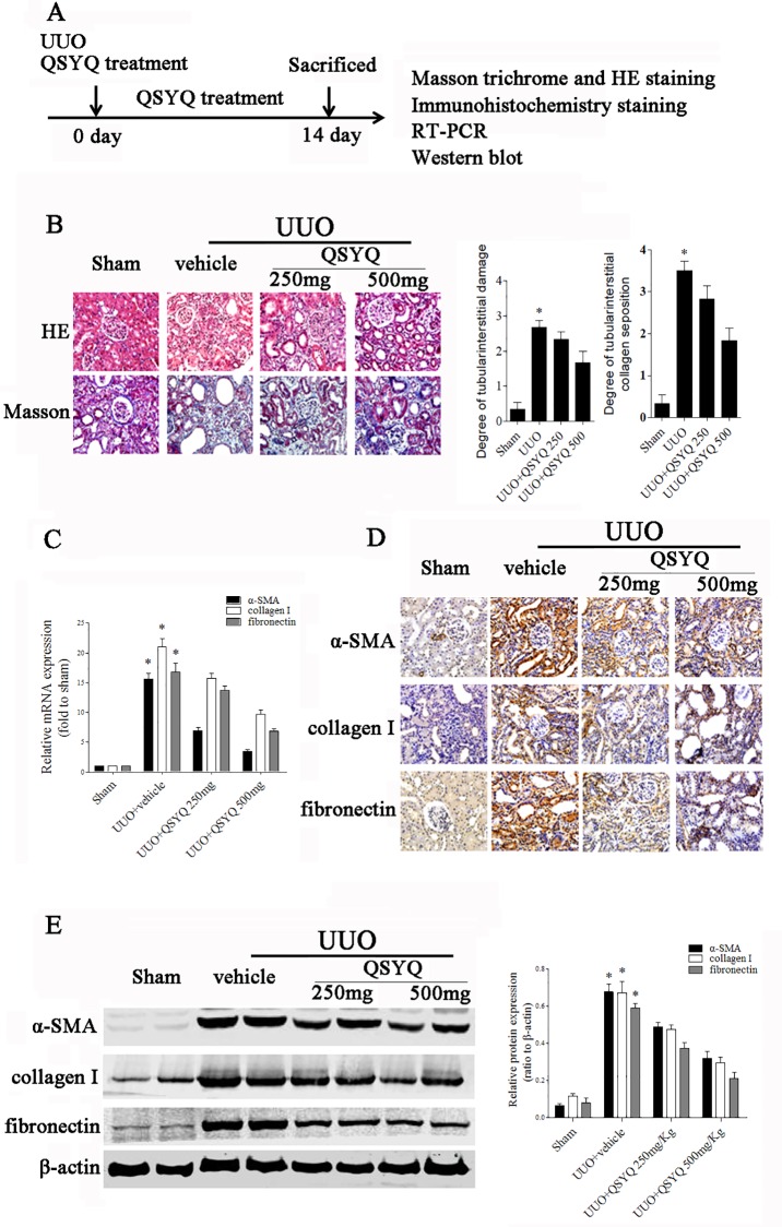
In the Experiment 1, the data showed that there had no significant difference on the levels of serum creatinine, urinary output and proteinuria between sham operation group and UUO group (S1 Table) and this could be attributed to the fact that the contralateral kidney maintains its function [26]. Also, the level of NGAL in the kidney of UUO model had no marked change compared to the sham operation group. Thus, these indicators might not reflect renal damage induced by UUO. And with HE and Masson trichrome, the result showed that marked interstitial inflammation and fibrosis occurred in UUO renal tissues stained QSYQ significantly reduced cell infiltration and interstitial fibrosis (Fig 1B) in rats with UUO. QSYQ also exerted dose-dependent inhibition of fibrotic gene expression, such as α-SMA, collagen I, and fibronectin in rats with UUO (Fig 1C–1E). Moreover, the data show the preventive effect of QSYQ in renal interstitial fibrosis.
Fig 1. QSYQ attenuated renal interstitial fibrosis in UUO.
Rats received vehicle or QSYQ at an oral dose of 250 or 500 mg/kg/d following UUO, and were sacrificed at day 14. (A) Schematic presentation of the experimental design. (B) HE and Masson trichrome staining in the obstructed kidney. (C) Real-time PCR of α-SMA, collagen I, and fibronectin. (D) Immunohistochemical staining of α-SMA, collagen I, and fibronectin. (E) Representative bands (two cases) of Western blot analyses for the expression of α-SMA, collagen I, and fibronectin in the obstructed kidneys. *p<10−8 vs sham operation group. ANOVA, p<10−8 in QSYQ-treated rats in B, C, and E; n = 6 for each group.
Delayed administration of QSYQ also attenuated renal fibrosis
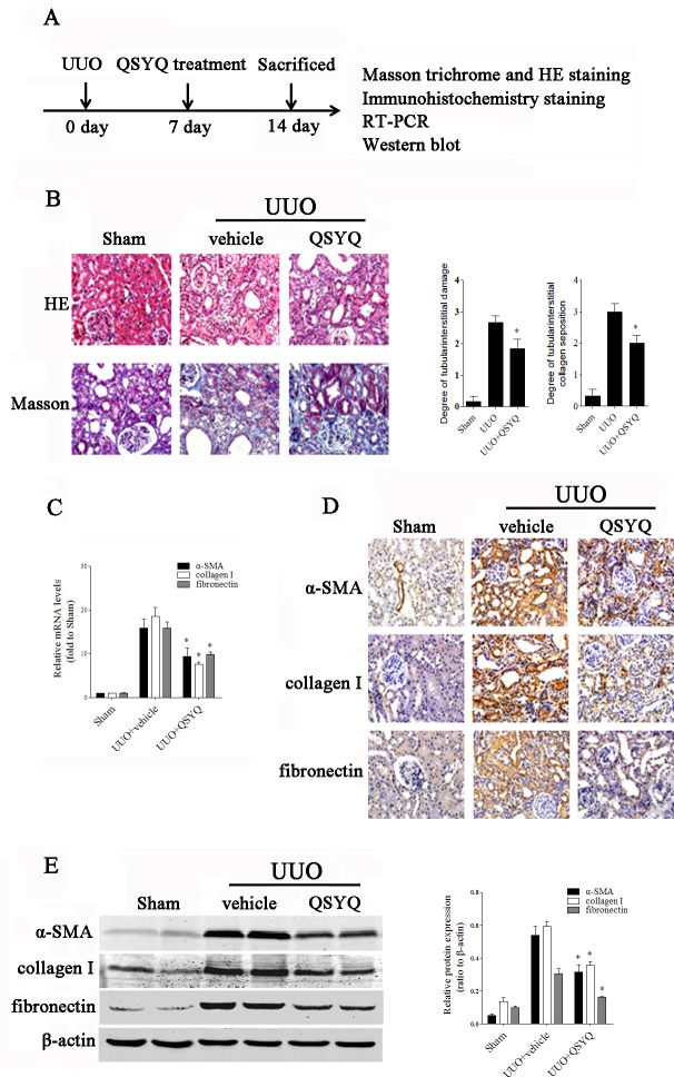
We also determined whether or not delayed administration of QSYQ was effective in renal interstitial fibrosis. Interestingly, delayed administration of QSYQ not only reduced inflammatory cell infiltration (Fig 2A), but also down-regulated the expression of α-SMA, collagen I, and fibronectin in rats with UUO (Fig 2B–2D), suggesting that QSYQ virtually hinder established renal fibrosis.
Fig 2. Late administration of QSYQ attenuated renal interstitial fibrosis in UUO.
Rats received vehicle or QSYQ at an oral dose of 500 mg/kg/d 7d after UUO, and were sacrificed at day 14. (A) Schematic presentation of the experimental design. (B) Representative micrographs of HE and Masson trichrome staining of rat kidney tissues in the groups, as indicated. *p<10−8 vs. vehicle. (C) Real-time PCR analyses of α-SMA, collagen I, and fibronectin in rat kidney tissues. *p = 0.035 vs. vehicle (α-SMA), p = 0.002 (collagen I) and p = 0.003 (fibronectin). (D) Immunohistochemical staining of α-SMA, collagen I, and fibronectin. (E) Western blot analyses of α-SMA, collagen I, and fibronectin in the obstructed kidneys. *p = 0.009 vs. vehicle (α-SMA), p<10−8 (collagen I), and p = 0.004 (fibronectin); n = 6 for each group. The Fig 2E fibronectin panel is excluded from this article's CC-BY license. See the accompanying retraction notice for more information.
QSYQ inhibited TGF-β1-induced fibrotic action in vitro
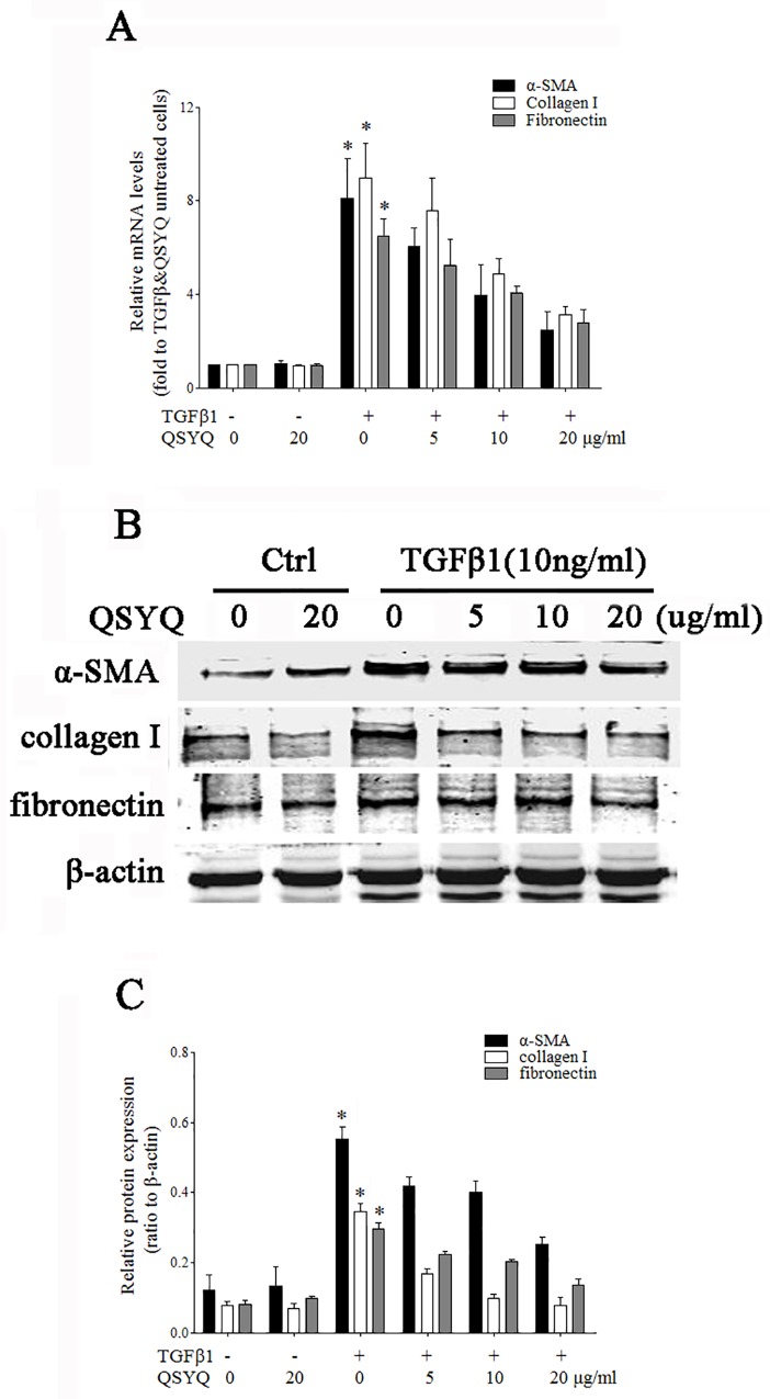
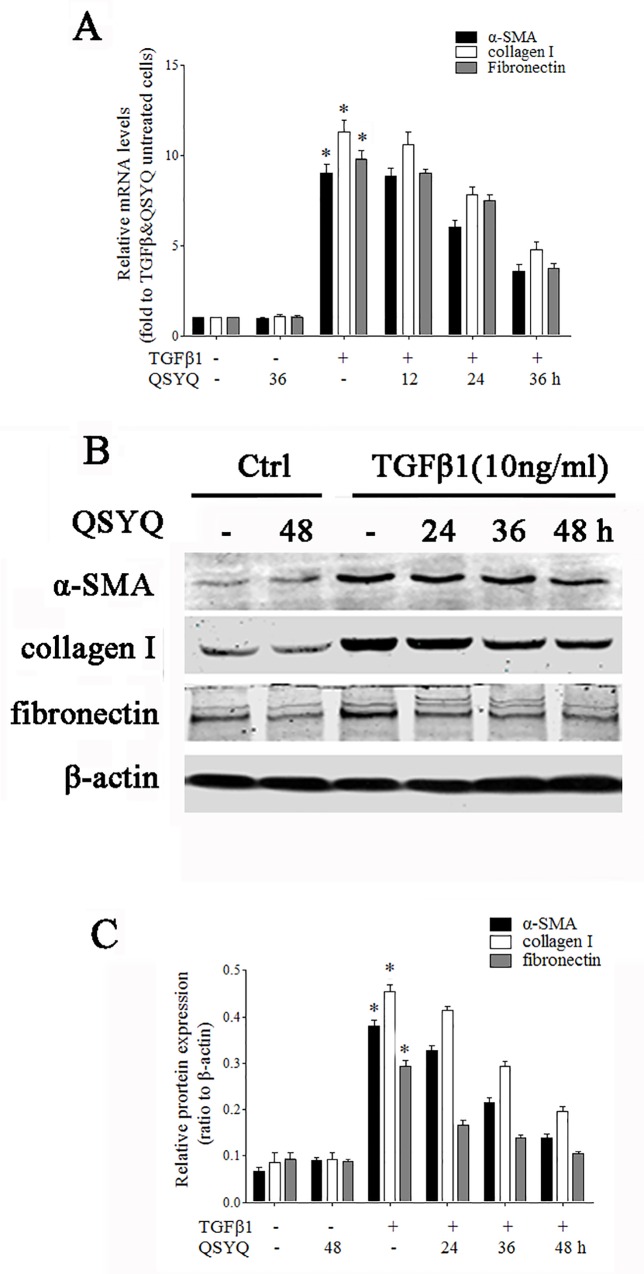
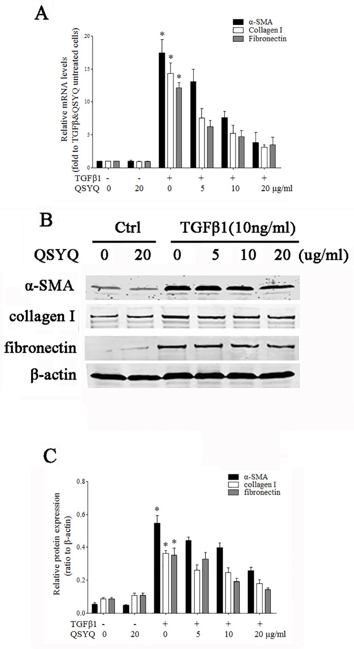
To further confirm the anti-fibrotic effect of QSYQ, we determined whether or not QSYQ could inhibit TGF-β1-induced fibrogenic action in vitro. As shown in Figs 3 and 4, TGF-β1 significantly up-regulated mRNA and protein expression of α-SMA, collagen I, and fibronectin in NRK52E cells. Treatment with QSYQ not only dose dependently (Fig 3) but also time dependently (Fig 4) down-regulated the expression of these fibrogenic genes at the mRNA (Figs 3A and 4A) and protein levels (Figs 3B and 4B). Similar results were obtained using NRK49F cells (Fig 5). Taken together, these data support the results obtained in UUO rats.
Fig 3. QSYQ dose-dependently inhibited the fibrotic activity mediated by TGF-β1 in NRK 52E cells.
NRK 52E cells were pre-incubated with QSYQ (5, 10, and 20 μg/ml) for 1h before TGF-β1 (10 ng/ml) treatment. Cells were harvested 36h after TGF-β1 stimulation for real-time PCR and 48h after TGF-β1 stimulation for Western blot analyses, respectively. (A) Real-time PCR analyses of α-SMA, collagen I, and fibronectin. *p = 0.002 vs. TGF-β1 and QSYQ untreated cells (α-SMA), p = 0.001 (collagen I) and p = <10−8 (fibronectin). ANOVA, p = 0.002 for QSYQ-treated cells (α-SMA), p = 0.001 (collagen I) and p = 0.002 (fibronectin). (B) Western blot analyses of α-SMA, collagen I, and fibronectin. *p = 0.002 vs. TGF-β1 and QSYQ untreated cells (α-SMA), p = 0.001 (collagen I) and p = 0.001 (fibronectin). ANOVA, p<10−8 for QSYQ-treated cells. Data are expressed as the mean ± SD of three independent experiments. The Fig 3B collagen I panel is excluded from this article's CC-BY license. See the accompanying retraction notice for more information.
Fig 4. QSYQ time-dependently inhibited the fibrotic activity mediated by TGF-β1 in NRK 52E cells.
NRK 52E cells were pre-incubated with TGF-β1 (10 ng/ml) and treated with QSYQ (20 μg/ml) 0, 12, and 24h after TGF-β1 incubation, respectively. Cells were harvested 36 h after TGF-β1 stimulation for real-time PCR and 48 h after TGF-β1 stimulation for Western blot analysis, respectively. (A) Real-time PCR analyses of α-SMA, collagen I, and fibronectin. (B) Western blot analyses of α-SMA, collagen I, and fibronectin. *p<10−8 vs TGF-β1 and QSYQ untreated cells. ANOVA, p<10−8 for QSYQ-treated cells in A and B. Data are expressed as the mean ± SD of three independent experiments. The Fig 4B collagen I and α-SMA panels are excluded from this article's CC-BY license. See the accompanying retraction notice for more information.
Fig 5. QSYQ inhibited the fibrotic activity mediated by TGF-β1 in NRK 49F cells.
NRK49F cells were pre-incubated with QSYQ (5, 10, and 20 μg/ml) for 1 h before TGF-β1 (10 ng/ml) treatment. Cells were harvested 36 h after TGF-β1 stimulation for real-time PCR and 48 h after TGF-β1 stimulation for Western blot analyses, respectively. (A) Real-time PCR analyses of α-SMA, collagen I, and fibronectin. *p = 0.001 vs TGF-β1 and QSYQ untreated cells. ANOVA, p = 0.001 for QSYQ-treated cells. (B) Western blot analyses of α-SMA, collagen I, and fibronectin. *p = 0.001 vs. TGF-β1 and QSYQ untreated cells (α-SMA), p = 0.001 (collagen I) and p = 0.003 (fibronectin). ANOVA, p = 0.001 for QSYQ-treated cells (α-SMA), p = 0.006 (collagen I) and p = 0.003 (fibronectin). Data are expressed as the mean ± SD of three independent experiments. The Fig 5B fibronectin panel is excluded from this article's CC-BY license. See the accompanying retraction notice for more information.
QSYQ blocked TGF-β1-induced β-catenin up-regulation and downstream gene transcription
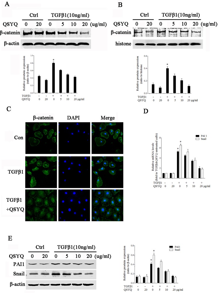
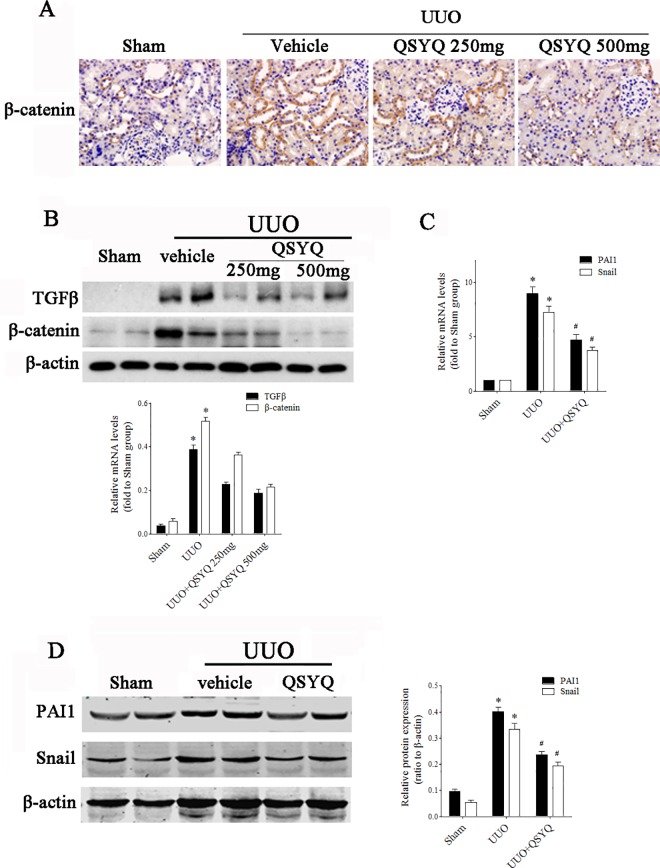
We next examined the potential mechanisms of the anti-fibrotic effect. Given the critical role of β-catenin activation in renal fibrosis, we reasoned that QYSQ might affect this protein. As shown in Fig 6, TGF-β1 significantly up-regulated β-catenin. Treatment with QSYQ inhibited the up-regulation of β-catenin in a dose-dependent fashion in the cytoplasm (Fig 6A) and nucleus (Fig 6B). Also, immunofluorescence staining revealed that pre-incubating NRK52E cells with QSYQ significantly reduced the TGF-β1-induced β-catenin nuclear translocation (Fig 6C). We further examined the effect of QSYQ on β-catenin driven gene transcription. As shown in sFig 6D and 6E, QSYQ inhibited β-catenin-driven PAI-1 and Snail expression in NRK52E cells in a dose-dependent fashion. The similar results were obtained from QSYQ treated UUO rats (Fig 7)
Fig 6. QSYQ blocked TGF-β1-induced β-catenin up-regulation and downstream gene transcription.
NRK52E cells were pre-incubated with or without QSYQ (5, 10, and 20 μg/ml) before treatment with TGF-β1 (10 ng/ml). (A) Cells were collected 24 h after treatment with TGF-β1 for total protein extraction, followed by immunobloting using antibodies against β-catenin. * p = 0.001 vs TGF-β1 and QSYQ untreated cells. ANOVA, p<10−8 for QSYQ-treated cells; (B) Cells were collected for nuclear protein extraction, followed by immunobloting using antibodies against β-catenin; * p = 0.002 vs TGF-β1 and QSYQ untreated cells. ANOVA, p = 0.001 for QSYQ-treated cells. (C) Immunofluorescence staining revealed that QSYQ treatment inhibited TGFβ1-induced nuclei translocation of β-catenin (800×). (D) Real-time PCR analyses of PAI 1 and Snail. *p<10−8 vs TGF-β1 and QSYQ untreated cells. ANOVA, p<10−8 for QSYQ-treated cells. (E) Cell lysates were collected for total protein extraction, followed by immunobloting using anti-PAI 1 and anti-Snail antibodies. *p = 0.002 vs TGF-β1 and QSYQ untreated cells (PAI 1), p = 0.001 (Snail). ANOVA, p<10−8 for QSYQ-treated cells. Data are expressed as the mean ± SD of three independent experiments.
Fig 7. QSYQ blocked β-catenin up-regulation and downstream gene transcription in UUO kidney.
Rats received vehicle or QSYQ at an oral dose of 250 or 500mg/kg/d following UUO, and were sacrificed at day 14. (A) Immunohistochemical staining of β-catenin. (B) Representative bands (two cases) of Western blot analyses for the expression of TGF-β and β-catenin in the obstructed kidneys. * p<10−8 vs sham operation groups. ANOVA, p<10−8 in QSYQ-treated rats. (C) Real-time PCR analyses of PAI1 and snail. (D) Western blot analyses of PAI 1 and snail. * p<10−8 vs sham operation groups. # p<10−8 vs. UUO groups (PAI 1) and p = 0.001 (Snail); n = 6 for each group.
Effect of QSYQ did not rely on the Smads, p38, PI3k, or ERK pathways
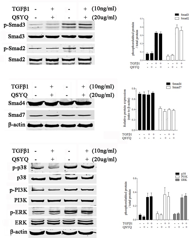
We further determined whether or not QSYQ affected other signaling pathways involving TGF-β1. As shown in Fig 8, TGF-β1 induced Smad2 and Smad3 phosphorylation in NRK52E cells. Treatment with QSYQ did not affect TGF-β1-induced Smad2 or Smad3 phosphorylation (Fig 6A). Furthermore, QSYQ did not affect Smad4 or Smad7 expression (Fig 6B). Interestingly, QSYQ did not affect p38, PI3K, or ERK phosphorylation (Fig 6C). These data suggested that the effect of QSYQ in renal fibrosis did not rely on the Smads, p38, PI3K, or ERK pathways.
Fig 8. Effect of QSYQ did not rely on Smads, p38, PI3K, or ERK pathways.
NRK 52E cells were pre-incubated with or without QSYQ (20 μg/ml) before treatment with TGF-β1 (10 ng/ml). Cells were collected for Western blot analyses. (A) Cell lysates were immunoblotted with antibodies against phosphorylated Smad2, Smad2, phosphorylated Smad3, and Smad3. (B) Cell lysates were immunoblotted with antibodies against Smad4 and Smad7. (C) Cell lysates were immunoblotted with antibodies against phosphorylated p38, p38, phosphorylated PI3K, PI3K, phosphorylated ERK, and ERK. Data are expressed as the mean ± SD of three independent experiments.
Discussion
In the current study, we demonstrated that QSYQ, a water-ethanol extract from R. astragali, S. miltiorrhiza, P. notoginseng, and rosewood, significantly attenuated renal interstitial fibrosis in rats with UUO. QSYQ might selectively inhibit TGF-β1-induced β-catenin up-regulation.
UUO is a typical animal model of renal interstitial fibrosis, in which inflammatory cell infiltration, tubular degeneration and atrophy, and interstitial fibrosis is observed 7 d after operation, while severe tubulo-interstitial injury occurs on d 14 after UUO [27]. The morphologic changes and accompanying α-SMA, collagen, and fibronectin up-regulation are widely used in detecting renal fibrosis [28–31]. Our in vivo data indicated that treatment with QSYQ results in impressive renal protection in rats with UUO; later administration of QSYQ was also effective in this established renal fibrosis model. In vitro data also provided similar results in epithelial and myofibroblast cells, two of the most important types of cells in renal interstitial cells [32–35], suggesting an inhibitory effect of QSYQ in renal interstitial fibrosis.
Because β-catenin has a significant role in mediating renal fibrosis [13–15], it might be essential that blocking β-catenin prevents renal fibrosis. Indeed, our study showed that QSYQ dramatically suppresses β-catenin up-regulation induced by TGF-β1. Treatment with QSYQ not only inhibited β-catenin-driven PAI-1 and Snail1 expression, but also inhibited fibrotic gene expression, including α-SMA, collagen I, and fibronectin in epithelial and myofibroblast cells. The inhibitory effect of QSYQ appears to be β-catenin-specific because QSYQ did not affect Smad2/3 phosphorylation or the expression of Smad4 or Smad7, or the activation of other downstream signaling pathways of TGF-β1, such as p38, ERK, and PI3K.
CKD is becoming a worldwide problem. However, there are few intervention strategies available that specifically target the pathogenesis of renal fibrosis. Given the critical role of TGF-β1 in renal fibrosis, the efforts for developing anti-fibrotic strategies are focusing on this signaling pathway. More and more molecules inhibiting the TGF-β pathway are under development, including neutralizing antibodies against TGF-β [36,37], soluble chimeric TGF-β1 receptor [38], small molecule inhibitors for TGF-β Receptors, such as ALK5 [39], selective Smad3 inhibitors [40], and GQ5 [41]. Selective β-catenin inhibitors, such as ICG-001, have been proven as an effective inhibitor of renal fibrosis [42,43]. Until now, these molecules are far from being ready for clinical application. In the present study, we found that QSYQ selectively inhibits β-catenin up-regulation and downstream fibrogenic action.
QSYQ is a complex extraction from a series of Chinese medicines and it contains many active components, including astragaloside, tanshinol, protocatechualdehyde, and ginsenosides Rg1 and Rb1 [44]. Astragaloside is the main effective component of Radix Astragali, and it can regulate the immune system, decrease glucose and inhibit the inflammation reaction [44]. As the main effective components of Radix Salviaemiltiorrhizae, Tanshinol and protocatechualdehydehave the effects on inhibition of thrombosis, and enhancement of the immune function[44]. Ginsenosides Rg1 and Rb1 are the main effective components of Radix Notoginseng, and their pharmacological actions include inhibiting platelet coagulation agents, reducing myocardial oxygen consumption, and so on[44]. However, the useful compounds were still unknown and the action of QSYQ on the renal interstitial fibrosis might contribute to the synergistic effect of many active components. In addition, few side effects associated with QSYQ have been reported. In a preliminary toxicity test, rats were treated with QSYQ via by oral gavage at a dose of 2.5 g/kg/day for 90 days. Blood examination and pathological observation demonstrated that QSYQ have nomarked toxicity. Thus, QSYQ might provide an alternative drug for treatment of CKD in the future.
Conclusions
In conclusion, we have identified a Chinese medicine, QSYQ, as a potent selective inhibitor of β-catenin. QSYQ, by blocking the activation of β-catenin, inhibits downstreamfibrogenic action and attenuates renal interstitial fibrosis in rats with UUO. Our study provides an incentive to conduct clinical trials to confirm the use of QSYQ in patients with CKD.
Supporting Information
Serum and urine were collected at 14 day after UUO procedure. Serum creatinine and 24h urine protein excretion were tested by automatic biomedical analyzer. Rat kidney NGALs were tested by rat NGAL ELISA kit.
(DOC)
Acknowledgments
This work was supported by the Nanfang Hospital Foundation of Southern Medical University (2012C08) to Dr. Ai.
Data Availability
All relevant data are within the paper and its Supporting Information files.
Funding Statement
This work was supported by the Nanfang Hospital Fundation, Southern Medical University (2012C08) to Dr. Ai. Tasly R&D Institute provided support in the form of salaries for author HZ, but did not have any additional role in the study design, data collection and analysis, decision to publish, or preparation of the manuscript. The specific role of this author is articulated in the ‘author contributions’ section.
References
- 1.Coresh J, Selvin E, Stevens LA, Manzi J, Kusek JW, Eggers P, et al. Prevalence of chronic kidney disease in the United States. JAMA. 2007; 298: 2038–2047. [DOI] [PubMed] [Google Scholar]
- 2.Crews DC, Plantinga LC, Miller ER 3rd, Saran R, Hedgeman E, Saydah SH, et al. Prevalence of chronic kidney disease in persons with undiagnosed or prehypertension in the United States. Hypertension. 2010; 55: 1102–1109. doi: 10.1161/HYPERTENSIONAHA.110.150722 [DOI] [PMC free article] [PubMed] [Google Scholar]
- 3.Tonelli M1, Wiebe N, Culleton B, House A, Rabbat C, Fok M, McAlister F, et al. Chronic kidney disease and mortality risk: a systematic review. J Am Soc Nephrol. 2006; 17: 2034–2047. [DOI] [PubMed] [Google Scholar]
- 4.Zeisberg M, Neilson EG. Mechanisms of tubulointerstitial fibrosis. J Am Soc Nephrol. 2010; 21: 1819–1834. doi: 10.1681/ASN.2010080793 [DOI] [PubMed] [Google Scholar]
- 5.Boor P, Ostendorf T, Floege J. Renal fibrosis: novel insights into mechanisms and therapeutic targets. Nat Rev Nephrol. 2010; 6: 643–656. doi: 10.1038/nrneph.2010.120 [DOI] [PubMed] [Google Scholar]
- 6.Liu Y. Renal fibrosis: new insights into the pathogenesis and therapeutics. Kidney Int. 2006; 69: 213–217. [DOI] [PubMed] [Google Scholar]
- 7.Wang W, Koka V, Lan HY. Transforming growth factor-beta and Smad signalling in kidney diseases. Nephrology (Carlton). 2005; 10: 48–56. [DOI] [PubMed] [Google Scholar]
- 8.Heldin CH, Miyazono K, ten Dijke P. TGF-beta signalling from cell membrane to nucleus through SMAD proteins. Nature. 1997; 390: 465–471. [DOI] [PubMed] [Google Scholar]
- 9.Yu L, Hebert MC, Zhang YE. TGF-beta receptor-activated p38 MAP kinase mediates Smad-independent TGF-beta responses. EMBO J. 2002; 21: 3749–3759. [DOI] [PMC free article] [PubMed] [Google Scholar]
- 10.Kolosova I, Nethery D, Kern JA. Role of Smad2/3 and p38 MAP kinase in TGF-beta1-induced epithelial-mesenchymal transition of pulmonary epithelial cells. J Cell Physiol. 2011; 226: 1248–1254. doi: 10.1002/jcp.22448 [DOI] [PMC free article] [PubMed] [Google Scholar]
- 11.Suwanabol PA, Seedial SM, Zhang F, Shi X, Si Y, Liu B, et al. TGF-beta and Smad3 modulate PI3K/Akt signaling pathway in vascular smooth muscle cells. Am J Physiol Heart Circ Physiol. 2012; 302: H2211–2219. doi: 10.1152/ajpheart.00966.2011 [DOI] [PMC free article] [PubMed] [Google Scholar]
- 12.Hough C, Radu M, Dore JJ. Tgf-beta induced Erk phosphorylation of smad linker region regulates smad signaling. PLOS ONE. 2012; 7: e42513. doi: 10.1371/journal.pone.0042513 [DOI] [PMC free article] [PubMed] [Google Scholar]
- 13.Schmidt-Ott KM, Barasch J. WNT/beta-catenin signaling in nephron progenitors and their epithelial progeny. Kidney Int. 2008; 74: 1004–1008. doi: 10.1038/ki.2008.322 [DOI] [PMC free article] [PubMed] [Google Scholar]
- 14.Yu J, Carroll TJ, Rajagopal J, Kobayashi A, Ren Q, McMahon AP. A Wnt7b-dependent pathway regulates the orientation of epithelial cell division and establishes the cortico-medullary axis of the mammalian kidney. Development. 2009; 136: 161–171. doi: 10.1242/dev.022087 [DOI] [PMC free article] [PubMed] [Google Scholar]
- 15.Surendran K, Schiavi S, Hruska KA. Wnt-dependent beta-catenin signaling is activated after unilateral ureteral obstruction, and recombinant secreted frizzled-related protein 4 alters the progression of renal fibrosis. J Am Soc Nephrol. 2005; 16: 2373–2384. [DOI] [PubMed] [Google Scholar]
- 16.Langham RG, Kelly DJ, Gow RM, Zhang Y, Cordonnier DJ, Pinel N, et al. Transforming growth factor-beta in human diabetic nephropathy: effects of ACE inhibition. Diabetes Care. 2006; 29: 2670–2675. [DOI] [PubMed] [Google Scholar]
- 17.Cassidy H, Radford R, Slyne J, O'Connell S, Slattery C, Ryan MP, et al. The role of MAPK in drug-induced kidney injury. J Signal Transduct. 2012; 2012: 463617. doi: 10.1155/2012/463617 [DOI] [PMC free article] [PubMed] [Google Scholar]
- 18.Hong C, Wang Y, Lou J, Liu Q, Qu H, Cheng Y. Analysis of myocardial proteomic alteration after Qishenyiqi formula treatment in acute infarcted rat hearts. Zhongguo Zhong Yao Za Zhi. 2009; 34: 1018–1021. [PubMed] [Google Scholar]
- 19.Li YC, Liu YY, Hu BH, Chang X, Fan JY, Sun K, et al. Attenuating effect of post-treatment with QiShen YiQi Pills on myocardial fibrosis in rat cardiac hypertrophy. Clin Hemorheol Microcirc. 2012; 51: 177–191. doi: 10.3233/CH-2011-1523 [DOI] [PubMed] [Google Scholar]
- 20.Lin SQ, Wei XH, Huang P, Liu YY, Zhao N, Li Q, et al. QiShenYiQi Pills(R) prevent cardiac ischemia-reperfusion injury via energy modulation. Int J Cardiol. 2013; 168: 967–974. doi: 10.1016/j.ijcard.2012.10.042 [DOI] [PubMed] [Google Scholar]
- 21.Shang H, Zhang J, Yao C, Liu B, Gao X, Ren M, et al. Qi-shen-yi-qi dripping pills for the secondary prevention of myocardial infarction: a randomised clinical trial. Evid Based Complement Alternat Med. 2013; 2013: 738391. doi: 10.1155/2013/738391 [DOI] [PMC free article] [PubMed] [Google Scholar]
- 22.Zeisberg M, Soubasakos MA, Kalluri R. Animal models of renal fibrosis. Methods Mol Med. 2005; 117: 261–272. [DOI] [PubMed] [Google Scholar]
- 23.Thornhill BA, Chevalier RL. Variable partial unilateral ureteral obstruction and its release in the neonatal and adult mouse. Methods Mol Biol. 2012; 886: 381–392. doi: 10.1007/978-1-61779-851-1_33 [DOI] [PubMed] [Google Scholar]
- 24.Radford MG Jr, Donadio JV Jr, Bergstralh EJ, Grande JP. Predicting renal outcome in IgA nephropathy. J Am Soc Nephrol. 1997; 8: 199–207. [DOI] [PubMed] [Google Scholar]
- 25.Lin SL, Chen RH, Chen YM, Chiang WC, Lai CF, Wu KD, et al. Pentoxifylline attenuates tubulointerstitial fibrosis by blocking Smad3/4-activated transcription and profibrogenic effects of connective tissue growth factor. J Am Soc Nephrol. 2005; 16: 2702–2713. [DOI] [PubMed] [Google Scholar]
- 26.Ning XH, Ge XF, Cui Y, An HX. Ulinastatin inhibits unilateral ureteral obstruction-induced renal interstitial fibrosis in rats via transforming growth factor beta (TGF-beta)/Smad signalling pathways. Int Immunopharmacol. 2013; 15: 406–413. doi: 10.1016/j.intimp.2012.12.019 [DOI] [PubMed] [Google Scholar]
- 27.Chevalier RL, Forbes MS, Thornhill BA. Ureteral obstruction as a model of renal interstitial fibrosis and obstructive nephropathy. Kidney Int. 2009; 75: 1145–1152. doi: 10.1038/ki.2009.86 [DOI] [PubMed] [Google Scholar]
- 28.Viedt C, Burger A, Hansch GM. Fibronectin synthesis in tubular epithelial cells: up-regulation of the EDA splice variant by transforming growth factor beta. Kidney Int. 1995; 48: 1810–1817. [DOI] [PubMed] [Google Scholar]
- 29.Strutz F, Zeisberg M. Renal fibroblasts and myofibroblasts in chronic kidney disease. J Am Soc Nephrol. 2006. l; 17: 2992–2998. [DOI] [PubMed] [Google Scholar]
- 30.Ina K, Kitamura H, Tatsukawa S, Fujikura Y. Significance of alpha-SMA in myofibroblasts emerging in renal tubulointerstitial fibrosis. Histol Histopathol. 2011; 26: 855–866. [DOI] [PubMed] [Google Scholar]
- 31.Ely JJ, Bishop MA, Lammey ML, Sleeper MM, Steiner JM, Lee DR. Use of biomarkers of collagen types I and III fibrosis metabolism to detect cardiovascular and renal disease in chimpanzees (Pan troglodytes). Comp Med. 2010; 60: 154–158. [PMC free article] [PubMed] [Google Scholar]
- 32.Chevalier RL. Molecular and cellular pathophysiology of obstructive nephropathy. Pediatr Nephrol. 1999; 13: 612–619. [DOI] [PubMed] [Google Scholar]
- 33.Grande MT, Lopez-Novoa JM. Fibroblast activation and myofibroblast generation in obstructive nephropathy. Nat Rev Nephrol. 2009; 5: 319–328. doi: 10.1038/nrneph.2009.74 [DOI] [PubMed] [Google Scholar]
- 34.Meran S, Steadman R. Fibroblasts and myofibroblasts in renal fibrosis. Int J Exp Pathol. 2011; 92: 158–167. doi: 10.1111/j.1365-2613.2011.00764.x [DOI] [PMC free article] [PubMed] [Google Scholar]
- 35.Moll S, Ebeling M, Weibel F, Farina A, Araujo Del Rosario A, Hoflack JC, et al. Epithelial cells as active player in fibrosis: findings from an in vitro model. PLOS ONE. 2013; 8: e56575. doi: 10.1371/journal.pone.0056575 [DOI] [PMC free article] [PubMed] [Google Scholar]
- 36.Sharma K, Jin Y, Guo J, Ziyadeh FN. Neutralization of TGF-beta by anti-TGF-beta antibody attenuates kidney hypertrophy and the enhanced extracellular matrix gene expression in STZ-induced diabetic mice. Diabetes. 1996; 45: 522–530. [DOI] [PubMed] [Google Scholar]
- 37.Komesli S, Vivien D, Dutartre P. Chimeric extracellular domain type II transforming growth factor (TGF)-beta receptor fused to the Fc region of human immunoglobulin as a TGF-beta antagonist. Eur J Biochem. 1998; 254: 505–513. [DOI] [PubMed] [Google Scholar]
- 38.Yang YA, Dukhanina O, Tang B, Mamura M, Letterio JJ, MacGregor J, et al. Lifetime exposure to a soluble TGF-beta antagonist protects mice against metastasis without adverse side effects. J Clin Invest. 2002; 109: 1607–1615. [DOI] [PMC free article] [PubMed] [Google Scholar]
- 39.Moon JA, Kim HT, Cho IS, Sheen YY, Kim DK. IN-1130, a novel transforming growth factor-beta type I receptor kinase (ALK5) inhibitor, suppresses renal fibrosis in obstructive nephropathy. Kidney Int. 2006; 70: 1234–1243. [DOI] [PubMed] [Google Scholar]
- 40.Jinnin M, Ihn H, Tamaki K. Characterization of SIS3, a novel specific inhibitor of Smad3, and its effect on transforming growth factor-beta1-induced extracellular matrix expression. Mol Pharmacol. 2006; 69: 597–607. [DOI] [PubMed] [Google Scholar]
- 41.Ai J, Nie J, He J, Guo Q, Li M, Lei Y, et al. GQ5 Hinders Renal Fibrosis in Obstructive Nephropathy by Selectively Inhibiting TGF-beta-Induced Smad3 Phosphorylation. J Am Soc Nephrol. 2015; 26: 1827–1838. doi: 10.1681/ASN.2014040363 [DOI] [PMC free article] [PubMed] [Google Scholar] [Retracted]
- 42.He W, Kang YS, Dai C, Liu Y. Blockade of Wnt/beta-catenin signaling by paricalcitol ameliorates proteinuria and kidney injury. J Am Soc Nephrol. 2011; 22: 90–103. doi: 10.1681/ASN.2009121236 [DOI] [PMC free article] [PubMed] [Google Scholar]
- 43.Hao S, He W, Li Y, Ding H, Hou Y, Nie J, et al. Targeted inhibition of beta-catenin/CBP signaling ameliorates renal interstitial fibrosis. J Am Soc Nephrol. 2011; 22: 1642–1653. doi: 10.1681/ASN.2010101079 [DOI] [PMC free article] [PubMed] [Google Scholar]
- 44.Lv S, Wu M, Li M, Wang Q, Wang X, Xu L, et al. Effect of QiShenYiQi Pill on Myocardial Collagen Metabolism in Rats with Partial Abdominal Aortic Coarctation. Evid Based Complement Alternat Med. 2015; 2015: 415068. doi: 10.1155/2015/415068 [DOI] [PMC free article] [PubMed] [Google Scholar]
Associated Data
This section collects any data citations, data availability statements, or supplementary materials included in this article.
Supplementary Materials
Serum and urine were collected at 14 day after UUO procedure. Serum creatinine and 24h urine protein excretion were tested by automatic biomedical analyzer. Rat kidney NGALs were tested by rat NGAL ELISA kit.
(DOC)
Data Availability Statement
All relevant data are within the paper and its Supporting Information files.


