Abstract
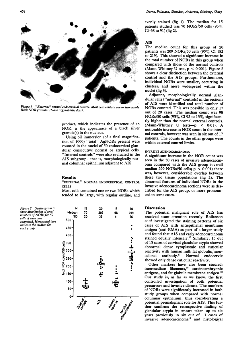
Silver binding nucleolar regions (AgNORs) were evaluated in normal endocervix, adenocarcinoma, and its potential precursor, adenocarcinoma in situ (AIS), in an attempt to increase an understanding of the natural history of cervical adenocarcinoma and to identify a marker for abnormal endocervical (atypical glandular) cells which could aid diagnosis and follow up of endocervical lesions. For every 50 cells the mean AgNOR counts were as follows: normal endocervical cells (n = 15) 79.8 (95% Cl 68-91); AIS (n = 20) 200.7 (95% Cl 182-219); and invasive adenocarcinoma (n = 30) 299 (271-328). There was no overlap between the groups of normal endocervical cells and invasive adenocarcinoma, but there was significant overlap between cases of invasive adenocarcinoma and carcinoma in situ. In six out of 17 cases with AIS, NOR count in adjacent morphologically normal glandular cells ("internal" controls) was increased when compared with the "external" (normal endocervical) control group. This suggests the presence of wider field changes not previously identified using routine histological methods. The findings suggest that AIS is a potential premalignant precursor of invasive adenocarcinoma, but that assessment of NORs is of no practical use in discriminating between the histological types of cervical carcinoma.
Full text
PDF


Images in this article
Selected References
These references are in PubMed. This may not be the complete list of references from this article.
- Boon M. E., Baak J. P., Kurver P. J., Overdiep S. H., Verdonk G. W. Adenocarcinoma in situ of the cervix: an underdiagnosed lesion. Cancer. 1981 Aug 1;48(3):768–773. doi: 10.1002/1097-0142(19810801)48:3<768::aid-cncr2820480318>3.0.co;2-l. [DOI] [PubMed] [Google Scholar]
- Boon M. E., Baak J. P., Kurver P. J., Overdiep S. H., Verdonk G. W. Adenocarcinoma in situ of the cervix: an underdiagnosed lesion. Cancer. 1981 Aug 1;48(3):768–773. doi: 10.1002/1097-0142(19810801)48:3<768::aid-cncr2820480318>3.0.co;2-l. [DOI] [PubMed] [Google Scholar]
- Brown L. J., Griffin N. R., Wells M. Cytoplasmic reactivity with the monoclonal antibody HMFG1 as a marker of cervical glandular atypia. J Pathol. 1987 Mar;151(3):203–208. doi: 10.1002/path.1711510308. [DOI] [PubMed] [Google Scholar]
- Burghardt E. Microinvasive carcinoma in gynaecological pathology. Clin Obstet Gynaecol. 1984 Apr;11(1):239–257. [PubMed] [Google Scholar]
- Chilvers C., Mant D., Pike M. C. Cervical adenocarcinoma and oral contraceptives. Br Med J (Clin Res Ed) 1987 Dec 5;295(6611):1446–1447. doi: 10.1136/bmj.295.6611.1446. [DOI] [PMC free article] [PubMed] [Google Scholar]
- Christopherson W. M., Nealon N., Gray L. A., Sr Noninvasive precursor lesions of adenocarcinoma and mixed adenosquamous carcinoma of the cervix uteri. Cancer. 1979 Sep;44(3):975–983. doi: 10.1002/1097-0142(197909)44:3<975::aid-cncr2820440327>3.0.co;2-7. [DOI] [PubMed] [Google Scholar]
- Clark A. H., Betsill W. L., Jr Early endocervical glandular neoplasia. II. Morphometric analysis of the cells. Acta Cytol. 1986 Mar-Apr;30(2):127–134. [PubMed] [Google Scholar]
- Crocker J., Nar P. Nucleolar organizer regions in lymphomas. J Pathol. 1987 Feb;151(2):111–118. doi: 10.1002/path.1711510203. [DOI] [PubMed] [Google Scholar]
- Crocker J., Skilbeck N. Nucleolar organiser region associated proteins in cutaneous melanotic lesions: a quantitative study. J Clin Pathol. 1987 Aug;40(8):885–889. doi: 10.1136/jcp.40.8.885. [DOI] [PMC free article] [PubMed] [Google Scholar]
- Dabbs D. J., Geisinger K. R., Norris H. T. Intermediate filaments in endometrial and endocervical carcinomas. The diagnostic utility of vimentin patterns. Am J Surg Pathol. 1986 Aug;10(8):568–576. doi: 10.1097/00000478-198608000-00007. [DOI] [PubMed] [Google Scholar]
- Davis J. R., Moon L. B. Increased incidence of adenocarcinoma of uterine cervix. Obstet Gynecol. 1975 Jan;45(1):79–83. [PubMed] [Google Scholar]
- Gallup D. G., Abell M. R. Invasive adenocarcinoma of the uterine cervix. Obstet Gynecol. 1977 May;49(5):596–603. [PubMed] [Google Scholar]
- Giri D. D., Nottingham J. F., Lawry J., Dundas S. A., Underwood J. C. Silver-binding nucleolar organizer regions (AgNORs) in benign and malignant breast lesions: correlations with ploidy and growth phase by DNA flow cytometry. J Pathol. 1989 Apr;157(4):307–313. doi: 10.1002/path.1711570407. [DOI] [PubMed] [Google Scholar]
- Husseinzadeh N., Shbaro I., Wesseler T. Predictive value of cone margins and post-cone endocervical curettage with residual disease in subsequent hysterectomy. Gynecol Oncol. 1989 May;33(2):198–200. doi: 10.1016/0090-8258(89)90551-9. [DOI] [PubMed] [Google Scholar]
- Ireland D., Hardiman P., Monaghan J. M. Adenocarcinoma of the uterine cervix: a study of 73 cases. Obstet Gynecol. 1985 Jan;65(1):82–85. [PubMed] [Google Scholar]
- Newbold K. M., Rollason T. P., Luesley D. M., Ward K. Nucleolar organiser regions and proliferative index in glandular and squamous carcinomas of the cervix. J Clin Pathol. 1989 Apr;42(4):441–442. doi: 10.1136/jcp.42.4.441-b. [DOI] [PMC free article] [PubMed] [Google Scholar]
- PEETE C. H., Jr, CARTER F. B., CHERNY W. B., PARKER R. T. FOLLOW-UP OF PATIENTS WITH ADENOCARCINOMA OF THE CERVIX AND CERVICAL STUMP. Am J Obstet Gynecol. 1965 Oct 1;93:343–356. doi: 10.1016/0002-9378(65)90063-3. [DOI] [PubMed] [Google Scholar]
- Qizilbash A. H. In-situ and microinvasive adenocarcinoma of the uterine cervix. A clinical, cytologic and histologic study of 14 cases. Am J Clin Pathol. 1975 Aug;64(2):155–170. doi: 10.1093/ajcp/64.2.155. [DOI] [PubMed] [Google Scholar]
- Rollason T. P., Byrne P., Williams A., Brown G. Expression of epithelial membrane and 3-fucosyl-N-acetyllactosamine antigens in cervix uteri with particular reference to adenocarcinoma in situ. J Clin Pathol. 1988 May;41(5):547–552. doi: 10.1136/jcp.41.5.547. [DOI] [PMC free article] [PubMed] [Google Scholar]
- Smith R., Crocker J. Evaluation of nucleolar organizer region-associated proteins in breast malignancy. Histopathology. 1988 Feb;12(2):113–125. doi: 10.1111/j.1365-2559.1988.tb01923.x. [DOI] [PubMed] [Google Scholar]
- Teshima S., Shimosato Y., Kishi K., Kasamatsu T., Ohmi K., Uei Y. Early stage adenocarcinoma of the uterine cervix. Histopathologic analysis with consideration of histogenesis. Cancer. 1985 Jul 1;56(1):167–172. doi: 10.1002/1097-0142(19850701)56:1<167::aid-cncr2820560126>3.0.co;2-t. [DOI] [PubMed] [Google Scholar]
- Vesterinen E., Forss M., Nieminen U. Increase of cervical adenocarcinoma: a report of 520 cases of cervical carcinoma including 112 tumors with glandular elements. Gynecol Oncol. 1989 Apr;33(1):49–53. doi: 10.1016/0090-8258(89)90602-1. [DOI] [PubMed] [Google Scholar]
- Wells M., Brown L. J. Glandular lesions of the uterine cervix: the present state of our knowledge. Histopathology. 1986 Aug;10(8):777–792. doi: 10.1111/j.1365-2559.1986.tb02578.x. [DOI] [PubMed] [Google Scholar]


