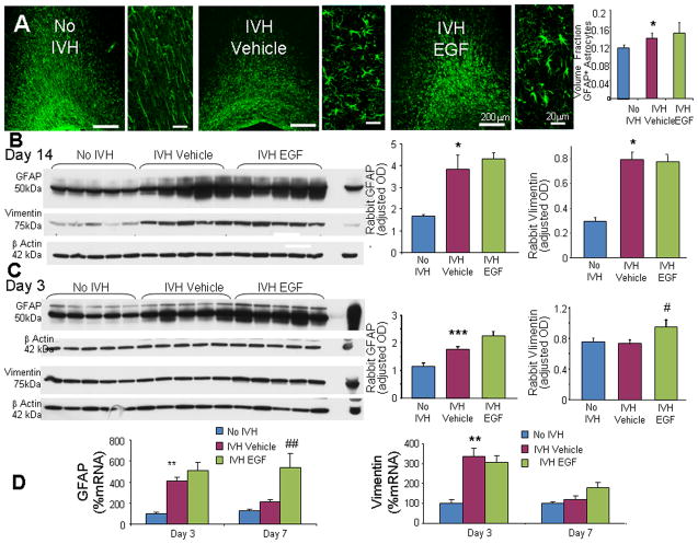
Fig. 6. rhEGF treatment promotes astrogliosis.
A) Representative immunofluorescence of cryosections from pups (D14) labeled with GFAP antibody are shown, as indicated. ‘V’ indicates ventricular side. Rectangular panels are high power view of the adjacent image. Note abundant hypertrophic astrocytes in vehicle and rhEGF treated pups with IVH. Scale bar, 200 μm and 20 μm, as indicated. Graph shows mean ± s.e.m. (n = 5 each). rhEGF treatment did not affect volume fraction of astroglial fibers compared to vehicle controls on stereological-analyses. B) Western blot analyses for GFAP and vimentin were performed in forebrain homogenates of pups (D 14). Adult rat brain was positive-control. Graph shows mean ± s.e.m. (n = 5 each). Values were normalized to β-actin. rhEGF treatment did not affect GFAP or vimentin expression in pups with IVH. C) Western blot analyses for GFAP and vimentin were performed in forebrain homogenates of pups (D 3). Adult rat brain was positive-control. Graph shows mean ± s.e.m. (n = 5 each). Values were normalized to β-actin. rhEGF treatment increased both GFAP and vimentin expression in pups with IVH. D) Data are mean ± s.e.m. GFAP mRNA expression increased with rhEGF treatment at D7, not D3. However, vimentin mRNA expression was not affected by rhEGF treatment at D 3 or 7. *P < 0.05, **P < 0.01, ***P < 0.001 for no IVH vs. IVH. #P < 0.05, ##P < 0.01for vehicle treated vs. rhEGF treated pups with IVH.

