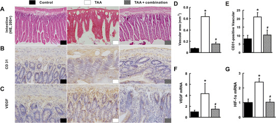
Fig. 3.

Inhibition of intestinal angiogenesis with the combination treatment. The increased intestinal angiogenesis in the TAA group was visualized by HE (a), IHC for CD31 (b) and IHC for VEGF (c). Intestinal vascular areas (d), number of intestinal CD31-postive vessels per fields (e), intestinal VEGF (f) and HIF-1α (g) mRNA quantified by qRT-PCR in the TAA group were the highest among three groups. *p < 0.05 versus control group; # p < 0.05 versus TAA group
