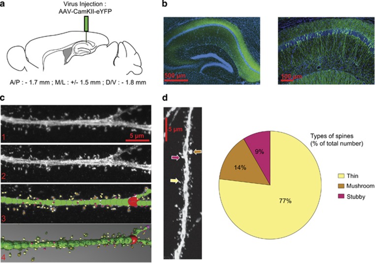
Figure 1.
Analysis of hippocampal CA1 dendritic spines. (a) Schematic representation of the site of AAV5-CamKII-eYFP injection. (b) Confocal image of the dorsal hippocampus 2 weeks following stereotaxic injection of AAV5-CamKII-eYFP (left panel) and a higher magnification of dorsal hippocampus showing expression of YFP in individual CA1 pyramidal neurons (right panel). (c) Example of image treatment and quantification of spines using NeuronStudio. (d) Quantification of types of spines found on hippocampal pyramidal neurons identifying thin spines (77%), mushroom (14%), and stubby spines (9%) expressed as % of total number of spines counted.

