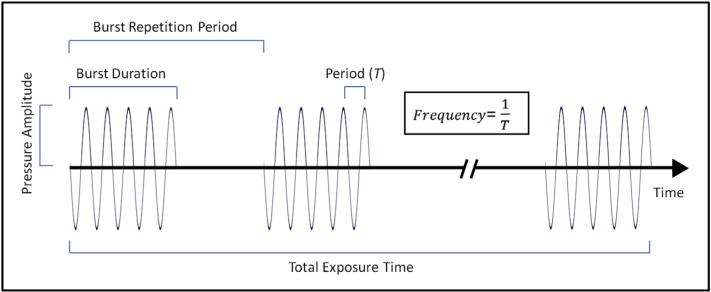
Fig. 4.
Parameters of an ultrasound wave. The ultrasound wave is depicted as a sinusoidal wave, with areas of compression being above the x-axis and areas of rarefaction below. The relationship between burst duration, burst repetition period, period, frequency, pressure amplitude, and total exposure time are displayed.

