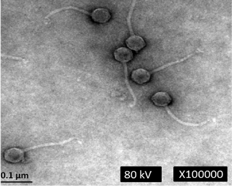
Figure 1. Transmission electron micrograph of phage phiC119 negatively stained with 2% unanyl acetate.

Phage phiC119 showing typical Siphoviridae morphology, which exhibit a noncontractile tail with a length of 168–172 nm. The icosahedral head of phiC119 has a length of 43–45 nm and a width of 7–9 nm. The bar indicates 100 nm.
