

Abstract
Antibiotics (antibacterial, antiviral, and antiparasitic) are class of drugs which result in either killing or inhibiting growth and multiplication of infectious organisms. Antibiotics are commonly prescribed by all specialties for treatment of infections. However, antibiotics have hitherto immunomodulatory and anti-inflammatory properties and can be exploited for various noninfectious dermatoses. Dermatologists routinely prescribe antibiotics in treatment of various noninfectious disorders. This study will review anti-inflammatory and immunomodulatory effects of antibiotics and their use in dermatology.
Keywords: Antibiotics, anti-inflammatory, dermatotherapeutics, inflammatory skin diseases, immunomodulation
Introduction
What was known?
Antibiotics are mainly considered as anti-bacterial agents used for infectious conditions
In dermatology antibiotics are being used for various infectious conditions.
Antibiotics are chemicals derived from microorganisms that have the capacity, in dilute solutions, to kill other microorganisms (bacteria, virus, fungi, and parasite) or inhibit their growth. In this study, antibiotics refer to collective term for antibacterial, antiviral, and antiparasitic agents. In routine clinical practice, antibiotics are chiefly used to eliminate various pathogens (bacteria, viruses, and parasites). Many antibiotics were later found to have anti-inflammatory properties apart from their antimicrobial action. We have discussed anti-inflammatory and anti-immunomodulatory effects of various antibacterial and antiparasitic drugs. Antiviral and antifungal drugs are seldom used for their anti-inflammatory properties.
Antibacterial Agents
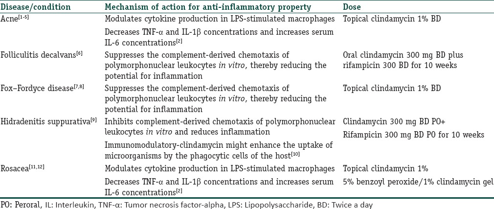
Clindamycin
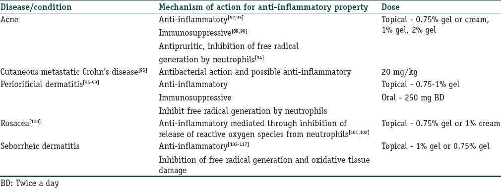
Clindamycin is a synthetic derivative of lincomycin and isolated from the Streptomyces species. The drug has broad-spectrum antibacterial action by binding irreversibly to 50S subunit of bacterial ribosome and thereby inhibiting bacterial protein synthesis. In dermatology, clindamycin is being used for several indications for its both antibacterial and anti-inflammatory properties [Table 1].
Table 1.
Indications of clindamycin as an anti-inflammatory agent
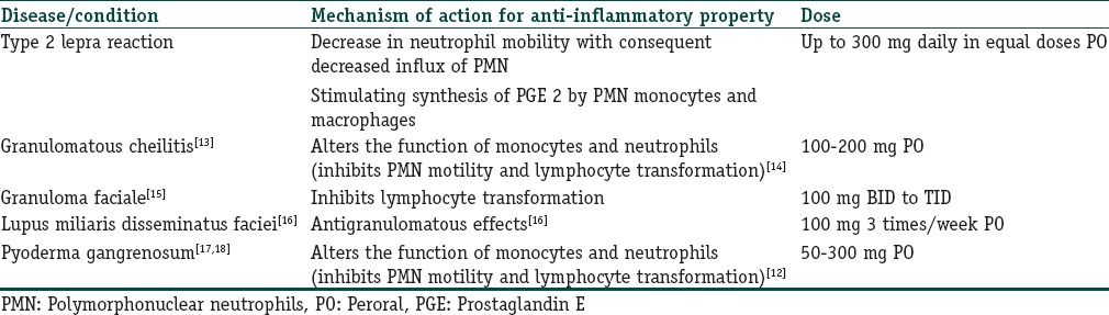
Clofazimine
Clofazimine is a iminophenazine dye known for its antimycobacterial properties. Its absorption is increased with food. It is highly lipophilic and concentrates in lipid-rich tissues. Because of slow elimination, the drug has long half-life of approximately 70 days. Metabolism of the drug occurs in liver and elimination occurs through sebum, sputum, tears, sweat, and urine. However, it also possesses good anti-inflammatory actions and is used in many dermatologic diseases for the same [Table 2].
Table 2.
Indications of clofazimine as an anti-inflammatory agent
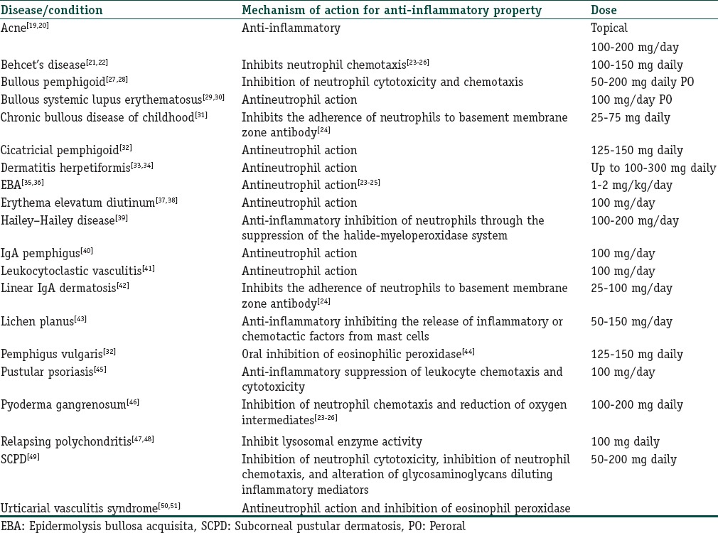
Dapsone
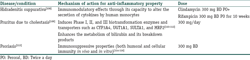
Dapsone (4,4’-diaminodiphenylsulfone) is an aniline derivative belonging to the group of synthetic sulfones. Dapsone is absorbed rapidly and nearly completely from the gastrointestinal tract. Peak plasma concentration is reached within 2–8 h after administration. The mean half-life of elimination is about 20–30 h. It is metabolized in liver by two distinct routes, N-acetylation and N-hydroxylation. It has dual functions of both antimicrobial/antiprotozoal effects and anti-inflammatory features similar to nonsteroidal anti-inflammatory drugs. Dapsone has been used as a treatment option in various dermatological conditions because of its anti-inflammatory effects [Table 3].
Table 3.
Indications of dapsone for its anti-inflammatory and immunomodulatory properties
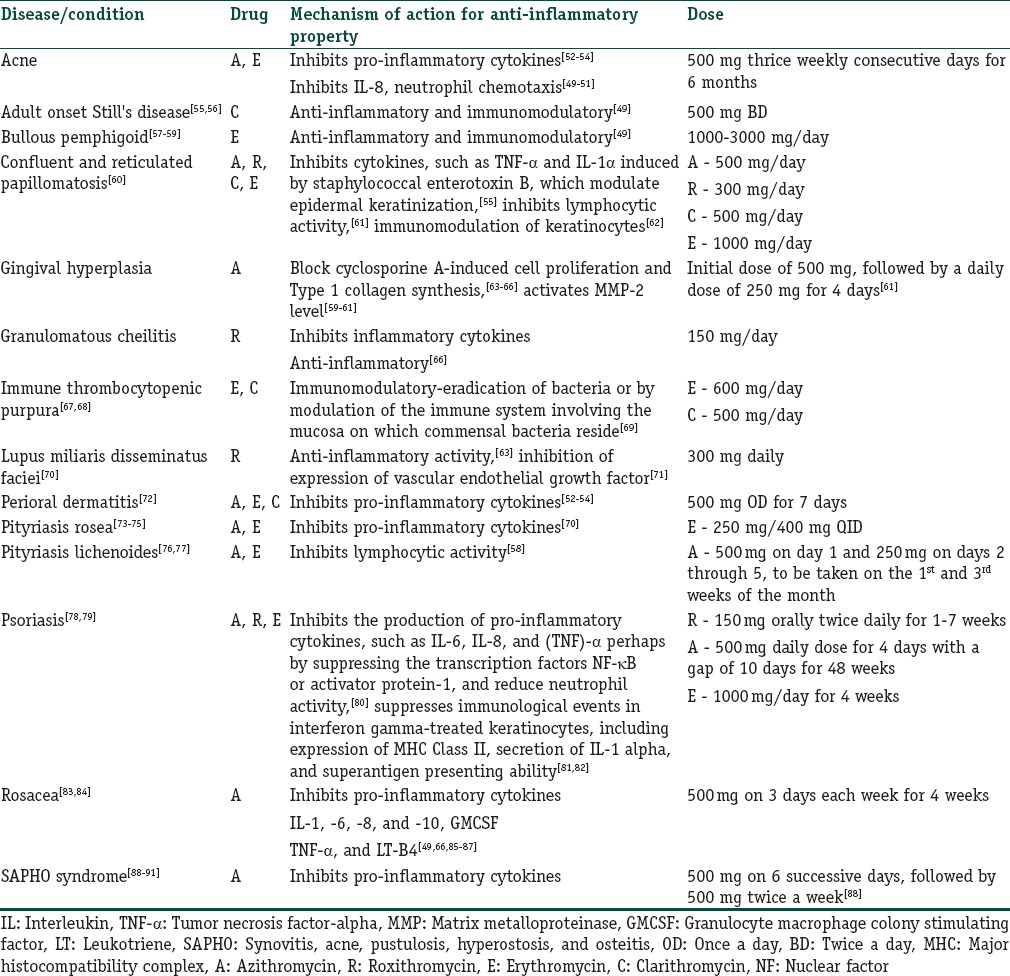
Macrolides
Macrolides contain a macrocyclic lactone ring structure. These are of actinomycetes or semisynthetic derivatives of same bacteria. They are bacteriostatic antibacterial agents which bind irreversibly to the large (50S) ribosomal subunit of bacteria, thereby inhibiting RNA-dependent protein synthesis. However, there have been many dermatological uses of macrolides for their immunomodulatory action. Azithromycin (A), roxithromycin (R), erythromycin (E), and clarithromycin (C) are commonly used in dermatology practice for their immunomodulatory and anti-inflammatory potential [Table 4].
Table 4.
Indications of macrolides in dermatological diseases for their its anti-inflammatory and immunomodulatory properties
Metronidazole
Metronidazole is a synthetic nitroimidazole antibacterial drug. It acts by DNA disruption and nucleic acid synthesis inhibition. It acts against anaerobic bacteria and protozoa. However, it has many actions other than its antibacterial action for which it is being used in different dermatological diseases [Table 5].
Table 5.
Indications of metronidazole for its anti-inflammatory properties
Rifampicin
Rifampicin (R) is a semisynthetic derivative of rifamycin B, an antimicrobial agent produced by Streptomyces mediterranei. It is a broad-spectrum antimicrobial and inhibits the growth of most Gram-positive bacteria, as well as many Gram-negative microorganisms. However, it has other properties besides antimicrobial action for which it has been used in various dermatological conditions [Table 6].
Table 6.
Indications of rifampicin for its anti-inflammatory and immunomodulatory properties
Tetracyclines
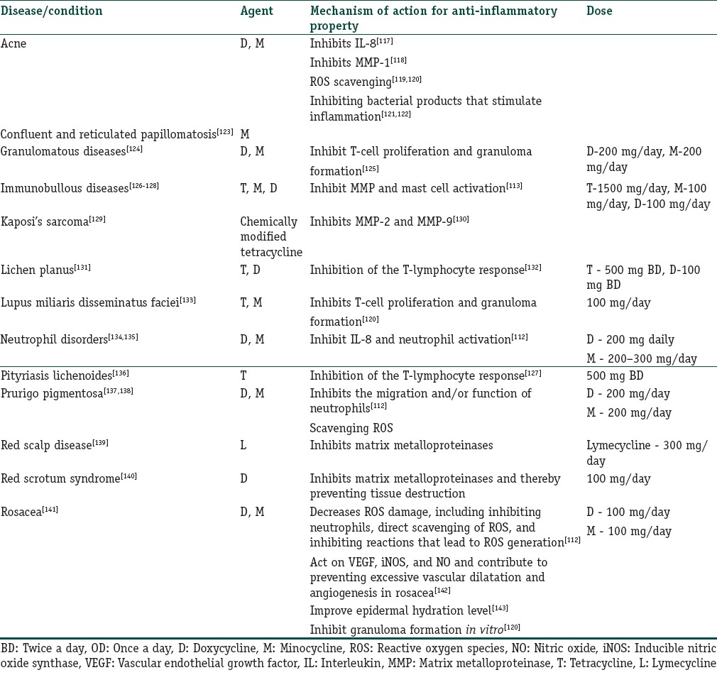
The tetracyclines are broad-spectrum antibiotics and comprise four main drugs (tetracycline [T], doxycycline [D], minocycline [M], and lymecycline [L]). Tetracycline group of antibacterial agents are indicated in a wide range of infections including Treponema pallidum (syphilis), Borrelia burgdorferi, Borrelia afzelii, Borrelia garinii (Lyme disease), Coxiella burnetii (Q fever), Rickettsia rickettsii (Rocky Mountain spotted fever), and Yersinia pestis (Plague). Their antibiotic effect is primarily exerted by binding to the 30S subunit of bacterial ribosomes, thereby halting protein synthesis. However, many tetracyclines have in addition anti-inflammatory properties. [Table 7] discusses the role of tetracyclines chiefly for their anti-inflammatory properties.
Table 7.
Indications of tetracyclines in dermatology for their its anti-inflammatory properties
Antimalarials
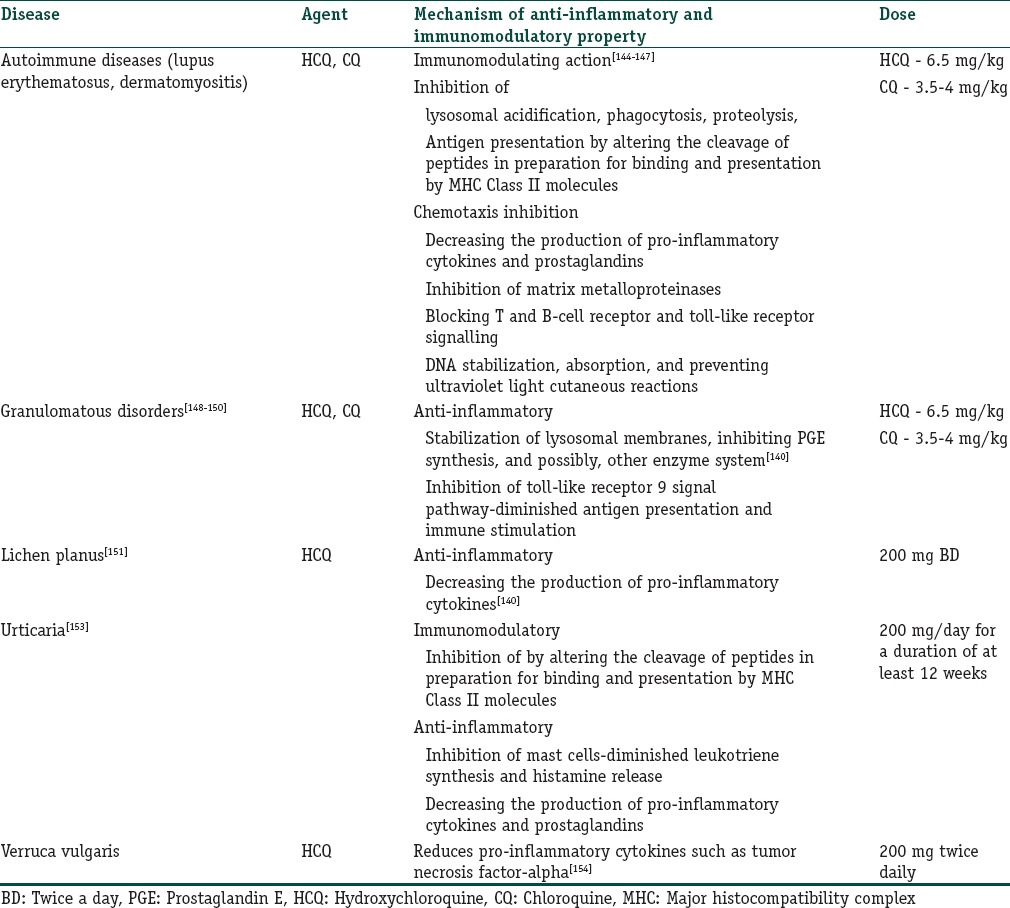
The parent molecule for the antimalarials is quinine. Among antimalarials, chloroquine (CQ) and hydroxychloroquine (HCQ) are used in various dermatological disorders. Both CQ and HCQ are alkylated 4-aminoquinolines. HCQ is a derivative of CQ and is nearly completely absorbed within 2–4 h of an oral dose and metabolized in liver by dealkylation. The drugs accumulate in thrombocytes, granulocytes, and erythrocytes; hence, their concentration in whole blood is 3–10 times higher than that of plasma. CQ has high affinity for melanin and gets accumulated in the eyes and the skin where the concentration is 100–200 times higher than that of plasma; in the epidermis, it is 3–7 times higher than that of the dermis. The maximum daily dosage is 3.5–4 mg/kg of body weight for CQ and 6–6.5 mg/kg body weight for HCQ. Various indications for antimalarials drug are shown in [Table 8].
Table 8.
Indications of antimalarials in dermatology
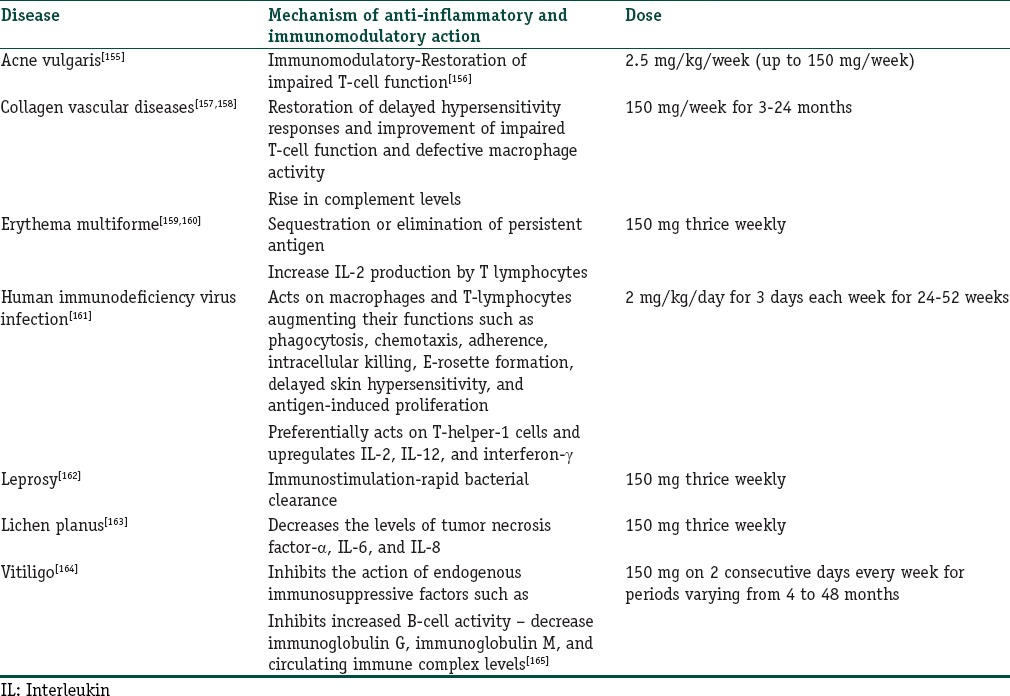
Levamisole
Levamisole is an anthelmintic agent with a wide range of immunomodulatory actions. It belongs to the class of imidazothiazole derivatives. It is water-soluble and gets rapidly absorbed from the gastrointestinal tract with peak blood levels achieved after 1.5–4 h. Metabolism of the drugs occurs mainly in liver and the plasma half-life is 16 h. Due to immunomodulatory properties, it has been widely used in various dermatological disorders. Usual dose of the drug is 150 mg/day for 2–4 days each week [Table 9].
Table 9.
Indications of levamisole in dermatology
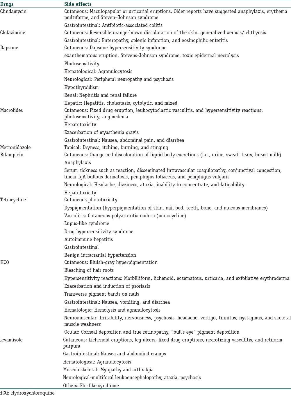
Side effects
All the above-discussed drugs have variety of side effects in the therapeutic dose range. The treating skin physician must be aware of commonly encountered side effects which can enable him or her to rationalize the treatment protocol and manage the side effects with due care [Table 10].
Table 10.
Commonly encountered side effects of various antibiotics
Conclusions
The study aims to highlight the role of various antibiotic drugs in the management of noninfectious diseases of skin and its appendages. In future, many more cutaneous diseases will be treated and managed with various antibiotics tapping their anti-inflammatory properties. We would like to highlight that in future, these antibiotics will be used albeit in continuous low-dose in various noninfectious dermatoses, thereby minimizing the incidence of side effects.165
Financial support and sponsorship
Nil.
Conflicts of interest
There are no conflicts of interest.
What is new?
Antibiotics refer to collective term for antibacterial, antiviral, and antiparasitic agents
Antibiotics have multifaceted actions besides killing the infectious organisms
Anti-inflammatory and immunomodulatory effects of antibiotics make them eligible to be used in various non-infectious conditions in dermatology
Anti-parasitic drugs are also used in dermatology for their anti-inflammatory and immunomodulatory properties.
References
- 1.Del Rosso JQ, Schmidt NF. A review of the anti-inflammatory properties of clindamycin in the treatment of acne vulgaris. Cutis. 2010;85:15–24. [PubMed] [Google Scholar]
- 2.Nakano T, Hiramatsu K, Kishi K, Hirata N, Kadota J, Nasu M. Clindamycin modulates inflammatory-cytokine induction in lipopolysaccharide-stimulated mouse peritoneal macrophages. Antimicrob Agents Chemother. 2003;47:363–7. doi: 10.1128/AAC.47.1.363-367.2003. [DOI] [PMC free article] [PubMed] [Google Scholar]
- 3.Thomas DR, Raimer S, Smith EB. Comparison of topical erythromycin 1.5 percent solution versus topical clindamycin phosphate 1.0 percent solution in the treatment of acne vulgaris. Cutis. 1982;29:624–5. 628-32. [PubMed] [Google Scholar]
- 4.Gold MH, Korotzer A. Sub-group analyses from a trial of a fixed combination of clindamycin phosphate 1.2% and benzoyl peroxide 3.75% gel for the treatment of moderate-to-severe acne vulgaris. J Clin Aesthet Dermatol. 2015;8:22–6. [PMC free article] [PubMed] [Google Scholar]
- 5.Webster G. Cutaneous safety and tolerability of a fixed combination clindamycin (1.2%) and benzoyl peroxide (3.75%) aqueous gel in moderate-to-severe acne vulgaris. J Clin Aesthet Dermatol. 2015;8:22–8. [PMC free article] [PubMed] [Google Scholar]
- 6.Powell JJ, Dawber RP, Gatter K. Folliculitis decalvans including tufted folliculitis: Clinical, histological and therapeutic findings. Br J Dermatol. 1999;140:328–33. doi: 10.1046/j.1365-2133.1999.02675.x. [DOI] [PubMed] [Google Scholar]
- 7.Yost J, Robinson M, Meehan SA. Fox-fordyce disease. Dermatol Online J. 2012;18:28. [PubMed] [Google Scholar]
- 8.George A, Bhatia A, Thomas E. Fox-fordyce disease: A report of 2 cases responding to topical clindamycin. Indian J Dermatol Venereol Leprol. 2015;81:87–8. doi: 10.4103/0378-6323.148597. [DOI] [PubMed] [Google Scholar]
- 9.van der Zee HH, Boer J, Prens EP, Jemec GB. The effect of combined treatment with oral clindamycin and oral rifampicin in patients with hidradenitis suppurativa. Dermatology. 2009;219:143–7. doi: 10.1159/000228337. [DOI] [PubMed] [Google Scholar]
- 10.Pasquale TR, Tan JS. Nonantimicrobial effects of antibacterial agents. Clin Infect Dis. 2005;40:127–35. doi: 10.1086/426545. [DOI] [PubMed] [Google Scholar]
- 11.Weinkle AP, Doktor V, Emer J. Update on the management of rosacea. Clin Cosmet Investig Dermatol. 2015;8:159–77. doi: 10.2147/CCID.S58940. [DOI] [PMC free article] [PubMed] [Google Scholar]
- 12.Breneman D, Savin R, VandePol C, Vamvakias G, Levy S, Leyden J. Double-blind, randomized, vehicle-controlled clinical trial of once-daily benzoyl peroxide/clindamycin topical gel in the treatment of patients with moderate to severe rosacea. Int J Dermatol. 2004;43:381–7. doi: 10.1111/j.1365-4632.2004.02283.x. [DOI] [PubMed] [Google Scholar]
- 13.Fdez-Freire LR, Serrano Gotarredona A, Bernabeu Wittel J, Pulpillo Ruiz A, Cabrera R, Navarrete Ortega M, et al. Clofazimine as elective treatment for granulomatous cheilitis. J Drugs Dermatol. 2005;4:374–7. [PubMed] [Google Scholar]
- 14.Arbiser JL, Moschella SL. Clofazimine: A review of its medical uses and mechanisms of action. J Am Acad Dermatol. 1995;32(2 Pt 1):241–7. doi: 10.1016/0190-9622(95)90134-5. [DOI] [PubMed] [Google Scholar]
- 15.Gómez-de la Fuente E, del Rio R, Rodriguez M, Guerra A, Rodriguez-Peralto JL, Iglesias L. Granuloma faciale mimicking rhinophyma: Response to clofazimine. Acta Derm Venereol. 2000;80:144. [PubMed] [Google Scholar]
- 16.Seukeran DC, Stables GI, Cunliffe WJ, Sheehan-Dare RA. The treatment of acne agminata with clofazimine. Br J Dermatol. 1999;141:596–7. doi: 10.1046/j.1365-2133.1999.03084.x. [DOI] [PubMed] [Google Scholar]
- 17.Goihman-Yahr M. Malignant pyoderma gangrenosum responding to clofazimine. Int J Dermatol. 1996;35:757–8. doi: 10.1111/j.1365-4362.1996.tb00660.x. [DOI] [PubMed] [Google Scholar]
- 18.Kaplan B, Trau H, Sofer E, Feinstein A, Schewach-Millet M. Treatment of pyoderma gangrenosum with clofazimine. Int J Dermatol. 1992;31:591–3. doi: 10.1111/j.1365-4362.1992.tb02727.x. [DOI] [PubMed] [Google Scholar]
- 19.Tan BB, Lear JT, Smith AG. Acne fulminans and erythema nodosum during isotretinoin therapy responding to dapsone. Clin Exp Dermatol. 1997;22:26–7. doi: 10.1046/j.1365-2230.1997.1830600.x. [DOI] [PubMed] [Google Scholar]
- 20.Prendiville JS, Logan RA, Russell-Jones R. A comparison of dapsone with 13-cis retinoic acid in the treatment of nodular cystic acne. Clin Exp Dermatol. 1988;13:67–71. doi: 10.1111/j.1365-2230.1988.tb00659.x. [DOI] [PubMed] [Google Scholar]
- 21.Sharquie KE, Najim RA, Abu-Raghif AR. Dapsone in Behçet's disease: A double-blind, placebo-controlled, cross-over study. J Dermatol. 2002;29:267–79. doi: 10.1111/j.1346-8138.2002.tb00263.x. [DOI] [PubMed] [Google Scholar]
- 22.Jacyk WK. Behçet's disease in South African blacks: Report of five cases. J Am Acad Dermatol. 1994;30(5 Pt 2):869–73. doi: 10.1016/s0190-9622(94)70102-4. [DOI] [PubMed] [Google Scholar]
- 23.Harvath L, Yancey KB, Katz SI. Selective inhibition of human neutrophil chemotaxis to N-formyl-methionyl-leucyl-phenylalanine by sulfones. J Immunol. 1986;137:1305–11. [PubMed] [Google Scholar]
- 24.Thuong-Nguyen V, Kadunce DP, Hendrix JD, Gammon WR, Zone JJ. Inhibition of neutrophil adherence to antibody by dapsone: A possible therapeutic mechanism of dapsone in the treatment of IgA dermatoses. J Invest Dermatol. 1993;100:349–55. doi: 10.1111/1523-1747.ep12471811. [DOI] [PubMed] [Google Scholar]
- 25.Booth SA, Moody CE, Dahl MV, Herron MJ, Nelson RD. Dapsone suppresses integrin-mediated neutrophil adherence function. J Invest Dermatol. 1992;98:135–40. doi: 10.1111/1523-1747.ep12555654. [DOI] [PubMed] [Google Scholar]
- 26.Debol SM, Herron MJ, Nelson RD. Anti-inflammatory action of dapsone: Inhibition of neutrophil adherence is associated with inhibition of chemoattractant-induced signal transduction. J Leukoc Biol. 1997;62:827–36. doi: 10.1002/jlb.62.6.827. [DOI] [PubMed] [Google Scholar]
- 27.Person JR, Rogers RS., 3rd Bullous pemphigoid responding to sulfapyridine and the sulfones. Arch Dermatol. 1977;113:610–5. [PubMed] [Google Scholar]
- 28.Jeffes EW, 3rd, Ahmed AR. Adjuvant therapy of bullous pemphigoid with dapsone. Clin Exp Dermatol. 1989;14:132–6. doi: 10.1111/j.1365-2230.1989.tb00910.x. [DOI] [PubMed] [Google Scholar]
- 29.Hall RP, Lawley TJ, Smith HR, Katz SI. Bullous eruption of systemic lupus erythematosus. Dramatic response to dapsone therapy. Ann Intern Med. 1982;97:165–70. doi: 10.7326/0003-4819-97-2-165. [DOI] [PubMed] [Google Scholar]
- 30.Burrows NP, Bhogal BS, Black MM, Rustin MH, Ishida-Yamamoto A, Kirtschig G, et al. Bullous eruption of systemic lupus erythematosus: A clinicopathological study of four cases. Br J Dermatol. 1993;128:332–8. doi: 10.1111/j.1365-2133.1993.tb00180.x. [DOI] [PubMed] [Google Scholar]
- 31.Rao CL, Hall RP. In: Linear immunoglobulin a dermatosis and chronic bullous disease of childhood. Fitzpatrick's Dermatology in General Medicine. Wolff K, Goldsmith LA, Katz SI, Gilchrest BA, Paller AS, Leffell DJ, editors. New York: McGraw Hill Medical; 2003. pp. 485–90. [Google Scholar]
- 32.Knudson RM, Kalaaji AN, Bruce AJ. The management of mucous membrane pemphigoid and pemphigus. Dermatol Ther. 2010;23:268–80. doi: 10.1111/j.1529-8019.2010.01323.x. [DOI] [PubMed] [Google Scholar]
- 33.Bolotin D, Petronic-Rosic V. Dermatitis herpetiformis. Part II. Diagnosis, management, and prognosis. J Am Acad Dermatol. 2011;64:1027–33. doi: 10.1016/j.jaad.2010.09.776. [DOI] [PubMed] [Google Scholar]
- 34.Antiga E, Caproni M. The diagnosis and treatment of dermatitis herpetiformis. Clin Cosmet Investig Dermatol. 2015;8:257–65. doi: 10.2147/CCID.S69127. [DOI] [PMC free article] [PubMed] [Google Scholar]
- 35.Kim JH, Kim YH, Kim SC. Epidermolysis bullosa acquisita: A retrospective clinical analysis of 30 cases. Acta Derm Venereol. 2011;91:307–12. doi: 10.2340/00015555-1065. [DOI] [PubMed] [Google Scholar]
- 36.Krähe J, Galal O, Kordass U, Bornemann P. Epidermolysis bullosa atrophicans gravis. Report of a therapeutic trial with dapsone. Monatsschr Kinderheilkd. 1988;136:140–2. [PubMed] [Google Scholar]
- 37.Fort SL, Rodman OG. Erythema elevatum diutinum. Response to dapsone. Arch Dermatol. 1977;113:819–22. [PubMed] [Google Scholar]
- 38.Cream JJ, Levene GM, Calnan CD. Erythema elevatum diutinum: An unusual reaction to streptococcal antigen and response to dapsone. Br J Dermatol. 1971;84:393–9. doi: 10.1111/j.1365-2133.1971.tb02522.x. [DOI] [PubMed] [Google Scholar]
- 39.Nanda KB, Saldanha CS, Jacintha M, Kamath G. Hailey-Hailey disease responding to thalidomide. Indian J Dermatol. 2014;59:190–2. doi: 10.4103/0019-5154.127684. [DOI] [PMC free article] [PubMed] [Google Scholar]
- 40.Beutner EH, Chorzelski TP, Wilson RM, Kumar V, Michel B, Helm F, et al. IgA pemphigus foliaceus. Report of two cases and a review of the literature. J Am Acad Dermatol. 1989;20:89–97. doi: 10.1016/s0190-9622(89)70014-1. [DOI] [PubMed] [Google Scholar]
- 41.Fredenberg MF, Malkinson FD. Sulfone therapy in the treatment of leukocytoclastic vasculitis. Report of three cases. J Am Acad Dermatol. 1987;16:772–8. doi: 10.1016/s0190-9622(87)70100-5. [DOI] [PubMed] [Google Scholar]
- 42.Rao CL, Hall RP. In: Linear immunoglobulin a dermatosis and chronic bullous disease of childhood. Fitzpatrick's Dermatology in General Medicine. Wolff K, Goldsmith LA, Katz SI, Gilchrest BA, Paller AS, Leffell DJ, editors. New York: McGraw Hill Medical; 2003. pp. 485–90. [Google Scholar]
- 43.Chopra A, Mittal RR, Kaur B. Dapsone versus corticosteroids in lichen planus. Indian J Dermatol Venereol Leprol. 1999;65:66–8. [PubMed] [Google Scholar]
- 44.Stendahl O, Molin L, Lindroth M. Granulocyte-mediated release of histamine from mast cells. Effect of myeloperoxidase and its inhibition by antiinflammatory sulfone compounds. Int Arch Allergy Appl Immunol. 1983;70:277–84. doi: 10.1159/000233335. [DOI] [PubMed] [Google Scholar]
- 45.Macmillan AL, Champion RH. Generalized pustular psoriasis treated with dapsone. Br J Dermatol. 1973;88:183–5. doi: 10.1111/j.1365-2133.1973.tb07523.x. [DOI] [PubMed] [Google Scholar]
- 46.Powell FC, Collins S. Pyoderma gangrenosum. Clin Dermatol. 2000;18:283–93. doi: 10.1016/s0738-081x(99)00119-4. [DOI] [PubMed] [Google Scholar]
- 47.Martin J, Roenigk HH, Lynch W, Tingwald FR. Relapsing polychondritis treated with dapsone. Arch Dermatol. 1976;112:1272–4. [PubMed] [Google Scholar]
- 48.Barranco VP, Minor DB, Soloman H. Treatment of relapsing polychondritis with dapsone. Arch Dermatol. 1976;112:1286–8. [PubMed] [Google Scholar]
- 49.Reed J, Wilkinson J. Subcorneal pustular dermatosis. Clin Dermatol. 2000;18:301–13. doi: 10.1016/s0738-081x(99)00121-2. [DOI] [PubMed] [Google Scholar]
- 50.Nishijima C, Hatta N, Inaoki M, Sakai H, Takehara K. Urticarial vasculitis in systemic lupus erythematosus: Fair response to prednisolone/dapsone and persistent hypocomplementemia. Eur J Dermatol. 1999;9:54–6. [PubMed] [Google Scholar]
- 51.Eiser AR, Singh P, Shanies HM. Sustained dapsone-induced remission of hypocomplementemic urticarial vasculitis – A case report. Angiology. 1997;48:1019–22. doi: 10.1177/000331979704801114. [DOI] [PubMed] [Google Scholar]
- 52.Labro MT. Anti-inflammatory activity of macrolides: A new therapeutic potential? J Antimicrob Chemother. 1998;41(Suppl B):37–46. doi: 10.1093/jac/41.suppl_2.37. [DOI] [PubMed] [Google Scholar]
- 53.Scaglione F, Rossoni G. Comparative anti-inflammatory effects of roxithromycin, azithromycin and clarithromycin. J Antimicrob Chemother. 1998;41(Suppl B):47–50. doi: 10.1093/jac/41.suppl_2.47. [DOI] [PubMed] [Google Scholar]
- 54.Kus S, Yucelten D, Aytug A. Comparison of efficacy of azithromycin vs. doxycycline in the treatment of acne vulgaris. Clin Exp Dermatol. 2005;30:215–20. doi: 10.1111/j.1365-2230.2005.01769.x. [DOI] [PubMed] [Google Scholar]
- 55.Thanou-Stavraki A, Aberle T, Aksentijevich I, Bane BL, Harley JB. Clarithromycin in adult-onset still's disease: A potentially useful therapeutic. J Clin Rheumatol. 2011;17:373–6. doi: 10.1097/RHU.0b013e3182320680. [DOI] [PubMed] [Google Scholar]
- 56.Saviola G, Benucci M, Abdi-Ali L, Baiardi P, Manfredi M, Bucci M, et al. Clarithromycin in adult-onset Still's disease: A study of 6 cases. Rheumatol Int. 2010;30:555–60. doi: 10.1007/s00296-009-1277-9. [DOI] [PubMed] [Google Scholar]
- 57.Mensing H, Krausse S. Erythromycin treatment for bullous pemphigoid. Med Klin. 1990;85:481–4. [PubMed] [Google Scholar]
- 58.Altomare G, Capella GL, Fracchiolla C, Frigerio E. Treatment of bullous pemphigoid with erythromycin: A reappraisal. Eur J Dermatol. 1999;9:83–5. [PubMed] [Google Scholar]
- 59.Fox BJ, Odom RB, Findlay RF. Erythromycin therapy in bullous pemphigoid: Possible anti-inflammatory effects. J Am Acad Dermatol. 1982;7:504–10. doi: 10.1016/s0190-9622(82)70133-1. [DOI] [PubMed] [Google Scholar]
- 60.Jang HS, Oh CK, Cha JH, Cho SH, Kwon KS. Six cases of confluent and reticulated papillomatosis alleviated by various antibiotics. J Am Acad Dermatol. 2001;44:652–5. doi: 10.1067/mjd.2001.112577. [DOI] [PubMed] [Google Scholar]
- 61.Tomazic J, Kotnik V, Wraber B. In vivo administration of azithromycin affects lymphocyte activity in vitro . Antimicrob Agents Chemother. 1993;37:1786–9. doi: 10.1128/aac.37.9.1786. [DOI] [PMC free article] [PubMed] [Google Scholar]
- 62.Kobayashi M, Shimauchi T, Hino R, Tokura Y. Roxithromycin downmodulates Th2 chemokine production by keratinocytes and chemokine receptor expression on Th2 cells: Its dual inhibitory effects on the ligands and the receptors. Cell Immunol. 2004;228:27–33. doi: 10.1016/j.cellimm.2004.03.011. [DOI] [PubMed] [Google Scholar]
- 63.Kim JY, Park SH, Cho KS, Kim HJ, Lee CK, Park KK, et al. Mechanism of azithromycin treatment on gingival overgrowth. J Dent Res. 2008;87:1075–9. doi: 10.1177/154405910808701110. [DOI] [PubMed] [Google Scholar]
- 64.Wahlstrom E, Zamora JU, Teichman S. Improvement in cyclosporine-associated gingival hyperplasia with azithromycin therapy. N Engl J Med. 1995;332:753–4. doi: 10.1056/NEJM199503163321116. [DOI] [PubMed] [Google Scholar]
- 65.Ramalho VL, Ramalho HJ, Cipullo JP, Azoubel R, Burdmann EA. Comparison of azithromycin and oral hygiene program in the treatment of cyclosporine-induced gingival hyperplasia. Ren Fail. 2007;29:265–70. doi: 10.1080/08860220701263580. [DOI] [PubMed] [Google Scholar]
- 66.Ianaro A, Ialenti A, Maffia P, Sautebin L, Rombolà L, Carnuccio R, et al. Anti-inflammatory activity of macrolide antibiotics. J Pharmacol Exp Ther. 2000;292:156–63. [PubMed] [Google Scholar]
- 67.Ohe M, Hashino S. Successful treatment with erythromycin for idiopathic thrombocytopenic purpura. Korean J Hematol. 2011;46:139–42. doi: 10.5045/kjh.2011.46.2.139. [DOI] [PMC free article] [PubMed] [Google Scholar]
- 68.Ohe M, Kohno M. Three cases of idiopathic thrombocytopenic purpura showing an increase in the platelet count following clarithromycin treatment. Rinsho Ketsueki. 2003;44:1044–6. [PubMed] [Google Scholar]
- 69.Tlaskalová-Hogenová H, Stepánková R, Hudcovic T, Tucková L, Cukrowska B, Lodinová-Zádníková R, et al. Commensal bacteria (normal microflora), mucosal immunity and chronic inflammatory and autoimmune diseases. Immunol Lett. 2004;93:97–108. doi: 10.1016/j.imlet.2004.02.005. [DOI] [PubMed] [Google Scholar]
- 70.Koizumi N, Hatamochi A, Shinkai H. Treatment of lupus miliaris disseminatus faciei with roxithromycin. Nishinihon J Dermatol. 2003;65:70–3. [Google Scholar]
- 71.Aoki D, Ueno S, Kubo F, Oyama T, Sakuta T, Matsushita K, et al. Roxithromycin inhibits angiogenesis of human hepatoma cells in vivo by suppressing VEGF production. Anticancer Res. 2005;25:133–8. [PubMed] [Google Scholar]
- 72.Goel NS, Burkhart CN, Morrell DS. Pediatric periorificial dermatitis: Clinical course and treatment outcomes in 222 patients. Pediatr Dermatol. 2015;32:333–6. doi: 10.1111/pde.12534. [DOI] [PubMed] [Google Scholar]
- 73.Ehsani A, Esmaily N, Noormohammadpour P, Toosi S, Hosseinpour A, Hosseini M, et al. The comparison between the efficacy of high dose acyclovir and erythromycin on the period and signs of pitiriasis rosea. Indian J Dermatol. 2010;55:246–8. doi: 10.4103/0019-5154.70672. [DOI] [PMC free article] [PubMed] [Google Scholar]
- 74.Miranda SB, Lupi O, Lucas E. Vesicular pityriasis rosea: Response to erythromycin treatment. J Eur Acad Dermatol Venereol. 2004;18:622–5. doi: 10.1111/j.1468-3083.2004.01021.x. [DOI] [PubMed] [Google Scholar]
- 75.Sharma PK, Yadav TP, Gautam RK, Taneja N, Satyanarayana L. Erythromycin in pityriasis rosea: A double-blind, placebo-controlled clinical trial. J Am Acad Dermatol. 2000;42(2 Pt 1):241–4. doi: 10.1016/S0190-9622(00)90132-4. [DOI] [PubMed] [Google Scholar]
- 76.Truhan AP, Hebert AA, Esterly NB. Pityriasis lichenoides in children: Therapeutic response to erythromycin. J Am Acad Dermatol. 1986;15:66–70. doi: 10.1016/s0190-9622(86)70144-8. [DOI] [PubMed] [Google Scholar]
- 77.Skinner RB, Levy AL. Rapid resolution of pityriasis lichenoides et varioliformis acuta with azithromycin. J Am Acad Dermatol. 2008;58:524–5. doi: 10.1016/j.jaad.2006.07.031. [DOI] [PubMed] [Google Scholar]
- 78.Ohshima A, Takigawa M, Tokura Y. CD8+ cell changes in psoriasis associated with roxithromycin-induced clinical improvement. Eur J Dermatol. 2001;11:410–5. [PubMed] [Google Scholar]
- 79.Saxena VN, Dogra J. Long-term oral azithromycin in chronic plaque psoriasis: A controlled trial. Eur J Dermatol. 2010;20:329–33. doi: 10.1684/ejd.2010.0930. [DOI] [PubMed] [Google Scholar]
- 80.Komine M, Tamaki K. An open trial of oral macrolide treatment for psoriasis vulgaris. J Dermatol. 2000;27:508–12. doi: 10.1111/j.1346-8138.2000.tb02217.x. [DOI] [PubMed] [Google Scholar]
- 81.Konno S, Adachi M, Asano K, Okamoto K, Takahashi T. Inhibition of human T-lymphocyte activation by macrolide antibiotic, roxithromycin. Life Sci. 1992;51:PL231–6. doi: 10.1016/0024-3205(92)90042-n. [DOI] [PubMed] [Google Scholar]
- 82.Wakita H, Tokura Y, Furukawa F, Takigawa M. The macrolide antibiotic, roxithromycin suppresses IFN-γ-mediated immunological functions of cultured normal human keratinocytes. Biol Pharm Bull. 1996;19:224–7. doi: 10.1248/bpb.19.224. [DOI] [PubMed] [Google Scholar]
- 83.Bakar O, Demirçay Z, Yuksel M, Haklar G, Sanisoglu Y. The effect of azithromycin on reactive oxygen species in rosacea. Clin Exp Dermatol. 2007;32:197–200. doi: 10.1111/j.1365-2230.2006.02322.x. [DOI] [PubMed] [Google Scholar]
- 84.Fernandez-Obregon A. Oral use of azithromycin for the treatment of acne rosacea. Arch Dermatol. 2004;140:489–90. doi: 10.1001/archderm.140.4.489. [DOI] [PubMed] [Google Scholar]
- 85.Rubin BK. Immunomodulatory properties of macrolides: Overview and historical perspective. Am J Med. 2004;117(Suppl 9A):2S–4S. doi: 10.1016/j.amjmed.2004.07.021. [DOI] [PubMed] [Google Scholar]
- 86.Levert H, Gressier B, Moutard I, Brunet C, Dine T, Luyckx M, et al. Azithromycin impact on neutrophil oxidative metabolism depends on exposure time. Inflammation. 1998;22:191–201. doi: 10.1023/a:1022340107017. [DOI] [PubMed] [Google Scholar]
- 87.Kadota J, Iwashita T, Matsubara Y, Ishimatsu Y, Yoshinaga M, Abe K, et al. Inhibitory effect of erythromycin on superoxide anion production by human neutrophils primed with granulocyte-colony stimulating factor. Antimicrob Agents Chemother. 1998;42:1866–7. doi: 10.1128/aac.42.7.1866. [DOI] [PMC free article] [PubMed] [Google Scholar]
- 88.Colina M, Lo Monaco A, Khodeir M, Trotta F. Propionibacterium acnes and SAPHO syndrome: A case report and literature review. Clin Exp Rheumatol. 2007;25:457–60. [PubMed] [Google Scholar]
- 89.Schaeverbeke T, Lequen L, de Barbeyrac B, Labbé L, Bébéar CM, Morrier Y, et al. Propionibacterium acnes isolated from synovial tissue and fluid in a patient with oligoarthritis associated with acne and pustulosis. Arthritis Rheum. 1998;41:1889–93. doi: 10.1002/1529-0131(199810)41:10<1889::AID-ART23>3.0.CO;2-F. [DOI] [PubMed] [Google Scholar]
- 90.Kirchhoff T, Merkesdal S, Rosenthal H, Prokop M, Chavan A, Wagner A, et al. Diagnostic management of patients with SAPHO syndrome: Use of MR imaging to guide bone biopsy at CT for microbiological and histological work-up. Eur Radiol. 2003;13:2304–8. doi: 10.1007/s00330-003-1849-0. [DOI] [PubMed] [Google Scholar]
- 91.Assmann G, Kueck O, Kirchhoff T, Rosenthal H, Voswinkel J, Pfreundschuh M, et al. Efficacy of antibiotic therapy for SAPHO syndrome is lost after its discontinuation: An interventional study. Arthritis Res Ther. 2009;11:R140. doi: 10.1186/ar2812. [DOI] [PMC free article] [PubMed] [Google Scholar]
- 92.Nishimuta K, Ito Y. Effects of metronidazole and tinidazole ointments on models for inflammatory dermatitis in mice. Arch Dermatol Res. 2003;294:544–51. doi: 10.1007/s00403-002-0381-4. [DOI] [PubMed] [Google Scholar]
- 93.Khodaeiani E, Fouladi RF, Yousefi N, Amirnia M, Babaeinejad S, Shokri J. Efficacy of 2% metronidazole gel in moderate acne vulgaris. Indian J Dermatol. 2012;57:279–81. doi: 10.4103/0019-5154.97666. [DOI] [PMC free article] [PubMed] [Google Scholar]
- 94.Akamatsu H, Oguchi M, Nishijima S, Asada Y, Takahashi M, Ushijima T, et al. The inhibition of free radical generation by human neutrophils through the synergistic effects of metronidazole with palmitoleic acid: A possible mechanism of action of metronidazole in rosacea and acne. Arch Dermatol Res. 1990;282:449–54. doi: 10.1007/BF00402621. [DOI] [PubMed] [Google Scholar]
- 95.Khan KJ, Ullman TA, Ford AC, Abreu MT, Abadir A, Marshall JK, et al. Antibiotic therapy in inflammatory bowel disease: A systematic review and meta-analysis. Am J Gastroenterol. 2011;106:661–73. doi: 10.1038/ajg.2011.72. [DOI] [PubMed] [Google Scholar]
- 96.Green B, Morrell DS. Persistent facial dermatitis: Pediatric perioral dermatitis. Pediatr Ann. 2007;36:796–8. doi: 10.3928/0090-4481-20071201-09. [DOI] [PubMed] [Google Scholar]
- 97.Vanderweil SG, Levin NA. Perioral dermatitis: It's not every rash that occurs around the mouth. Dermatol Nurs. 2009;21:317–20, 353. [PubMed] [Google Scholar]
- 98.Boeck K, Abeck D, Werfel S, Ring J. Perioral dermatitis in children – Clinical presentation, pathogenesis-related factors and response to topical metronidazole. Dermatology. 1997;195:235–8. doi: 10.1159/000245950. [DOI] [PubMed] [Google Scholar]
- 99.Rodriguez-Caruncho C, Bielsa I, Fernandez-Figueras MT, Ferrándiz C. Childhood granulomatous periorificial dermatitis with a good response to oral metronidazole. Pediatr Dermatol. 2013;30:e98–9. doi: 10.1111/pde.12110. [DOI] [PubMed] [Google Scholar]
- 100.Schmadel LK, McEvoy GK. Topical metronidazole: A new therapy for rosacea. Clin Pharm. 1990;9:94–101. [PubMed] [Google Scholar]
- 101.Del Rosso JQ, Baum EW. Comprehensive medical management of rosacea: An interim study report and literature review. J Clin Aesthet Dermatol. 2008;1:20–5. [PMC free article] [PubMed] [Google Scholar]
- 102.Del Rosso JQ. A status report on the medical management of rosacea: Focus on topical therapies. Cutis. 2002;70:271–5. [PubMed] [Google Scholar]
- 103.Zip CM. Innovative use of topical metronidazole. Dermatol Clin. 2010;28:525–34. doi: 10.1016/j.det.2010.03.015. [DOI] [PubMed] [Google Scholar]
- 104.Miyachi Y, Imamura S, Niwa Y. Anti-oxidant action of metronidazole: A possible mechanism of action in rosacea. Br J Dermatol. 1986;114:231–4. doi: 10.1111/j.1365-2133.1986.tb02802.x. [DOI] [PubMed] [Google Scholar]
- 105.Bikowski J. Facial seborrheic dermatitis: A report on current status and therapeutic horizons. J Drugs Dermatol. 2009;8:125–33. [PubMed] [Google Scholar]
- 106.McFalda WL, Roebuck HL. Rational management of papulopustular rosacea with concomitant facial seborrheic dermatitis: A case report. J Clin Aesthet Dermatol. 2011;4:40–2. [PMC free article] [PubMed] [Google Scholar]
- 107.Seckin D, Gurbuz O, Akin O. Metronidazole 0.75% gel vs. ketoconazole 2% cream in the treatment of facial seborrheic dermatitis: A randomized, double-blind study. J Eur Acad Dermatol Venereol. 2007;21:345–50. doi: 10.1111/j.1468-3083.2006.01927.x. [DOI] [PubMed] [Google Scholar]
- 108.Mendonça CO, Griffiths CE. Clindamycin and rifampicin combination therapy for hidradenitis suppurativa. Br J Dermatol. 2006;154:977–8. doi: 10.1111/j.1365-2133.2006.07155.x. [DOI] [PubMed] [Google Scholar]
- 109.Mela M, Mancuso A, Burroughs AK. Review article: Pruritus in cholestatic and other liver diseases. Aliment Pharmacol Ther. 2003;17:857–70. doi: 10.1046/j.1365-2036.2003.01458.x. [DOI] [PubMed] [Google Scholar]
- 110.LeCluyse EL. Pregnane X receptor: Molecular basis for species differences in CYP3A induction by xenobiotics. Chem Biol Interact. 2001;134:283–9. doi: 10.1016/s0009-2797(01)00163-6. [DOI] [PubMed] [Google Scholar]
- 111.Marschall HU, Wagner M, Zollner G, Fickert P, Diczfalusy U, Gumhold J, et al. Complementary stimulation of hepatobiliary transport and detoxification systems by rifampicin and ursodeoxycholic acid in humans. Gastroenterology. 2005;129:476–85. doi: 10.1016/j.gastro.2005.05.009. [DOI] [PubMed] [Google Scholar]
- 112.Khurana S, Singh P. Rifampin is safe for treatment of pruritus due to chronic cholestasis: A meta-analysis of prospective randomized-controlled trials. Liver Int. 2006;26:943–8. doi: 10.1111/j.1478-3231.2006.01326.x. [DOI] [PubMed] [Google Scholar]
- 113.Kazandjieva J, Kamarashev J, Hinkov G, Tsankov N. Rifampicin und psoriasis. Akt Dermatol. 1997;23:78–81. [Google Scholar]
- 114.Paunescu E. In vivo and in vitro suppression of humoral and cellular immunological response by rifampicin. Nature. 1970;228:1188–90. doi: 10.1038/2281188a0. [DOI] [PubMed] [Google Scholar]
- 115.Nilsson BS. Rifampicin: An immunosuppressant? Lancet. 1971;2:374. doi: 10.1016/s0140-6736(71)90087-0. [DOI] [PubMed] [Google Scholar]
- 116.Dajani BM, Canady MS, Thompson JS, Kasik JE. Rifampicin: An immunosuppressant? Lancet. 1972;2:1094. doi: 10.1016/s0140-6736(72)92390-2. [DOI] [PubMed] [Google Scholar]
- 117.Monk E, Shalita A, Siegel DM. Clinical applications of non-antimicrobial tetracyclines in dermatology. Pharmacol Res. 2011;63:130–45. doi: 10.1016/j.phrs.2010.10.007. [DOI] [PubMed] [Google Scholar]
- 118.Griffin MO, Fricovsky E, Ceballos G, Villarreal F. Tetracyclines: A pleitropic family of compounds with promising therapeutic properties. Review of the literature. Am J Physiol Cell Physiol. 2010;299:C539–48. doi: 10.1152/ajpcell.00047.2010. [DOI] [PMC free article] [PubMed] [Google Scholar]
- 119.Sarici G, Cinar S, Armutcu F, Altinyazar C, Koca R, Tekin NS. Oxidative stress in acne vulgaris. J Eur Acad Dermatol Venereol. 2010;24:763–7. doi: 10.1111/j.1468-3083.2009.03505.x. [DOI] [PubMed] [Google Scholar]
- 120.Skidmore R, Kovach R, Walker C, Thomas J, Bradshaw M, Leyden J, et al. Effects of subantimicrobial-dose doxycycline in the treatment of moderate acne. Arch Dermatol. 2003;139:459–64. doi: 10.1001/archderm.139.4.459. [DOI] [PubMed] [Google Scholar]
- 121.Webster GF, Leyden JJ, McGinley KJ, McArthur WP. Suppression of polymorphonuclear leukocyte chemotactic factor production in Propionibacterium acnes by subminimal inhibitory concentrations of tetracycline, ampicillin, minocycline, and erythromycin. Antimicrob Agents Chemother. 1982;21:770–2. doi: 10.1128/aac.21.5.770. [DOI] [PMC free article] [PubMed] [Google Scholar]
- 122.Bahrami F, Morris DL, Pourgholami MH. Tetracyclines: Drugs with huge therapeutic potential. Mini Rev Med Chem. 2012;12:44–52. doi: 10.2174/138955712798868977. [DOI] [PubMed] [Google Scholar]
- 123.Rao TN, Guruprasad P, Sowjanya CH, Nagasridevi I. Confluent and reticulated papillomatosis: Successful treatment with minocycline. Indian J Dermatol Venereol Leprol. 2010;76:725. doi: 10.4103/0378-6323.72480. [DOI] [PubMed] [Google Scholar]
- 124.Bachelez H, Senet P, Cadranel J, Kaoukhov A, Dubertret L. The use of tetracyclines for the treatment of sarcoidosis. Arch Dermatol. 2001;137:69–73. doi: 10.1001/archderm.137.1.69. [DOI] [PubMed] [Google Scholar]
- 125.Webster GF, Toso SM, Hegemann L. Inhibition of a model of in vitro granuloma formation by tetracyclines and ciprofloxacin. Involvement of protein kinase C. Arch Dermatol. 1994;130:748–52. [PubMed] [Google Scholar]
- 126.McCarty M, Fivenson D. Two decades of using the combination of tetracycline derivatives and niacinamide as steroid-sparing agents in the management of pemphigus: Defining a niche for these low toxicity agents. J Am Acad Dermatol. 2014;71:475–9. doi: 10.1016/j.jaad.2014.04.040. [DOI] [PubMed] [Google Scholar]
- 127.Le Saché-de Peufeilhoux L, Raynaud E, Bouchardeau A, Fraitag S, Bodemer C. Familial benign chronic pemphigus and doxycycline: A review of 6 cases. J Eur Acad Dermatol Venereol. 2014;28:370–3. doi: 10.1111/jdv.12016. [DOI] [PubMed] [Google Scholar]
- 128.Shan XF, Zhang FR, Tian HQ, Wang N, Zhou SJ, Wang GJ. A case of linear IgA dermatosis successfully treated with tetracycline and niacinamide. Int J Dermatol. 2016;55:e216–7. doi: 10.1111/ijd.12524. [DOI] [PubMed] [Google Scholar]
- 129.Richards C, Pantanowitz L, Dezube BJ. Antimicrobial and non-antimicrobial tetracyclines in human cancer trials. Pharmacol Res. 2011;63:151–6. doi: 10.1016/j.phrs.2010.10.008. [DOI] [PubMed] [Google Scholar]
- 130.Dezube BJ, Krown SE, Lee JY, Bauer KS, Aboulafia DM. Randomized phase II trial of matrix metalloproteinase inhibitor COL-3 in AIDS-related Kaposi's sarcoma: An AIDS malignancy consortium study. J Clin Oncol. 2006;24:1389–94. doi: 10.1200/JCO.2005.04.2614. [DOI] [PubMed] [Google Scholar]
- 131.Hantash BM, Kanzler MH. The efficacy of tetracycline antibiotics for treatment of lichen planus: An open-label clinical trial. Br J Dermatol. 2007;156:758–60. doi: 10.1111/j.1365-2133.2006.07733.x. [DOI] [PubMed] [Google Scholar]
- 132.Kloppenburg M, Verweij CL, Miltenburg AM, Verhoeven AJ, Daha MR, Dijkmans BA, et al. The influence of tetracyclines on T cell activation. Clin Exp Immunol. 1995;102:635–41. doi: 10.1111/j.1365-2249.1995.tb03864.x. [DOI] [PMC free article] [PubMed] [Google Scholar]
- 133.Esteves T, Faria A, Alves R, Marote J, Viana I, Vale E. Lupus miliaris disseminatus faciei: A case report. Dermatol Online J. 2010;16:10. [PubMed] [Google Scholar]
- 134.Joshi RK, Atukorala DN, Abanmi A, al Khamis O, Haleem A. Successful treatment of Sweet's syndrome with doxycycline. Br J Dermatol. 1993;128:584–6. doi: 10.1111/j.1365-2133.1993.tb00242.x. [DOI] [PubMed] [Google Scholar]
- 135.Berth-Jones J, Tan SV, Graham-Brown RA, Pembroke AC. The successful use of minocycline in pyoderma gangrenosum – A report of seven cases and review of the literature. J Dermatol Treat. 1989;1:23–5. [Google Scholar]
- 136.Piamphongsant T. Tetracycline for the treatment of pityriasis lichenoides. Br J Dermatol. 1974;91:319–22. doi: 10.1111/j.1365-2133.1974.tb12902.x. [DOI] [PubMed] [Google Scholar]
- 137.Kim TI, Choi JW, Jeong KH, Shin MK, Lee MH. Pustular prurigo pigmentosa treated with doxycycline. J Dermatol. 2016 doi: 10.1111/1346-8138.13307. Epub ahead of print. [DOI] [PubMed] [Google Scholar]
- 138.Matsumoto C, Kinoshita M, Baba S, Suzuki H, Kanematsu S, Kanematsu N. Vesicular prurigo pigmentosa cured by minocycline. J Eur Acad Dermatol Venereol. 2001;15:354–6. [PubMed] [Google Scholar]
- 139.Oberholzer PA, Nobbe S, Kolm I, Kerl K, Kamarachev J, Trüeb RM. Red scalp disease – A rosacea-like dermatosis of the scalp. Successful therapy with oral tetracycline? Dermatology. 2009;219:179–81. doi: 10.1159/000228319. [DOI] [PubMed] [Google Scholar]
- 140.Wollina U. Red scrotum syndrome. J Dermatol Case Rep. 2011;5:38–41. doi: 10.3315/jdcr.2011.1072. [DOI] [PMC free article] [PubMed] [Google Scholar]
- 141.Baldwin HE. Diagnosis and treatment of rosacea: State of the art. J Drugs Dermatol. 2012;11:725–30. [PubMed] [Google Scholar]
- 142.Perret LJ, Tait CP. Non-antibiotic properties of tetracyclines and their clinical application in dermatology. Australas J Dermatol. 2014;55:111–8. doi: 10.1111/ajd.12075. [DOI] [PubMed] [Google Scholar]
- 143.Ní Raghallaigh S, Powell FC. Epidermal hydration levels in patients with rosacea improve after minocycline therapy. Br J Dermatol. 2014;171:259–66. doi: 10.1111/bjd.12770. [DOI] [PubMed] [Google Scholar]
- 144.Ben-Zvi I, Kivity S, Langevitz P, Shoenfeld Y. Hydroxychloroquine: From malaria to autoimmunity. Clin Rev Allergy Immunol. 2012;42:145–53. doi: 10.1007/s12016-010-8243-x. [DOI] [PMC free article] [PubMed] [Google Scholar]
- 145.Abarientos C, Sperber K, Shapiro DL, Aronow WS, Chao CP, Ash JY. Hydroxychloroquine in systemic lupus erythematosus and rheumatoid arthritis and its safety in pregnancy. Expert Opin Drug Saf. 2011;10:705–14. doi: 10.1517/14740338.2011.566555. [DOI] [PubMed] [Google Scholar]
- 146.Sisó A, Ramos-Casals M, Bové A, Brito-Zerón P, Soria N, Muñoz S, et al. Previous antimalarial therapy in patients diagnosed with lupus nephritis: Influence on outcomes and survival. Lupus. 2008;17:281–8. doi: 10.1177/0961203307086503. [DOI] [PubMed] [Google Scholar]
- 147.Costedoat-Chalumeau N, Dunogué B, Morel N, Le Guern V, Guettrot-Imbert G. Hydroxychloroquine: A multifaceted treatment in lupus. Presse Med. 2014;43(6 Pt 2):e167–80. doi: 10.1016/j.lpm.2014.03.007. [DOI] [PubMed] [Google Scholar]
- 148.Carlin MC, Ratz JL. A case of generalized granuloma annulare responding to hydroxychloroquine. Cleve Clin J Med. 1987;54:229–32. doi: 10.3949/ccjm.54.3.229. [DOI] [PubMed] [Google Scholar]
- 149.Babuna G, Buyukbabani N, Yazganoglu KD, Baykal C. Effective treatment with hydroxychloroquine in a case of annular elastolytic giant cell granuloma. Indian J Dermatol Venereol Leprol. 2011;77:110–1. doi: 10.4103/0378-6323.74988. [DOI] [PubMed] [Google Scholar]
- 150.Jones E, Callen JP. Hydroxychloroquine is effective therapy for control of cutaneous sarcoidal granulomas. J Am Acad Dermatol. 1990;23(3 Pt 1):487–9. doi: 10.1016/0190-9622(90)70246-e. [DOI] [PubMed] [Google Scholar]
- 151.Atzmony L, Reiter O, Hodak E, Gdalevich M, Mimouni D. Treatments for cutaneous lichen planus: A systematic review and meta-analysis. Am J Clin Dermatol. 2016;17:11–22. doi: 10.1007/s40257-015-0160-6. [DOI] [PubMed] [Google Scholar]
- 152.van Loosdregt J, Spreafico R, Rossetti M, Prakken BJ, Lotz M, Albani S. Hydroxychloroquine preferentially induces apoptosis of CD45RO effector T cells by inhibiting autophagy: A possible mechanism for therapeutic modulation of T cells. J Allergy Clin Immunol. 2013;131:1443–6.e1. doi: 10.1016/j.jaci.2013.02.026. [DOI] [PMC free article] [PubMed] [Google Scholar]
- 153.Reeves GE, Boyle MJ, Bonfield J, Dobson P, Loewenthal M. Impact of hydroxychloroquine therapy on chronic urticaria: Chronic autoimmune urticaria study and evaluation. Intern Med J. 2004;34:182–6. doi: 10.1111/j.1444-0903.2004.00532.x. [DOI] [PubMed] [Google Scholar]
- 154.Ochsendorf FR. Use of antimalarials in dermatology. J Dtsch Dermatol Ges. 2010;8:829–44. doi: 10.1111/j.1610-0387.2010.07490.x. [DOI] [PMC free article] [PubMed] [Google Scholar]
- 155.Rassai S, Mehri M, Yaghoobi R, Sina N, Mohebbipour A, Feily A. Superior efficacy of azithromycin and levamisole vs. azithromycin in the treatment of inflammatory acne vulgaris: An investigator blind randomized clinical trial on 169 patients. Int J Clin Pharmacol Ther. 2013;51:490–4. doi: 10.5414/CP201861. [DOI] [PubMed] [Google Scholar]
- 156.De Cree J, De Cock W, Verhaegen H. Levamisole treatment of inflammatory acne. Restoration of impaired T-cell function accompanied by clearing of the lesions. Biomedicine. 1979;31:95–9. [PubMed] [Google Scholar]
- 157.Scherak O, Smolen J, Kolarz G, Kojer M, Menzel J. Clinical experience with levamisole treatment of patients with systemic lupus erythematosus (author's transl) Wien Klin Wochenschr. 1979;91:758–62. [PubMed] [Google Scholar]
- 158.Rovenský J, Cebecauer L, Zitnan D, Lukác J, Ferencík M. Levamisole treatment of systemic lupus erythematosus. Arthritis Rheum. 1982;25:470–1. doi: 10.1002/art.1780250420. [DOI] [PubMed] [Google Scholar]
- 159.Lozada F, Spitler L, Silverman S., Jr Clinical and immunologic responses to levamisole in 13 patients with erythema multiforme. Int J Immunopharmacol. 1980;2:63–8. [Google Scholar]
- 160.Lozada F. Levamisole in the treatment of erythema multiforme: A double-blind trial in fourteen patients. Oral Surg Oral Med Oral Pathol. 1982;53:28–31. doi: 10.1016/0030-4220(82)90482-0. [DOI] [PubMed] [Google Scholar]
- 161.Castro Garzón M, Mubita M, Kachinka L. Levamisole treatment in HIV-infected Zambian children. Lancet. 1992;340:1099–100. doi: 10.1016/0140-6736(92)93121-3. [DOI] [PubMed] [Google Scholar]
- 162.Kar HK, Bhatia VN, Kumar CH, Sirumban P, Roy RG. Evaluation of levamisole, an immunopotentiator, in the treatment of lepromatous leprosy. Indian J Lepr. 1986;58:592–600. [PubMed] [Google Scholar]
- 163.Lu SY, Chen WJ, Eng HL. Response to levamisole and low-dose prednisolone in 41 patients with chronic oral ulcers: A 3-year open clinical trial and follow-up study. Oral Surg Oral Med Oral Pathol Oral Radiol Endod. 1998;86:438–45. doi: 10.1016/s1079-2104(98)90370-5. [DOI] [PubMed] [Google Scholar]
- 164.Khondker L, Khan SI. Efficacy of levamisole for the treatment of slow spreading vitiligo. Mymensingh Med J. 2013;22:761–6. [PubMed] [Google Scholar]
- 165.Renoux G. Modulation of immunity by levamisole. J Pharmacol Ther. 1978;2:288–96. [Google Scholar]