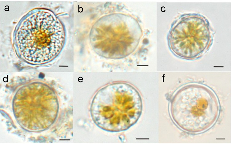
Figure 3.
Resting cysts and pellicle cysts of Alexandrium minutum. Resting cyst from a sediment trap (a), pellicle cyst with only a thin pellicle layer (b), pellicle cyst with the theca of the vegetative cell remaining (c), pellicle cyst with uncondensed cytoplasm (d), pellicle cyst with condensed cytoplasm (e), and double-walled resting cyst from the sediment (f). Scale bar: 10 µm. Adapted from [31].

