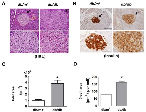
Figure 1. Histology and immunohistochemical staining for insulin in pancreatic islets of db/db diabetic mice.

Hematoxylin and eosin staining A. and immunohistochemical staining for insulin B. in pancreatic sections from db/db and db/m+ mice were shown. Original magnification, ×400, scale bar: 100 μm; x1000, scale bar: 50 μm. The arrows showed that indicated areas have enlarged scales. Moreover, the islet area C. and β-cell area D. in islets of db/db and db/m+ mice with 6 random areas per section was determined by ImageJ software. Data are presented as mean ± SEM. *P < 0.05, db/db versus db/m+ mice.
