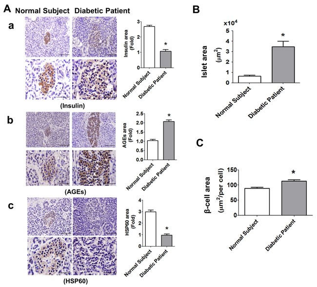
Figure 9. Immunohistochemical staining for insulin, AGEs, and HSP60, in the pancreatic islets of normal subject and diabetic patient.

The immunohistochemical staining for insulin (A-a), AGEs (A-b), and HSP60 (A-c) were performed on the pancreatic sections (islet areas) of normal subject and diabetic patient. Original magnification, ×400, scale bar: 100 μm; x1000, scale bar: 50 μm. Moreover, the islet area B. and β-cell area C. in islets of normal subject and diabetic patient with 6 random areas per section was determined by ImageJ software. Data are presented as mean ± SEM. *P < 0.05, diabetic patient versus normal subject.
