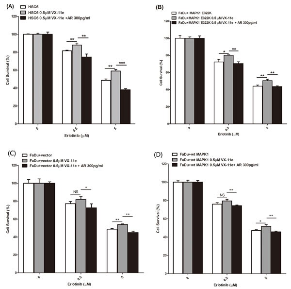
Figure 4. MAPK1 inhibition decreased sensitivity to erlotinib in MAPK1E322K mutant HNSCC cells, while exogenous AREG restored erlotinib sensitivity.

HSC-6 cells and FaDu cells engineered to express MAPK1E322K were pretreated with DMSO, VX-11e (0.5 μM) or combination of VX-11e (0.5 μM) and AREG (300 pg/ml) for 24 hours. Cells were then treated with two concentrations of erlotinib or a combination of VX-11e (0.5μM) and erlotinib or the triple combination of VX-11e (0.5 μM) and AREG (300pg/ml) and erlotinib for 48 hours. Cell survival was measured by crystal violet dye extraction growth assay and plotted relative to DMSO vehicle control (erlotinib alone) or VX-11e control (combination of VX-11e and erlotinib) or VX-11e and AREG control (combination of VX-11e, AREG and erlotinib). A. MAPK1 inhibition by VX-11e decreases sensitivity to erlotinib in HSC-6 cells, while exogenous AREG restored sensitivity. B. MAPK1 inhibition by VX-11e decreased sensitivity to erlotinib in FaDu cells engineered to express MAPK1E322K, while exogenous AREG restored its sensitivity. Decreased sensitivity to erlotinib with MAPK1 inhibition was attenuated in vector control transfected FaDu cells C. and FaDu cells engineered to express MAPK1WT D. (n = 3. *p < 0.05, **p < 0.01, ***p < 0.001). Similar results were obtained with triplicate wells in three independent experiments.
