Abstract
The “pontine warning syndrome” is characterized by recurrent episodes of motor hemiparesis, dysarthria and horizontal gaze palsy associated with basilar artery branch infarction. We report a case of a patient who presented with recurrent, self-limited episodes of locked-in syndrome, related to a bilateral pontine infarction. As far as we know, this clinical presentation as a subtype of pontine warning syndrome has never been described. We discuss the case, the differential diagnosis of the neuroimaging and the possible underlying mechanism.
Keywords: Stroke, locked-in, capsular warning syndrome, pontine warning syndrome
Case report
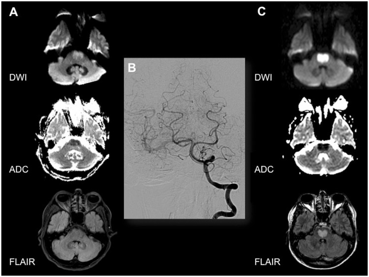
A 49-year-old male, current smoker, was transferred to our center because of an acute onset of generalized weakness, six hours after the last time seen asymptomatic. At admission, neurological examination showed an alert patient with anarthria, quadriplegia and bilateral extensor plantar response. Corneal reflexes were present and there was no gaze palsy, scoring 26 on the National Institute of Health Stroke Scale (NIHSS). Blood analysis, heart rate and blood pressure were unremarkable. An acute brain magnetic resonance imaging (MRI) revealed a bilateral anterior pontine hyperintensity in the diffusion-weighted image (DWI) sequence, with slight hypointensity in the apparent diffusion coefficient (ADC) map. Fluid-attenuated inversion recovery (FLAIR) sequence was normal and magnetic resonance angiography (MRA) showed patency of vertebrobasilar arteries (Figure 1(a)). Because of absence of FLAIR demarcation, intravenous tissue plasminogen activator (tPA) was administered. Fifteen minutes after starting tPA, the patient experienced a complete and rapid clinical improvement (NIHSS 0). One hour later, he suffered a new episode of quadriplegia and anarthria with preserved consciousness. Facing severe clinical worsening, a conventional angiography was performed to completely rule out a vertebrobasilar occlusion or stenosis suitable for endovascular treatment (Figure 1(b)). Afterwards, the patient presented a new improvement, being admitted asymptomatic at the intensive care unit. Within the following 24 hours, he experienced three similar self-limited episodes, lasting minutes. In the last one, he suffered a respiratory failure requiring endotracheal intubation. Blood, urine and cerebrospinal fluid tests were unremarkable. Electrocardiographic monitoring for 26 days showed sinus rhythm and transthoracic echocardiography were normal. Drug screening test for cocaine, cannabis and amphetamines was negative. Electroencephalography did not show seizure activity. A follow-up MRI three days later confirmed a bilateral medial anterior pontine ischemic infarction (“heart shaped”) (Figure 1(c)). Treatment with acetylsalicylic acid and statins was started. The patient was discharged to a rehabilitation center one month later, remaining the locked-in syndrome. Transesophageal echocardiography and screening for prothrombotic states were not performed during admission and not requested during the follow-up because of poor outcome and lack of therapeutic prospects.
Figure 1.
Neuroimaging evolution of the patient. (a) Baseline MRI. Mild restriction in DWI in the central and anterior pons, with normal FLAIR sequence. (b) Cerebral angiography. Sequence performed from the left vertebral artery, in posteroanterior projection, showing patency of basilar and bilateral posterior cerebral arteries. (c) Seventy-two-hour MRI. Established bilateral medial anterior pontine DWI restriction, with typical “heart appearance” shape in FLAIR sequence, compatible with ischemic infarction. MRI: magnetic resonance imaging; DWI: diffusion-weighted imaging; FLAIR: fluid-attenuated inversion recovery.
Discussion
The “capsular warning syndrome”1 is a crescendo transient ischemic attack characterized by three or more episodes affecting the face, arm and leg, without cortical symptoms. This syndrome is usually associated with lacunar infarction in the internal capsule. In 2008, Saposnik et al.2 proposed the term “pontine warning syndrome,” referring to recurrent and stereotyped episodes of motor hemiparesis, dysarthria and horizontal gaze palsy associated with a high risk of imminent basilar artery branch infarction. As far as we know, a fluctuating locked-in syndrome as a subtype of pontine warning syndrome has never been described.
Although ischemic stroke was initially suspected in our case, the atypical presentation led us to contemplate other pathologies affecting the pons. A central pontine myelinolysis (CPM) was considered because similar early changes in DWI sequences and ADC maps have been described.3 However, our patient had no risk factors for CPM as hyponatremia, malnutrition or alcoholism. Moreover, on the MRI follow-up, hyperintensity in T2 sequences is described but usually have a characteristic “trident-shaped” form,3 different from the “heart-shaped” image of our case. A diffuse brainstem glioma was also taken into account, but symptoms were not progressive and no contrast enhancement was observed in T1 sequences. Wernicke’s encephalopathy4 may affect the pons but MRI usually shows concomitant involvement of the thalamus, hypothalamus or mammillary bodies, none of which were present in our patient. Finally, infectious and demyelinating disorders, like multiple sclerosis or Behçet’s disease,5 were also ruled out.
Bilateral anteromedial pontine infarction is rarely reported.6 The typical imaging feature is the “heart appearance” shape, as a result of a bilateral paramedian and short circumferential branches infarction. Kumral et al.7 reported 150 pontine ischemic strokes, describing 9% of them as having bilateral involvement, half of them with heart appearance. They hypothesized microatheromatosis in the basilar artery branches as the most probable etiology of infarction.
The presented case was a diagnostic challenge for us. Although the first MRI was compatible with an ischemic infarction, the presence of five self-limited episodes of locked-in syndrome with no arterial occlusion raised a doubt about a vascular etiology. However, the clinical and neuroimaging evolution ensured that we were facing a stroke. Microatheromatosis affecting the perforator branches of the basilar artery was postulated as the underlying mechanism. In conclusion, we report an unusual clinical presentation of a pontine warning syndrome in which bilateral compromise of the pons led to fluctuating locked-in syndrome.
Funding
This research received no specific grant from any funding agency in the public, commercial, or not-for-profit sectors.
Conflicts of interest
The authors received no financial support for the research, authorship, and/or publication of this article.
References
- 1.Donnan GA, O’Malley HM, Quang L, et al. The capsular warning syndrome: Pathogenesis and clinical features. Neurology 1993; 43: 957–962. [DOI] [PubMed] [Google Scholar]
- 2.Saposnik G, Noel de Tilly L, Caplan LR. Pontine warning syndrome. Arch Neurol 2008; 65: 1375–1377. [DOI] [PubMed] [Google Scholar]
- 3.Ruzek KA, Campeau NG, Miller GM. Early diagnosis of central pontine myelinolysis with diffusion-weighted imaging. AJNR Am J Neuroradiol 2004; 25: 210–213. [PMC free article] [PubMed] [Google Scholar]
- 4.Sechi G, Serra A. Wernicke’s encephalopathy: New clinical settings and recent advances in diagnosis and management. Lancet Neurol 2007; 6: 442–455. [DOI] [PubMed] [Google Scholar]
- 5.Al-Araji A, Kidd DP. Neuro-Behçet’s disease: Epidemiology, clinical characteristics and management. Lancet Neurol 2009; 8: 192–204. [DOI] [PubMed] [Google Scholar]
- 6.Sen D, Arorar V, Adlakha S, et al. The “heart appearance” sign in bilateral pontine infarction. J Stroke Cerebrovasc Dis 2015; 24: e21–e24. [DOI] [PubMed] [Google Scholar]
- 7.Kumral E, Bayülkem G, Evyapan D. Clinical spectrum of pontine infarction. Clinical-MRI correlations. J Neurol 2002; 249: 1659–1670. [DOI] [PubMed] [Google Scholar]


