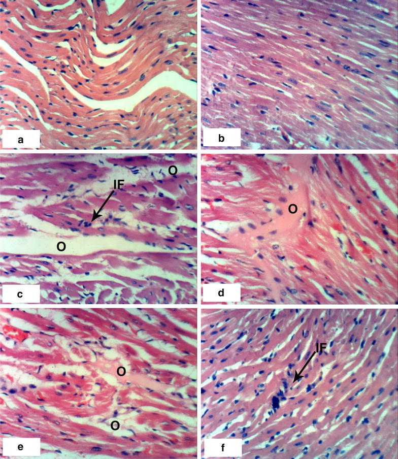
Fig. 1.

Photomicrographs of H and E stained heart sections of normal and latex treated rats. Sections of control rats administered 1 % CMC for 4 weeks (a) and 8 weeks (b) showing normal myocytes of heart. c Section of rat treated with 1/20 LD50 of latex for 4 weeks showing intermuscular odema (O) associated with inflammatory cell infiltration (IF), d, e sections of rats treated with 1/20 LD50 of latex for 8 weeks showing intermuscular odema (O), f section of rat treated with 1/10 LD50 of latex for 4 weeks showing inflammatory cell infiltration (IF) (×400)
