

Introduction
Central venous cannulation is routinely performed in operation theatres and intensive care units. The requirement of catheter in central vein is increasing as large numbers of extensive surgical procedures are being undertaken. In USA approximately 5 million central venous catheters are inserted annually [1]. Any patient staying for more than few days in the intensive care units invariably needs central venous catheter for the purpose of central venous pressure monitoring, vasopressor infusion, parenteral nutrition, or haemodialysis.
Routinely, central venous catheters are inserted percutaneously as a blind procedure using anatomical landmarks. Due to underlying anatomical variations this may result in multiple punctures and injury to nearby arteries, nerves, and pleura. Life-threatening complications like pneumothorax and arterial bleeding are known to occur. Mechanical complications are reported to occur in 5 to 19% of patients [2]. In 1984, two-dimensional ultrasound and Doppler techniques were first reported for internal jugular vein (IJV) cannulation. The recent development of portable lightweight ultrasound machines designed specifically for central venous cannulations has made them practical for routine clinical use. The needle can now be inserted under ultra sound guidance, making the procedure extremely safe.
Equipment
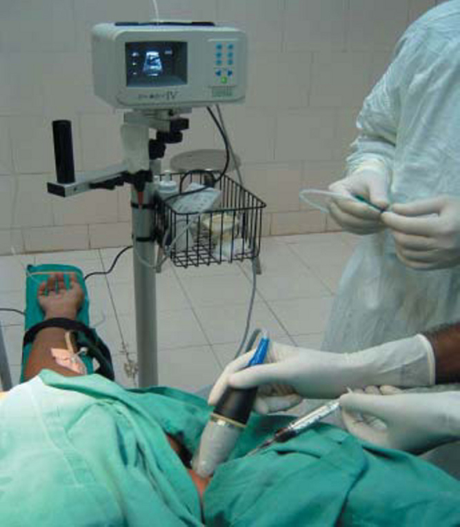
For ultra sonic guidance we use light weight, portable, high-resolution ultrasound scanner specifically designed for central venous cannulation. For superficial imaging two standoff probes are available in frequencies of 9.0 MHz and 7.5 MHz. The probe has needle hook guide to which the short end of the needle guide is clipped. The equipment and probe with needle guide is shown in Fig. 1. The needle guides direct a needle so that it will cross the ultrasound beam at a predetermined depth that lies along the path of the dot markers. The ultrasound system with associated probes provides ultrasound imaging of vascular structure, various organs and structures of body. Each probe has two scan depths (2 and 4 cm) that we can select using the ultrasound scanner depth selection button. The image produced is a truncated sector scan, trapezoid in shape.
Fig 1.

Ultrasound scanner with probe and needle guide.
Technique
Depth of internal jugular vein from skin is 1-3 cm in adult patient. Right internal jugular vein is our first choice because of higher success rate. Patient is positioned in mild Trendelenberg position (10 to 15 degree) and the head is turned to other side. After usual aseptic precaution puncture site is infiltrated with 2 % lignocaine at the apex of triangle formed by the two heads of sternocleidomastoid muscle and the clavicle (central approach). On linear probe of 9.0 MHz ultrasonic coupling gel is applied on the acoustic window of the probe head and is covered with sterile transparent plastic sheath then needle guide is clipped. The probe is placed at the apex of triangle to obtain transverse view of carotid artery and right internal jugular vein.
The internal carotid artery is seen as a thick walled, noncompressible, round pulsating hypoechoic structure of about 1.0 to 1.5 cm diameter. Posterolateral to the artery, the internal jugular vein (IJV) is seen as an oval shaped thin walled non-pulsating hypoechoic structure, easily compressible by probe, about 1.5 to 2.0 cm in diameter beneath the sternocleidomastoid muscle (Fig. 2). IJV image is positioned in the centre of acoustic window; an introducer needle with attached syringe is inserted under the probe at an angle of 450. The distortion of the vein during needle pressure on the skin, movement of needle and needle indenting and entering the vein can be easily appreciated. Once the tip of needle is in vein blood is aspirated, free flow of blood confirms the correct needle placement (Fig. 3). Subsequently guide wire is inserted and CVP catheter is passed using Seldinger's technique. The average time taken to puncture the vein is 70 sec and this time decreases with increased experience. The overall success rate is 98%, complication and failure rate is 2%. During IJV cannulation, ultrasound guidance reduces the number of mechanical complications, the number of catheter placement failures, and the time required for insertion [3]. However, its use during subclavian venous catheterization has had mixed results in clinical trials [4] probably for anatomical reasons. The fixed anatomical relation between the subclavian vein and the clavicle makes ultrasound-guided catheter insertion more difficult and less reliable than landmark-based insertion. Factors that may complicate cannulation using the anatomic landmark technique include obesity, neck deformity or rigidity, previous surgery at the cannulation site, IJV thrombosis, hypovolemia, or the inability to lie flat.
Fig 2.

Ultrasound image of right internal jugular vein and common carotid artery under sternocleidomastoid muscle.
Fig 3.
Aspiration of dark blood confirms correct placement of needle.
Conclusion
We conclude that use of ultrasound technique reduces complications and time to insertion of central venous catheter. As per the recommendation of National Institute for Clinical Excellence the use of ultrasound is mandatory for every central venous cannulation [5]. However this recommendation has been questioned in subsequent meta-analysis [6].
References
- 1.Raad I. Intravascular catheter related infections. Lancet. 1998;351:893–898. doi: 10.1016/S0140-6736(97)10006-X. [DOI] [PubMed] [Google Scholar]
- 2.Merrer J, De Jonghe B, Golliot F. Complications of femoral and subclavian venous catheterization in critically ill patients: a randomized controlled trial. JAMA. 2001;286:700–707. doi: 10.1001/jama.286.6.700. [DOI] [PubMed] [Google Scholar]
- 3.Teichgraber UK, Benter T, Gebel M, Mann MP. A sonographically guided technique for central venous access. AJR Am J Roentgenol. 1997;169:731–733. doi: 10.2214/ajr.169.3.9275887. [DOI] [PubMed] [Google Scholar]
- 4.Randolph AG, Cook DJ, Gonzales CA, Pribble CG. Ultrasound guidance for placement of central venous catheters: a meta- analysis of the literature. Crit Care Med. 1996;24:2053–2058. doi: 10.1097/00003246-199612000-00020. [DOI] [PubMed] [Google Scholar]
- 5.National Institute for Clinical Excellence. Guidance on the use of ultrasound locating devices for placing central venous catheters. NICE Technical report No. 49; 2002.
- 6.Hind D, Calvert N, Mc Williams R, Davidson A, Paisley S, Beverly C, Thomas S. Ultrasonic locating devices for central venous cannulation: Meta-analysis. BMJ. 2003;327:361–364. doi: 10.1136/bmj.327.7411.361. [DOI] [PMC free article] [PubMed] [Google Scholar]