Abstract
Zephycandidine A (1), the first naturally occurring imidazo[1,2-f]phenanthridine alkaloid, was isolated from Zephyranthes candida (Amaryllidaceae). The structure of 1 was elucidated by spectroscopic analyses and NMR calculation, and a plausible biogenetic pathway for zephycandidine A (1) was proposed. Zephycandidine A (1) exhibited significant cytotoxicity against five cancer cell lines with IC50 values ranging from 1.98 to 7.03 μM with selectivity indices as high as 10 when compared to the normal Beas-2B cell. Further studies suggested that zephycandidine A (1) induces apoptosis in leukemia cells by the activation of caspase-3, upregulation of Bax, downregulation of Bcl-2, and degradation of PARP expression. In addition, zephycandidine A (1) showed acetylcholinesterase (AChE) inhibitory activity, and the docking studies of zephycandidine A (1) and galanthamine (2) with AChE revealed that interactions with W286 and Y337 are necessary.
Amaryllidaceae alkaloids are an important resource for new drug discovery, among them, galanthamine, a well-known acetylcholinesterase (AChE) inhibitor, is a first-line treatment of Alzheimer’s disease. More than 500 amaryllidace alkaloids belonging to 18 framework types have been reproted to possess AChE inhibitory, analgesic, antibacterial, antifungal, antimalarial, antitumor, and antiviral activities1. Zephyranthes candida (Lindl.) Herb., an amaryllidaceous bulbous herb, is widely cultured as an ornamental flower, and the whole plants are used as a folk medicine in China to treat infantile convulsions, epilepsy, and tetanus2. In 1990, Pettit group reported the principal cytostatic component, trans-dihydronarciclasine, from the extract of Z. candida collected in China3. In a previous study on the cytotoxic alkaloids of Z. candida collected in June 2010 at Shiyan, China, total 15 alkaloids were isolated, and some of them exhibited signficant cytotoxicity against five cancer cell lines4. Among them, new alkaloid N-methylhemeanthidine chloride showed potent inhibitory activities against pancreatic cancer in vitro and in vivo, and the mechanism was found to be via down-regulating AKT activation5.
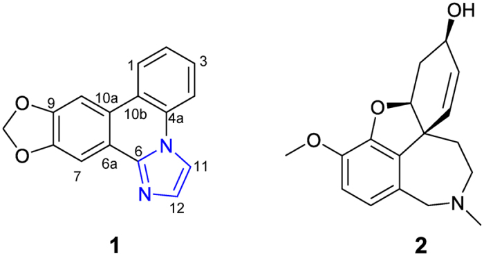
In order to search for more active alkaloids from Z. candida, the whole plants were re-collected at the same place on October 20116. Further study on this collection led to the isolation of a new minor alkaloid with a rare imidazo[1,2-f]phenanthridine framework, which was named zephycandidine A (1) (Fig. 1). Phenanthridine alkaloids are very common in the Amaryllidaceae plants1, however, there is no report of the presence of the imidazo[1,2-f]phenanthridine type alkaloids, although imidazo [1,2-f] phenanthridine was synthesized as a synthon by Cronin group in 20067. Zephycandidine A (1) represents the first example of a naturally occurring imidazo[1,2-f]phenanthridine type alkaloid. In this paper, we describe the isolation, structure determination, plausible biosynthetic pathway, cytotoxicity and inducing apoptosis in leukemia cells, AChE inhibitory activity, and the docking study with AChE of zephycandidine A (1).
Figure 1. Chemical structures of zephycandidine A (1) and galanthamine (2).
Results and Discussion
Structural elucidation of zephycandidine A (1)
Zephycandidine A (1) was isolated as colorless oil. The molecular formula C16H10N2O2 was determined by the HRESIMS ion at m/z 263.0789 [M + H]+ (calcd for 263.0821) in conjunction with the 13C NMR data, requiring thirteen degrees of unsaturation. The 1H and 13C NMR data of 1 (Table 1) were very similar to those of trisphaeridine, which was co-isolated from this plant4. The major differences between them were that the methine (δH 9.07, s, H-6; δC 152.0, C-6) at C-6 in trisphaeridine is replaced by a quaternary olefinic carbon (δC 143.8, C-6) in 1, and there are two additional olefinic methines (δH 7.52, d, J = 1.4 Hz, H-11; 8.32, d, J = 1.4 Hz, H-12; δC 113.8, C-11; 131.5, C-12) in 1. The trisphaeridine unit accounts for eleven degrees of unsaturation, two additional olefinic methines account for one degree of unsaturation, and the remaining one degree of unsaturation suggested the presence of additional ring in 1. Due to the small coupling constant J = 1.4 Hz between H-11 and H-12, as well as the observed the correlations of H-11 to H-12 in 1H-1H COSY spectrum, H-11 and H-12 should be in a meta or ortho position of a five-membered ring. In consideration of the molecular formula and chemical shifts, there are two possible structures 1a and 1b (Fig. 2) for 1. However, there were no obvious HMBC correlations from H-11 and H-12 to C-4a or C-6a. So, it is impossible to verify the structure of 1 by the HMBC analysis.
Table 1. 1H (400 MHz) and 13C NMR (100 MHz) data for zephycandidine A (1) (Methanol-d 4) and DFT calculation of 13C NMR for 1a and 1b.
| position | 1 |
1a |
1b |
|||
|---|---|---|---|---|---|---|
| δH (mult, J) | δC | δC(cal.) | δC(cor.) | δC(cal.) | δC(cor.) | |
| 1 | 8.47, dd, (8.2, 1.2) | 125.4 | 128.6 | 125.4 | 128.6 | 126.2 |
| 2 | 7.58, ddd, (8.2, 7.4, 1.0) | 126.9 | 129.1 | 125.9 | 129.8 | 127.7 |
| 3 | 7.67, ddd, (8.4, 7.4, 1.2) | 129.8 | 131.8 | 128.9 | 131.9 | 130.3 |
| 4 | 8.14, dd, (8.4, 1.0) | 117.5 | 120.9 | 116.9 | 120.3 | 115.8 |
| 4a | 132.3 | 135.4 | 132.9 | 134.4 | 133.5 | |
| 6 | 143.8 | 145.5 | 144.0 | 132.8 | 131.5 | |
| 6a | 119.8 | 125.0 | 121.4 | 125.2 | 121.9 | |
| 7 | 7.88, s | 103.2 | 107.8 | 102.5 | 107.3 | 99.5 |
| 8 | 150.7 | 151.8 | 150.9 | 151.9 | 155.4 | |
| 9 | 151.6 | 152.3 | 151.4 | 151.3 | 154.6 | |
| 10 | 7.99, s | 103.1 | 106.8 | 101.4 | 107.4 | 99.6 |
| 10a | 125.5 | 128.1 | 124.8 | 126.1 | 123.1 | |
| 10b | 123.1 | 127.3 | 123.9 | 127.9 | 125.3 | |
| 11 | 7.52, d, (1.4) | 113.8 | 116.8 | 112.4 | 131.5 | 129.8 |
| 12 | 8.32, d, (1.4) | 131.5 | 134.7 | 132.1 | 125.3 | 122.1 |
| OCH2O | 6.16, s | 103.7 | 111.8 | 106.9 | 111.8 | 105.2 |
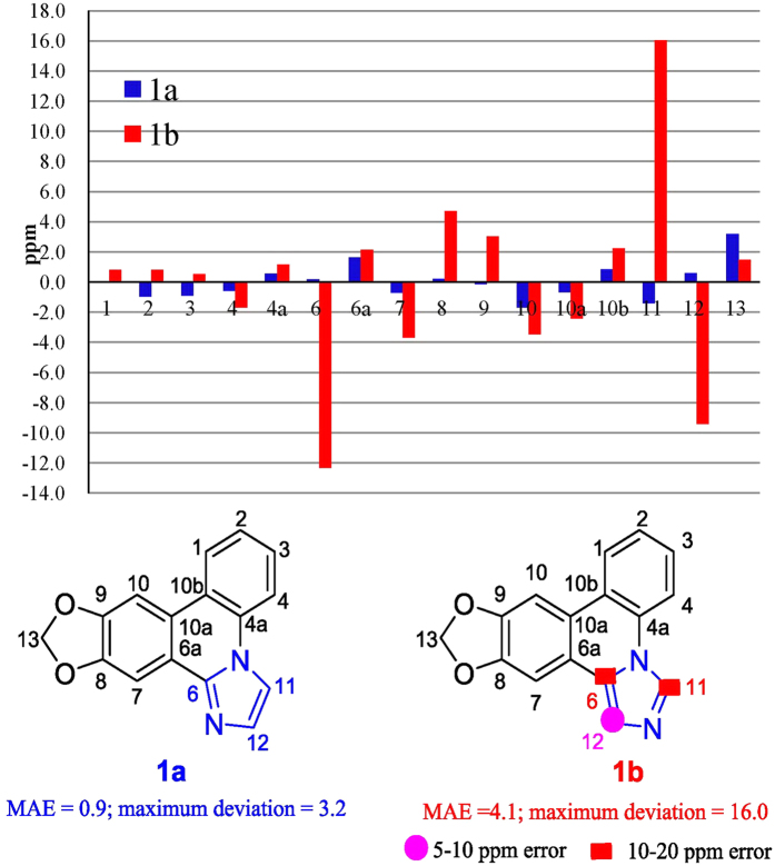
Figure 2. Structures and differences in ppm between calculated and experimental 13C NMR shifts for 1a and 1b.
To determine the structure of 1, the 13C NMR shifts of two possible structures 1a and 1b (Table 1) were calculated with the GIAO option using the B3LYP/6-31G* DFT method8. According to the method of P. Crews et al.9, the corrected 13C NMR shifts for 1a and 1b (Table 1) were calculated, and then, the differences between the experimental and corrected 13C NMR shifts for 1a and 1b were analysed (Fig. 2). As shown in Fig. 2, the MAE (mean absolute error) and MD (maximum deviation)9 for 1a were 0.9 and 3.2 ppm, respectively, which are acceptable to meet MAE < 2.2 and MD < 59. However, the MAE and MD for 1b were 4.1 and 16.0 ppm, respectively, which are far higher than standard of MAE < 2.2 and MD < 5. Especially, the absolute error of atoms at C-6, C-11, and C-12 were much higher than 5 ppm. Thus, the chemical structure of 1 should be 1a, instead of 1b.
Furthermore, a weak correlation between H-12 and H-11 was finally observed in the NOESY spectrum of 1 when the acquisition time for the NOESY data was increased much more, which further supported the structure of 1a. Therefore, the structure of zephycandidine A (1) was assigned to [1,3]dioxolo[4,5-j]imidazo[1,2-f]phenanthridine.
Proposed biosynthetic pathway for zephycandidine A (1)
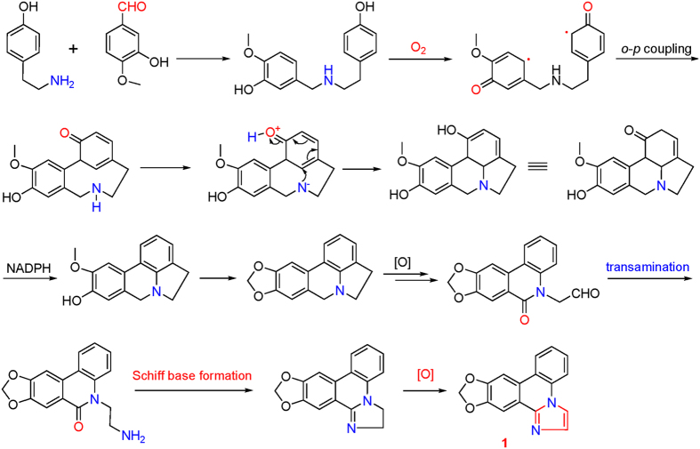
Although the imidazo-phenanthridine synthon had been produced in a five-step one pot reaction by Cronin group7, the biosynthetic pathway of 1 in Z. candida (Fig. 3) could be traced back to tyramine and isovanillin to form a benzylphenethylamine alkaloid, following by the oxidative coupling of phenols to form a lycorine-type alkaloid. After oxidation, transamination, intramolecular Schiff base formation, and final oxidation, the lycorine-type alkaloid intermediate was transformed to be the imidazo[1,2-f]phenanthridine alkaloid 1.
Figure 3. Plausible biogenetic pathway for zephycandidine A (1).
Cytotoxity of zephycandidine A (1)
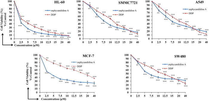
Cytotoxity of zephycandidine A (1) against five human cancer cell lines, human myeloid leukemia HL-60, lung cancer A549, breast cancer MCF-7, colon cancer SW480, and hepatocellular carcinoma SMMC-7721, together with one non-cancerous cell line human bronchial epithelial cells Beas-2B, were evaluated using the MTT (3-(4,5-dimethylthiazol-2-yl)-2,5-diphenyltetrazolium bromide) method10. DDP (cis-platin) and DMSO were used as the positive and negative controls, respectively. The results were showed in Fig. 4 and Table 2. Zephycandidine A (1) showed more potent cytotoxicity against five human cancer cell lines than the positive control (cis-platin) with IC50 values of 1.98, 6.49, 3.44, 6.27, and 7.03 μM, respectively. More interestingly, 1 showed weak cytotoxicity against the normal Beas-2B cell line (IC50 = 20.08 μM) with the selectivity indices as high as 10.1.
Figure 4. Cytotoxity of zephycandidine A (1) against five human cancer cell lines (human myeloid leukemia HL-60, hepatocellular carcinoma SMMC-7721, lung cancer A549, breast cancer MCF-7, and colon cance4r SW480).
Cells were treated with various concentrations of zephycandidine A for 48 h and cell viability was determined by MTT assays using DDP as positive control. Data are presented as the means ± SD of three experiments, P values were derived from Student’s t-test (*P < 0.05, **P < 0.01, ***P < 0.001), compared to the control group.
Table 2. Cytotoxicity of zephycandidine A (1) (IC50 in μM) against five cancer cell lines and a non-cancerous human Beas-2B cell linea.
| Compound | HL-60 | A549 | MCF-7 | SW480 | SMMC-7721 | BEAS-2B | Highest index of selectivity |
|---|---|---|---|---|---|---|---|
| 1 | 1.98 | 6.49 | 3.44 | 6.27 | 7.03 | 20.08 | 10 |
| cis-platin | 3.12 | 8.95 | 18.48 | 11.37 | 9.82 | 13.30 | 4.3 |
aThe “Highest index of selectivity” is the ratio of the IC50 value for the Beas-2B cell line over the lowest cancer cell IC50 value. cis-Platin was used as a positive control.
Morphological changes of HL-60 cells treated with zephycandidine A (1)
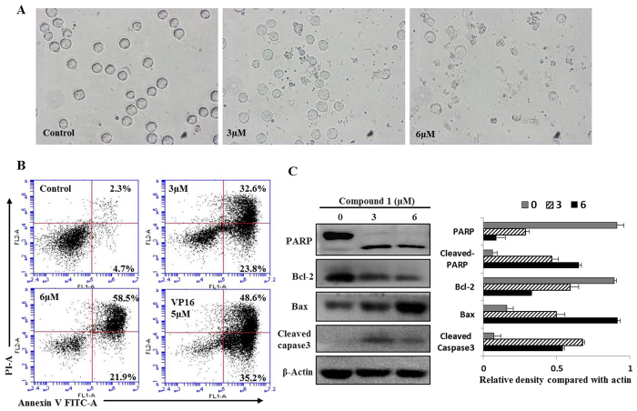
To further analysis the anticancer effects of zephycandidine A (1), we used HL-60 as a model. The morphological changes and cytoclasis of HL-60 cells were examined under an inverted phase contrast microscope. As shown in Fig. 5A, cell shrinkage and abnormal morphological features could be observed in 3.0 and 6.0 μM zephycandidine A (1)-treated groups.
Figure 5. Zephycandidine A (1) induces apoptosis in leukemia cells.
(A) Morphological changes of HL-60 cells after treated with zephycandidine A (1) for 48 h. Phase-contrast microscopic view, magnification 400 × . (B) Apoptosis was determined by Annexin V-FITC and PI staining using flow cytometric analysis. Cells in the lower right quadrant indicate early apoptotic cells, and cells in the upper right quadrant indicate late apoptotic cells. (C) Western blot analysis for PARP, Bcl-2, Bax and caspase-3 levels, β-Actin used as a loading control. The relative density of each protein was detected by Image J, data are presented as the means of three experiments, bars, SD.
Zephycandidine A (1) causes HL-60 cells death by apoptosis
To ensure whether zephycandidine A (1) meditated inhibition of growth and proliferation was associated with apoptosis, the flow cytometry apoptosis assay was performed using Annexin V and PI staining11. After treatment at various concentrations (0, 3.0, and 6.0 μM) for 48 h, the apoptosis of the cells were detected. Compared with the untreated cells, the treatment of 6 μM zephycandidine A (1) can result by up to 80.4% apoptosis incidence (Fig. 5B). As a positive control, the treatment of VP16 (etoposide phosphate, 5 μM) leads to 83.8% apoptosis incidence (Fig. 5B). These results demonstrate that zephycandidine A (1) induced apoptosis of HL-60 cells.
Zephycandidine A (1) changes the expression of apoptotic-related proteins
As we know, among the caspase-family, caspase-3 is considered to be a critical effector, and the activation of caspase-3 could result in PARP degradation12. Bcl-2 family also plays a central regulatory role in the mitochondrial pathway of apoptosis, the balance between Bcl-2 and Bax is critical for cell survival and death13.
HL-60 cells were treated with zephycandidine A (1) as describe above. Changes of apoptosis-related protein, including Bax, Bcl-2, cleaved caspase-3 and PARP, were assessed by western blot analysis. β-actin was used as an internal control, and relative optical density of proteins were compared with β-actin. As shown in Fig. 5C, zephycandidine A (1) treatment altered the expression levels of PARP and cleaved-caspase 3, it also down regulated Bcl-2 and upregulated Bax. Taken together, these data suggest that zephycandidine A (1)-induced apoptosis in leukemia cells is mediated by the activation of caspase-3, upregulation of Bax, downregulation of Bcl-2 and degradation of PARP.
Zephycandidine A (1) exhibites acetylcholinesterase inhibitory activity
Acetylcholinesterase (AChE) activity was measured using a 96-well microplate reader based on Ellman’s method14 with slight modification15. As showed in Fig. 6, zephycandidine A (1) showed AChE inhibitory activity in a dose-dependent manner, and the IC50 value was calculated to be 127.99 μM. While, the positive control, galanthamine (2), exhibited potent AChE inhibitory activity with an IC50 value of 1.02 μM.
Figure 6. AChE inhibitory activities of zephycandidine A (1) and galanthamine (2).
Docking studies on zephycandidine A (1) and galanthamine (2) with AChE
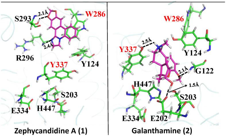
To study the relationship of the structures and the AChE inhibitory activities, the binding modes of zephycandidine A (1) and galanthamine (2) with AChE have been obtained by our comprehensive docking studies (Fig. 7). There are two binding sites in the gorge of AChE, i.e., the catalytic binding site in the bottom of gorge and the peripheral anionic site at the lip of the gorge. As shown in Fig. 7, galanthamine (2) binds at the catalytic binding site, whereas zephycandidine A (1) binds at the peripheral anionic site. Galanthamine (2) is stabilized by forming three hydrogen bonds with E202, Y337 and G122, respectively. In the peripheral anionic site, zephycandidine A (1) binds the enzyme primarily by the π–π stacking interaction with W286 and by two hydrogen bonds with S293 and R296, respectively.
Figure 7. The binding modes of zephycandidine A (1) and galanthamine (2) in AChE.
The ligand atoms are shown in purple stick. The relevant AChE residues are shown in green sticks. The oxygen, nitrogen, and hydrogen atoms are colored in red, blue, and white, respectively.
Extensive studies on the binding of ligands with AChE shows that W286 in peripheral anionic site and Y337 in catalytic binding site play an important role more than stabilizing the binding with the ligands. It has been shown that the loss of interaction with W286 resulted in the loss of binding with AChE, while the ligand is still able to bind butyrylcholinesterase (BuChE)16. BuChE has similar but larger gorge than AChE and does not have tryptophan residue in the peripheral anionic site. It is thus suggested that interaction with W286 is the key for ligands to bind AChE. Similarly, Y337 is characterized as “swinging gate” of the catalytic binding site17. It hinders the ligands from entering the catalytic binding site, but stabilizes the ligand after it moves into the catalytic site18. Recent crystal structures also show there is hydrogen bond between Y337 and quaternary ammonium of ligands19. Clearly, W286 and Y337 play an important role in recognizing the ligands. Unable to bind these two residues is very likely unable to enter the peripheral anionic site or the catalytic binding site.
As discussed above, zephycandidine A (1) and galanthamine (2) interact with the key W286 and Y337 residues, respectively, suggesting that zephycandidine A (1) and galanthamine (2) do bind AChE. This explains why the inhibition activities against AChE were observed for zephycandidine A (1) and galanthamine (2). The potential AChE inhibitors are required to interact with W286 to bind at peripheral anionic site, or to interact with both W286 and Y337 to bind at catalytic binding site. This suggested that without obvious interaction with these two residues, no matter how tight binding it shows, the ligand just cannot move into the gorge and therefore express no or few inhibitory activity against AChE.
In conclusion, an Amaryllidaceae alkaloid with a rare imidazo[1,2-f]phenanthridine framework, zephycandidine A (1), was isolated from Z. candida. Zephycandidine A (1), [1,3]dioxolo[4,5-j]imidazo[1,2-f]phenanthridine, represents the first example of a naturally occurring imidazo[1,2-f]phenanthridine type alkaloid. Zephycandidine A (1) exhibited cytotoxicity against five cancer cell lines with IC50 values ranging from 1.98 to 7.03 μM with selectivity indices as high as 10 when compared to the normal Beas-2B cells. More importantly, zephycandidine A (1) induced apoptosis in leukemia cells by the activation of caspase-3, upregulation of Bax, downregulation of Bcl-2 and degradation of PARP expression. Zephycandidine A (1) showed AChE inhibitory activity with IC50 value of 127.99 μM, and the docking studies of 1 with AChE revealed that interactions with W286 and Y337 are necessary.
Methods
General experimental procedures
The UV and FT-IR spectra were determined using Varian Cary 50 and Bruker Vertex 70 instruments, respectively. NMR spectra were recorded with a Bruker AM-400 spectrometer for 1H NMR, 13C NMR, COSY, HSQC, HMBC, and NOESY experiments. Chemical shift values are given in δ (ppm) based on the peak signals of the solvent methanol-d4 (δH 3.31 and δC 49.15) as references, and coupling constants are reported in Hz. High-resolution electrospray ionization mass spectra (HRESIMS) were carried out in the positive-ion mode on a Thermo Fisher LC-LTQ-Orbitrap XL spectrometer. The OD values were measured on a Bio-Rad 680 microtiter plate reader. HPLC was performed on an Agilent 2100 quaternary system with a UV detector using a reversed-phased C18 column (5 μm, 10 × 250 mm, Weltch Ultimate XB-C18). Column chromatography was performed using Sephadex LH-20 (GE Healthcare Bio-Sciences AB, Sweden), silica gel (Qingdao Marine Chemical Inc., China), and ODS (50 μm, YMC Co. Ltd., Japan). Thin-layer chromatography (TLC) was performed with silica gel 60 F254 (Yantai Chemical Industry Research Institute) and RP-C18 F254 plates (Merck, Germany).
Plant material
The whole plants of Zephyranthes candida were collected at Shiyan, Hubei Province, People’s Republic of China, in October, 2011. The plant material was identified by Professor Changgong Zhang of the School of Pharmacy, Tongji Medical College, Huazhong University of Science and Technology. A voucher specimen (No. 20111001) has been deposited at the Hubei Key Laboratory of Natural Medicinal Chemistry and Resource Evaluation, School of Pharmacy, Tongji Medical College, Huazhong University of Science and Technology.
Extraction and isolation
The whole dried plants of Z. candida (10 kg) were extracted four times with 25 L each of 95% aqueous EtOH with 2% HCl at room temperature. The filtrates were combined and concentrated under vacuum to afford 627 g of crude extract, which was then partitioned between CHCl3 and adjusted the pH = 2 with dilute HCl, then extracted the aqueous phase with CHCl3, followed by re-extracting the aqueous phase three additional times with CHCl3 (3 L). After the aqueous phase was adjusted to pH 10 with an ammonia solution, it was partitioned between CHCl3 (4 × 1.5 L) for the second time. On evaporation, the CHCl3 phase at pH = 10 (7.6 g) was subjected to silica gel CC (CHCl3–MeOH, 1:0, 50:1, 30:1, 20:1, 10:1) to afford 5 fractions (Fr. 1–5). Fr.2 was fractionated on MPLC with RP-18 CC (MeOH–H2O, 60:40, 90:10) to give four subfractions (Fr. 2A–Fr.2D). Fr.2C was subjected to separation over silica gel using a gradient system of petroleum ether–acetone (10:1–2:1) to provide Fr.2CA. Compound 1 (3.5 mg, tR 23.8 min) were purified from the Fr.2CA, using semi-preparative RP HPLC (MeOH–H2O–Et2NH, 80:20:0.2).
Zephycandidine A (1)
colorless oil; UV (MeOH) λmax (log ε) 207 (4.63), 220 (4.48), 256 (3.63), 297 (4.18) nm; IR (KBr) νmax 2920, 2851, 1622, 1486, 1383, 1258, 1208, 1037, 886, 829, 750, 721 cm−1; 1H NMR data, see Table 1; 13C NMR data, see Table 1; HRESIMS m/z 263.0789 [M + H]+ (calcd for C16H11N2O2, 263.0821).
Cytotoxicity assay
Cytotoxity of the compound against five human cancer cell lines, human myeloid leukemia HL-60, lung cancer A549, breast cancer MCF-7, colon cancer SW480, and hepatocellular carcinoma SMMC-7721, together with one non-cancerous cell line human bronchial epithelial cells Beas-2B, was evaluated using the MTT method10. Briefly, the cells were grown in RPMI 1640 (Hyclone) medium supplemented with 10% heat-inactivated newborn calf serum, penicillin (50 units/mL), and streptomycin (50 g/mL) in a humidified incubator under 5% CO2 at 37 °C. Cell suspensions (200 μL, containing 5000–10000 cells per well) were placed into 96 well microplates and allowed to adhere for 24 h before drug addition, while suspended cells were seeded just before drug addition. A 100 μL aliquot of the test compounds at concentrations ranging from 2.5 to 40 μM was added to each well. The medium was replaced with one containing the test compounds, and the cells were further cultured at 37 °C. After incubation for 48 h, 20 μL of MTT (5 mg/mL) solution was added to each well and the cells were incubated under the same conditions for 4 h until a purple precipitate was visible. DMSO (200 mL) was added and the optical density was measured at 570 nm in a microplate reader (Bio-Tek Synergy HT). DDP (cis-platin) and DMSO were used as the positive and negative controls, respectively.
Cell morphological assessment
The cell morphological assessment was performed after HL-60 cells were treated with 0, 3 and 6 μM of zephycandidine A (1) for 48 hours. Cell morphological changes were observed with inverted light microscopy (NIKON, Tokyo, Japan).
Flow cytometry analysis of cell apoptosis
For identification of the apoptotic induction effect of zephycandidine A (1), a FITC-labeled Annexin V/PI apoptosis detection Kit (Keygen, Nanjing, China) was used according to the manufacturer’s instructions. Briefly, HL-60 cells were exposure to vehicle control (DMSO, < 0.1%), zephycandidine A (1) (3 and 6 μM) for 48 h, VP16 was used as a positive control20. After that cells were harvested and washed with PBS and resuspended in binding buffer, and then, AnnexinV-FITC and PI were added. After staining 15 minutes, the cells were immediately analyzed using flow cytometry (Becton Dickinson, USA).
Western blot analysis
Western blot analysis was conducted to analysis the protein expression. Briefly, cells were incubated with vehicle control and zephycandidine A (1) for 48 hours, and then cells were lysed in a radio immune-precipitation assay buffer. Protein concentrations were determined and equalized before loading. Samples containing equal amounts of protein were denatured and subjected to electrophoresis in 10% SDS-PAGE gels followed by transfer to PVDF membrane and probed with specific antibodies, including PARP, Bcl-2, Bax, Cleaved Caspase 3 and β-Actin (Cell Signaling Technology, Inc.). Blots bands were visualized using the horseradish peroxidase conjugated secondary antibodies and chemiluminescent substrate.
Acetylcholinesterase inhibitory activity assay
AChE activity was measured using a 96-well microplate reader based on Ellman’s method14 with slight modification15. The enzyme hydrolyzes the substrate acetylthiocholine resulting in the product thiocholine which reacts with Ellman’s reagent (DTNB) to produce a yellow compound 5-thio-2-nitrobenzoate which can be detected at 405 nm. All solutions were brought to room temperature prior to use. In a typical assay, 25 μL of 15 mM ATCI (dissolved in water), 125 μL of 3 mM DTNB (50 mM Tris–HCl, pH 7.6, containing 0.1 M NaCl and 0.02 M MgCl2•6 H2O), 50 μL of buffer (50 mM Tris–HCl, pH 7.6, containing 0.1% BSA), 25μL of sample were added in the 96-well plates and the absorbance was measured at 405 nm every minute for six times. Then, 25 μL of 0.22 U/ml of enzyme was added. Read the absorbance again every minute for fifteen times. The rates of reaction were calculated by Microplate Manager software version 4.0. Any increase in absorbance due to the spontaneous hydrolysis of substrate was corrected by subtracting the rate of the reaction before adding the enzyme from the rate after adding the enzyme. Percentage of inhibition was calculated by comparing the rates for the sample to the blank (10% MeOH in buffer). The inhibition rate of zephycandidine A (1) against AChE was initially evaluated at the concentration of 200 μM. Based on the inhibition rate at 200 μM, the test concentrations of the active compounds were further optimized for IC50 calculation.
NMR data calculations
The 3D structure of the compound 1a and 1b were optimized in methanol by using Gaussian09 at the B3LYP/6-31G* level. Both optimized structures (Tables S1 and S2, Supplementary Information) were then further used as the input structures for NMR calculation. The NMR calculations were performed by using Gaussian09 at the B3LYP/6-31G* level. Finally, the relative errors between the computed (Tables S3–S6, Supplementary Information) and measured NMR were calculated21,22 (Figures S1–S6, Supplementary Information).
Docking Studies
The docking studies were performed by using Autodock 4.223 in combination with MM/GBSA method implemented in AMBER 1124. The basic idea of this combination method is to replace the scoring function used in Autodock by the more expensive but more accurate MM/GBSA energy. Therefore, only 1 generation was evolved within the Genetic Algorithm, and the wide spread docking poses were produced as many as 10,000 by the Autodock software. All conformations were subjected to MM/GBSA calculations, and then were ranked according the much more reliable MM/GBSA results.
To evaluate the accuracy of our docking method, five crystal structures23,25 (PDB IDs: 4EY5, 4EY6, 4EY7, 4M0E, 4M0F) with ligands in complex with human AChE were used for re-docking tests. The initial coordinates of ligands were not taken from the crystal structures, but generated by BALLON software26 in order to avoid the bias from Autodock software to the initial structure. The top 1 ranked binding structures obtained in our docking method are all having RMSD less than 2 Å as compared to the binding poses in crystal structures, indicating that our docking method is very reliable in reproducing the correct binding poses for AChE inhibitors.
Additional Information
How to cite this article: Zhan, G. et al. Zephycandidine A, the First Naturally Occurring Imidazo[1,2-f ]phenanthridine Alkaloid from Zephyranthes candida, Exhibits Significant Anti-tumor and Anti-acetylcholinesterase Activities. Sci. Rep. 6, 33990; doi: 10.1038/srep33990 (2016).
Supplementary Material
Acknowledgments
We thank the staff at Analysis and Measurement Centre of Huazhong University of Science and Technology for the IR analyses. This work was supported financially by the National Natural Science Foundation of China (31170323, 81001368, and 81503305), Wuhan Youth Chenguang Program of Science and Technology (201271031389), Shandong Provincial Natural Science Foundation, China (ZR2014HL102), China Postdoctoral Science Foundation (2015M582227), and the National Science and Technology Project of China (2013ZX09103001-020).
Footnotes
Author Contributions G.Z., J.Z. and X.Q. isolated and identified the natural products, G.Z. and X.Q. tested the cytotoxicity and AChE inhibitory activity, Q.T. studied the apoptosis, J.L. and B.S. calculated the NMR data and studied the docking with AChE, G.Y. designed this project. G.Z., G.Y., J.L. and Q.T. wrote the manuscript. All authors reviewed the manuscript.
References
- Jin Z. Amaryllidaceae and Sceletium alkaloids. Nat. Prod. Rep. 26, 363–381 (2009). [DOI] [PubMed] [Google Scholar]
- State Administration of Traditional Chinese Medicine. Zhonghuabencao. Shanghai Science and Technology Publishing Company: Shanghai ; pp 7268–7268 (1999). [Google Scholar]
- Pettit G. R., Cragg G. M., Singh S. B., Duke J. A. & Doubek D. L. Antineoplastic agents, 162. Zephyranthes candida. J. Nat. Prod. 53, 176–178 (1990). [DOI] [PubMed] [Google Scholar]
- Luo Z. W. et al. Cytotoxic alkaloids from the whole plants of Zephyranthes candida. J. Nat. Prod. 75, 2113–2120 (2012). [DOI] [PMC free article] [PubMed] [Google Scholar]
- Guo G. L. et al. N-methylhemeanthidine chloride, a novel Amaryllidaceae alkaloid, inhibits pancreatic cancer cell proliferation via down-regulating AKT activation. Toxicol. Appl. Pharm. 280, 475–483 (2014). [DOI] [PubMed] [Google Scholar]
- Zhan G. et al. Galanthamine, plicamine, and secoplicamine alkaloids from Zephyranthes candida and their anti-acetylcholinesterase and anti-inflammatory activities. J. Nat. Prod. 79, 760–766 (2016). [DOI] [PubMed] [Google Scholar]
- Parenty A. D. et al. Discovery of an imidazo-phenanthridine synthon produced in a five-step one-pot reaction leading to a new family of heterocycles with novel physical properties. Chem. Commun. 11, 1194–1196 (2006). [DOI] [PubMed] [Google Scholar]
- Rychnovsky S. D. Predicting NMR spectra by computational methods: structure revision of hexacyclinol. Org. Lett. 8, 2895–2898 (2006). [DOI] [PubMed] [Google Scholar]
- White K. N. et al. Structure revision of spiroleucettadine, a sponge alkaloid with a bicyclic core meager in H-atoms. J. Org. Chem. 73, 8719–8722 (2008). [DOI] [PMC free article] [PubMed] [Google Scholar]
- Alley M. C. et al. Feasibility of drug screening with panels of human tumor cell lines using a microculture tetrazolium assay. Cancer Res. 48, 589–601 (1988). [PubMed] [Google Scholar]
- Vermes I., Haanen C., Steffens-Nakken H. & Reutelingsperger C. A novel assay for apoptosis. Flow cytometric detection of phosphatidylserine expression on early apoptotic cells using fluorescein labelled Annexin V. J. Immunol. Methods. 184, 39–51 (1995). [DOI] [PubMed] [Google Scholar]
- Ghavami S. et al. Apoptosis and cancer: mutations within caspase genes. J. Med. Genet. 46, 497–510 (2009). [DOI] [PubMed] [Google Scholar]
- Ouyang L. et al. Programmed cell death pathways in cancer: a review of apoptosis, autophagy and programmed necrosis. Cell Proliferat. 45(6), 487–498 (2012). [DOI] [PMC free article] [PubMed] [Google Scholar]
- Ellman G. L., Courtney K. D. & Featherstone R. M. A new and rapid colorimetric determination of acetylcholinesterase activity. Biochem. Pharmacol. 7, 88–95 (1961). [DOI] [PubMed] [Google Scholar]
- Rhee I. K., van de Meent M., Ingkaninan K. & Verpoorte R. Screening for acetylcholinesterase inhibitors from Amaryllidaceae using silica gel thin-layer chromatography in combination with bioactivity staining. J. Chromatogr. A. 915(1), 217–223 (2001). [DOI] [PubMed] [Google Scholar]
- Savini L. et al. Specific targeting of acetylcholinesterase and butyrylcholinesterase recognition sites. Rational design of novel, selective, and highly potent cholinesterase inhibitors. J. Med. Chem. 46(1), 1–4 (2003). [DOI] [PubMed] [Google Scholar]
- Kryger G., Silman I. & Sussman J. L. Structure of acetylcholinesterase complexed with E2020 (Aricept): implications for the design of new anti-Alzheimer drugs. Structure. 7(3), 297–307 (1999). [DOI] [PubMed] [Google Scholar]
- Bai F. et al. Free energy landscape for the binding process of Huperzine A to acetylcholinesterase. P. Natl. Acad. Sci. USA. 110(11), 4273–4278 (2013). [DOI] [PMC free article] [PubMed] [Google Scholar]
- Cheung J. et al. Structures of human acetylcholinesterase in complex with pharmacologically important ligands. J. Med. Chem. 55(22), 10282–10286 (2012). [DOI] [PubMed] [Google Scholar]
- Bonhoure E. et al. Overcoming MDR-associated chemoresistance in HL–60 acute myeloid leukemia cells by targeting shingosine kinase-1. Leukemia 20(1), 95–102 (2005). [DOI] [PubMed] [Google Scholar]
- Frisch M. J. et al. Gaussian 09, Revision D.01, Gaussian, Inc. Wallingford CT (2009). [Google Scholar]
- Tomasi J., Mennucci B. & Cammi R. Quantum mechanical continuum solvation models. Chem. Rev. 105(8), 2999–3093 (2005). [DOI] [PubMed] [Google Scholar]
- Morris G. M. et al. AutoDock4 and AutoDockTools4: Automated docking with selective receptor flexibility. J. Comput. Chem. 30(16), 2785–2791 (2009). [DOI] [PMC free article] [PubMed] [Google Scholar]
- Case D. A. et al. AMBER 11, University of California, San Francisco (2010). [Google Scholar]
- Cheung J., Gary E. N., Shiomi K. & Rosenberry T. L. Structures of human acetylcholinesterase bound to dihydrotanshinone I and territrem B show peripheral site flexibility. ACS Med. Chem. Lett. 4(11), 1091–1096 (2013). [DOI] [PMC free article] [PubMed] [Google Scholar]
- Vainio M. J. & Johnson M. S. Generating conformer ensembles using a multiobjective genetic algorithm. J. Chem. Inf. Model. 47(6), 2462–2474 (2007). [DOI] [PubMed] [Google Scholar]
Associated Data
This section collects any data citations, data availability statements, or supplementary materials included in this article.


