
Figure 8. Pair-rule gene expression is perturbed from gastrulation onwards in opa mutant embryos.
Pair-rule gene expression in wild-type and opa mutant embryos at late cellularisation, late gastrulation, and early germband extension. During cellularisation, pair-rule gene expression in opa mutant embryos is very similar to wild-type. Expression from gastrulation onwards is severely abnormal; in particular, note that single-segment patterns do not emerge. All panels show a lateral view, anterior left, dorsal top.

Figure 8—figure supplement 1. Pair-rule gene expression in opa mutant embryos at cellularisation.

Relative expression patterns of pair-rule genes in wild-type and opa mutant embryos at late cellularisation. All images are double in situs for odd and one other pair-rule gene. Individual channels are shown to the right of each double-channel image (odd on the left, other pair-rule genes on the right). All panels show a lateral view, anterior left, dorsal top.
Figure 8—figure supplement 2. Pair-rule gene expression in opa mutant embryos at gastrulation.

Relative expression patterns of pair-rule genes in wild-type and opa mutant embryos at gastrulation. All images are double in situs for odd and one other pair-rule gene. Individual channels are shown to the right of each double-channel image (odd on the left, other pair-rule genes on the right). All panels show a lateral view, anterior left, dorsal top.
Figure 8—figure supplement 3. Pair-rule gene expression in opa mutant embryos at early germband extension.

Relative expression patterns of pair-rule genes in wild-type and opa mutant embryos at early germband extension. All images are double in situs for odd and one other pair-rule gene. Individual channels are shown to the right of each double-channel image (odd on the left, other pair-rule genes on the right). All panels show a lateral view, anterior left, dorsal top.
Figure 8—figure supplement 4. The transition to single-segment periodicity does not occur in opa mutant embryos.

Comparison of early phase 3 segmentation gene expression in wild-type and opa mutant embryos. The horizontal axis represents an idealised portion of the AP axis (~12 nuclei across). The grey vertical lines demarcate a double parasegment repeat (~8 nuclei across), of an odd- followed by an even-numbered parasegment (see Figure 6). The pattern of protein (intense colour) and transcript expression (paler colour) of the pair-rule genes, and the segment-polarity genes en and wg, are shown for each genotype. Wild-type patterns are the same as in Figure 6B. Transcript distributions for opa mutant embryos were inferred from our double in situ data, while protein distributions were extrapolated from transcript data. Fading expression of Eve and Runt is represented by lighter sections at the posterior of the stripes. In opa mutant embryos, expression of eve and runt fades prematurely, while the expression of odd, prd and slp remains double segmental. Only the even-numbered stripes of wg emerge, with en expression delayed until mid-germband extension (Benedyk et al., 1994; Figure 10). Stronger expression in the posterior of the Eve stripes in opa mutants is inferred from the observation that the eve stripes remain broad at a time when they would have already narrowed in wild-type (compare panels A and F in Appendix 2—figure 2, or see Figure 8—figure supplement 5). For simplicity, the low-level or residual expression of eve and runt observed in opa mutant embryos is not included in the schematic. See text for further details.
Figure 8—figure supplement 5. Opa activates the eve “'late' element.

eve and odd expression in wild-type and opa mutant embryos at various timepoints spanning mid-phase 2 (mid-cellularisation, top row) to late phase 3 (onset of germband extension, bottom row). In opa mutant embryos, eve stripes are initially expressed normally (row 1), but fail to narrow and refine at the end of cellularisation (row 3), and largely fade away at gastrulation (row 4). Residual eve expression persists in some stripes into germband extension (bottom row) in opa mutant embryos, particularly in ventral regions. Individual channels are shown to the right of the double-channel images. All panels show a lateral view, anterior left, dorsal top. Embryo morphology and the pattern of odd expression in the head were used for staging.
Figure 8—figure supplement 6. 'Late' eve expression is observed in cells that do not express prd.
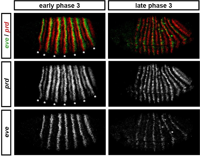
eve and prd expression in wild-type embryos during phase 3. During early phase 3 (left), eve is strongly expressed in stripes ~2 cells wide. These stripes only partially overlap with the 'P' stripes of prd expression (asterisks), meaning that the eve 'late' element is active in many cells that have never expressed prd. eve expression is largely lost from non-prd expressing cells by the end of gastrulation (late phase 3, right), indicating that Prd protein may nevertheless be required for the maintenance of eve late element expression. Individual channels are shown below each double-channel image. All panels show a lateral view, anterior left, dorsal top.