

Introduction
Diffuse hyperdensity affecting the Circle of Willis, dural sinuses and other vessels is sometimes encountered on non-enhanced computed tomography (NECT) scans of brain. Polycythemia produces this appearance and it is associated with an increase in circulating red cell mass. This condition is often complicated by both arterial and venous thrombosis, hence an early diagnosis becomes imperative. Since many polycythemic patients are asymptomatic until late in disease or present with nonspecific symptoms, NECT brain done for some nonspecific neurologic abnormality can give a clue to the diagnosis. We report this case for its rarity and unique characteristic appearance of polycythemia on unenhanced CT brain.
Case Report
A seven year old, female child with history of headache for the last three months which did not respond to medication underwent NECT brain to rule out the possibility of any organic pathology. There was no history of trauma. NECT brain revealed diffusely hyperdense vasculature of the brain including large and small branches of Circle of Willis (Fig.1), dural venous sinuses including transverse sinus and deep cerebral veins (Fig. 2) and cortical veins draining in to superior sagittal sinus (Fig. 3), with HU values ranging from 50–55. There was no evidence of any focal or diffuse parenchymal abnormality in the supratentorial or infratentorial compartment. Based on the these findings, the possibility of polycythemia was suggested. The diagnosis was confirmed following laboratory tests which revealed increased red cell mass in the form of raised hemoglobin (18 gm/dL%) and raised hematocrit (75%).
Fig. 1.

NECT axial image of brain shows diffuse hyperdensity in the branches of the Circle of Willis, along tentorium cerebelli and sigmoid sinuses
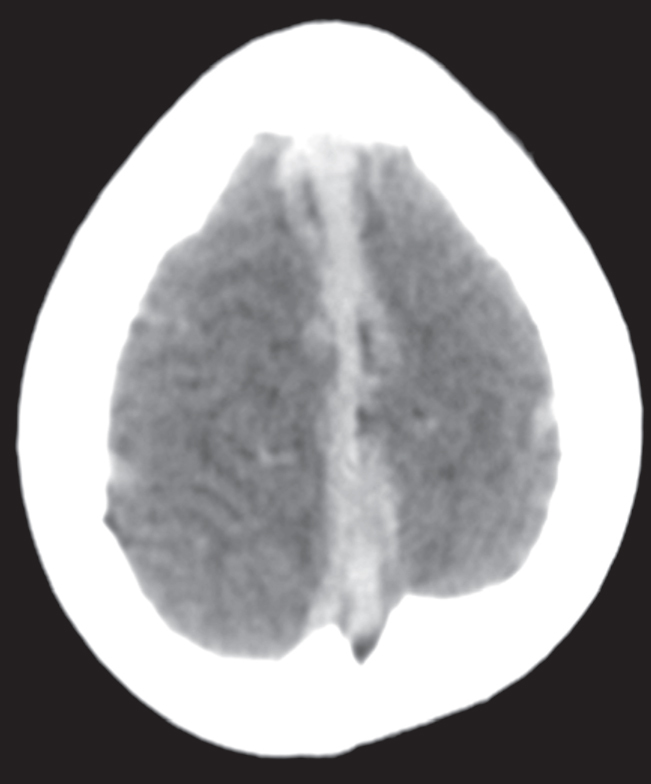
Fig. 2.

NECT axial image of brain shows diffuse hyperdensity in the deep veins, dural venous sinuses (straight and transverse sinus) and small vasculature of cerebral parenchyma
Fig. 3.
NECT axial image of brain shows diffuse hyperdensity in the superior sagittal sinus and draining cortical veins
Discussion
On CT studies only one factor i.e. electron density, determines image contrast and there is a linear relationship between CT attenuation and hematocrit, hemoglobin concentration and protein content [1]. Flowing blood at a hematocrit of 45% measures approximately 40 HU. Hematocrit ranges from 42-52% in normal adults and 37-47% in normal neonates. Therefore, large vessels and the dural venous sinuses appear isodense or minimally hyperdense in the normal adult as the normal adult gray matter measures approximately 39 HU [2].
In patients with a hematocrit percentage exceeding 60%, both Circle of Willis and dural sinuses are hyperdense on NECT scans of brain. A linear relationship exists between the hemoglobin level and the contrast of the dural sinuses compared with the gray matter, suggesting that increased density of cerebral vessels on NECT is a sign of a high hemoglobin level [3].
Polycythemia is defined as an increase in the circulating red cell mass. It is often detected as an incidental finding by increased hemoglobin or hematocrit. Concern that the hemoglobin may be abnormally high is usually triggered at 17g/dL for males and 15 g/dL for females. Hematocrit > 60% in male and 55% in female are almost invariably associated with increase in red cell mass [4]. Patients with polycythemia may be asymptomatic or experience symptoms related to an increase in red cell mass including hyperviscosity and thrombosis (both arterial and venous). Manifestations range from digital ischemia to Budd-Chiari syndrome, while abdominal thromboses are common. Neurologic symptoms include vertigo, tinnitus, headache and visual disturbances. Cardiovascular abnormalities are related to hypoxia and in late stages there is development of cor pulmonale due to pulmonary arterial hypertension [4].
NECT brain is usually performed as the first investigation in patients with a recent persisting headache [5]. Besides polycythemia, other differential diagnoses for similar appearance are normal neonatal brain, diffuse brain edema, dural venous thromboses and acute subarachnoid or subdural hemorrhage [1, 2, 3]. In premature and term newborn infants, the dural sinuses and the other blood vessels of the brain may appear falsely “hyperdense” on NECT as compared to the brain parenchyma. This apparent hyperdensity is due to the combination of low-density unmyelinated brain and physiologic polycythemia [1].
Unclotted dural sinuses, vein of Galen, internal cerebral veins, Circle of Willis and main arterial branches appear hyperdense in patients with diffuse cerebral parenchymal hypodensity due to edema, ischemia or injury [2]. Normal appearance of the cerebellum in such cases (white cerebellum or reverse cerebellar sign) along with the relevant history of neurologic deficit or trauma usually clinches the diagnosis.
Selective venous hyperdensity is necessary for a positive diagnosis of the dural venous sinus thrombosis, the so-called “cord sign” [1]. Since the hematocrit of an acute retracted thrombus/clot is very high (up to 90%) and the globin (protein) component of the hemoglobin have high-density mass, fresh blood clots typically appear hyperdense on CT when compared to normal brain [1]. Some cases of polycythemia may present with hyperdense venous sinuses only [6]. Magnetic resonance (MR) imaging and time-of-flight venography can confirm the presence or absence of venous sinus thrombosis in such conditions. Recently, CT venography using multidetector CT has been recommended in cases of cerebral venous thrombosis especially when the MR findings are inconclusive or MR is unavailable/contraindicated [7]. Acute subarachnoid or subdural hemorrhage layered along the falx or tentorium cerebelli may give rise to false hyperdensity to the dural venous sinuses.
The presence of sulcal bleed or subdural collections elsewhere, parenchymal hypodensities and history of trauma or hypertension usually helps in making the correct diagnosis [1].
To summarize, diffuse hyperdensity of the cerebral vasculature including the arteries and veins on NECT scan is a rare phenomenon seen characteristically in polycythemia. The knowledge of this phenomenon can help the radiologist in guiding the clinicians towards early diagnosis of this condition as it is associated with high morbidity.
Conflicts of Interest
None identified
References
- 1.Osborn AG. Diagnostic Neuroradiology. 2nd. Indian reprint; Mosby: 2007. p. 158. [Google Scholar]; Diagnostic Neuroradiology. 2nd. Indian reprint; Mosby: 2007. p. 388. [Google Scholar]
- 2.Grossman CB. Magnetic Resonance Imaging and Computed Tomography of the Head and Spine. 2nd ed. Williams and Wilkins; USA: 1996. pp. 277–280. [Google Scholar]
- 3.Ben Salem D, Osseby GV, Rezaizadeh-Bourdariat K. Spontaneous hyperdense intracranial vessels seen on CT scan in polycythemia cases. J Radiol. 2003;84:605–608. [PubMed] [Google Scholar]
- 4.Adamson JW, Longo DL. Anemia and Polycythemia. In: Kasper DL, Braunwald E, Fauci AS, Hauser SL, Longo DL, Jameson JL, editors. Harrison's Principle of Internal Medicine. 16th ed. McGraw-Hill; USA: 2005. pp. 335–336. [Google Scholar]
- 5.Cumurciuc R, Crassard I, Sarov M, Valade D, Bousser MG. Headache as the only neurological sign of cerebral venous thrombosis: A series of 17 cases. Journal of Neurology, Neurosurgery and Psychiatry. 2005;76:1084–1087. doi: 10.1136/jnnp.2004.056275. [DOI] [PMC free article] [PubMed] [Google Scholar]
- 6.Healy JF, Nichols C. Polycythemia mimicking venous sinus thrombosis. AJNR. 2002;23:1402–1403. [PMC free article] [PubMed] [Google Scholar]
- 7.Rodallec MH, Krainik A, Feydy A. Cerebral Venous Thrombosis and Multidetector CT Angiography: Tips and Tricks. Radiographics. 2006;26:S5–S18. doi: 10.1148/rg.26si065505. [DOI] [PubMed] [Google Scholar]