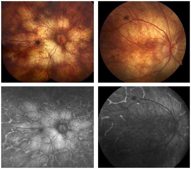
Figure 5. Widefield autofluorescence imaging demonstrates extensive RPE loss that is worst in the posterior pole corresponding to areas of atrophy.
(A) Montage of color fundus photographs demonstrating patchy RPE atrophy extending beyond the mid-periphery of two patients (LC03 and LC14) with advanced LCHADD. (B) Corresponding widefield fundus autofluorescence images from the same patients from the same year (LC03) and three years later (LC14) demonstrating extensive RPE loss in the posterior pole. There is a network of residual RPE hyperautofluorescence around well-demarcated nummular regions of RPE loss in the periphery. The diffusely bright signal in areas of RPE loss is thought to be most consistent with scleral autofluorescence, but may represent artifact from wavelength overlap between exciting and detected light.

