ABSRTRACT
The latest studies suggest that prophylactic chemotherapy or adjuvant chemotherapy for early stage breast cancer may increase the leukemia risk in patients. For patients with a low risk for breast cancer recurrence, physicians who make the choice for adjuvant therapy should consider the risk of its long-term side effects. Is the occurrence of lymphatic system cancer and leukemia after breast cancer treatment associated with chemotherapy? Can these types of leukemia be classified as therapy-related leukaemias? We believe that there may be correlations between any diseases, butwe cannot rush to conclusions or dismiss a correlation because we understand little about the diseases themselves.In this paper, we present a case of secondary diffuse large B-cell lymphoma and leukemia in patients after breast cancer chemotherapy, it is undeniable that this is a special event. For two distinct tumouroccurrences at different times, we cannot give a clear explanation because of thechanges in the genes that might link them together and we hope to attract the attention of other clinicians.
KEYWORDS: Breast cancer, bone marrow puncture, lymphoma, lymphoma cell leukemia, therapy-related leukaemias
Introduction
With the recent progress in research and the clinical applications of endocrine therapy and molecular targeted therapy,the survival of breast cancer patients has been significantly prolonged.However, we are finding that the long-term side effects caused by chemotherapy at an early stage are also graduallybecoming more prominent. The latest research by Professor Wolff Antonio has shown that prophylactic chemotherapy or adjuvant chemotherapy inpatients with early stage breast cancer can increase the risk of leukemia. This study reported that the 10-year risk of lymphoid and myeloid cancer for such patients was 0.5%, approximately twicethe risk observed in previous studies. However, thefive-year (recurrence) risk after treatment did not decrease.1 Therapy-related leukemia (TRL) has gradually become a clinical concern. However, there may be another form of leukemia onsite thatis due to malignant lymphoma leukemia. For such patients, if not diagnosed early, it is difficult to determine whether they have a lymphoma transformed toleukaemia ora leukemia invading into the lymphatic system. In this paper, a clinical case of secondary lymphoma and lymphosarcoma after breast cancer chemotherapy is reported, and its characteristics and pathogenesis are explored. We would like to draw the attention of other clinicians tothese treatment-related side effects.
Case report
A 56-year-old female patient was admitted to the first Affiliated Hospital of Dalian Medical University in September 2007 for a non-tender firm mass in the right breast. Physical examination revealed a palpable and immobile mass in the upper outer quadrant of the right breast with a diameter of approximately 3.0cm. It was not adhered to the skin, and there was no nipple retraction, axillary lymph node enlargement, or masses in the contralateral breast. Ultrasound showed a 3.0cm×2.5 cm uneven hypoechoic area in the upper outer quadrant of the right breast. Therefore, we suspected that the mass was a breast tumor. The patient was eligible for surgery and had no surgical contraindications. We performed a radical mastectomy of the right breast. Pathological examination revealed a grade II-III invasive ductal carcinoma of the right breast with no lymph node metastasis and no vascular blockade by tumourcells. Immunohistochemistry revealed that the tumor was estrogen receptor(ER)2+, progesterone receptor(PR)+ and human epidermal growth factor receptor(HER)-2-. The patient was diagnosed withstage IIAbreast cancer of the right breast. The patient received adjuvant EC (Epirubicin, EPI, Cyclophosphamide, CTX) followed by multiple rounds of T (Docetaxel, DOC), repeated for fourrounds. After chemotherapy, the patient received 2.5 mg of oralletrozole dailyas endocrine therapy.
In January 2011, a palpable mass was found in the left neck area. We measured the patient's carcinoembryonic antigen (CEA), cancer antigen (CA)-125, and CA-153 level, and they were all normal. Computed tomography (CT) showed no obvious abnormality in the lungs or mediastinum. There were multiple small lymph nodes in the neck region. There was no clear sign of a breast cancer recurrence or metastasis. The patient's surgeons recommended follow-up for observation. The patient self-administered an anti-inflammatory medication, and the lymph nodes got smaller. Therefore, hersymptomswere not considered serious.
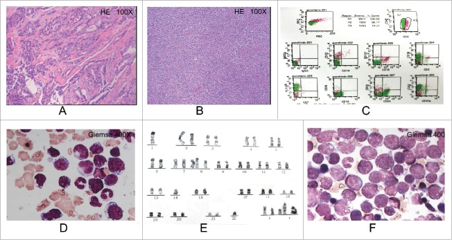
Unforturnately, in May 2011, the mass in the neck mass again increased in size,along with swollen masses in the bilateral axillary and inguinal regions. CT revealed multiple swollen lymph nodes in the chest and neck. There was confluence between mediastinal lymph nodes. Follow-up testing of the breast tumourmarkersreturned normal results. Blood routine: White blood cell 12. 73×109 /L,Lymphocyte2.69×109/L,Hemoglobin 132g /L,Platelet 293×109 /L, Lactate dehydrogenase452 IU/L, Positron emission tomography (PET)-CT examination revealed radionuclide accumulation foci in the neck, supraclavicular, axillary, mediastinal, intra-abdominal and groin areas. The largest standardized uptake value(SUV) was 6.5. We preliminarily diagnosed the patient with a lymphoma. Pathological examination of a biopsy of a left inguinal lymph node revealed a diffuse large B-cell lymphoma, Immunohistochmeistryshowed CD3(-),Ki-67>50%,CD43(-),CyclinD1(-),CD20(+),CD5(-),CD79a(+),CD21 (+),CD10(+),mum-1(-),bcl-6(+),bcl-2(+).the patient wasdiagnosed with astage IIIB non-Hodgkin's lymphoma. We initially planned for R-CHOP chemotherapy,but because the patient was allergic to rituximab, twocycles of CHOP(CTX1100 mg IV d1; EPI120 mg IV d1; Vincristine, VCR2mg IV d1;Prednisone,PDN 100 mg PO Qd d1-5 q21d) chemotherapy were used,and the patientachieved partial remission (PR). The patient began to experience a fever prior to the third cycle of chemotherapy. Her body temperature fluctuated between 38 and 40°C, and blood abnormalities emerged, White blood cell 57.12×10⁹/L, Lymphocyte10.54×109 /L, Hemoglobin108g/L, Platelet 168×109/L. We carried out a bone marrow biopsy to confirm the diagnosis. Cytological immunotyping examination of the bone marrow morphology showed bone marrow hyperplasia with abnormal lymphoid tissue accounting for 50%, uniform sizes, minimal cytoplasm, blue-colouredcytoplasmwith vacuoles, visibly twisted and folded nuclei, coarse nuclear chromatin and unclear nucleoli. We considered a diagnosis of lymphoma cell leukemia. Immunotyping showed R2 accounting for 80.77% of the nucleated cells, the expression of CD10(20.91%),CD22(82.24%),CD20(69.85%),CD19(63.50%)and HLA-DR(91.39%), and the absence of CD34,CD117,CD13,CD33andCD14 expression. Chromosomaltyping showed 46,-X, -1, +7,8q+, -14,-22,+mar2,+mar3,+mar4[5]/46 and xx[5],suggesting complex chromosomal aberrations. The patient was diagnosed with astage IIB diffuse large B-cell lymphoma and lymphoma cell leukemia.
After confirming the diagnosis, the patient received DOCPL(CTX800mg IV d1;Pirarubicin, THP30mg IV d1-3;VCR2mgIV d1, PDN60mg PO Qd d1-5;Lasparaginase,LASPAR 10000U IV Qod×5;q21d ) chemotherapy for a first cycle and with a total of twocycles planned. Due to disease progression, we replaced DOCPL with CTOAP(CTX 1000mg IV d1, THP 40mg IV d1-3;VCR 2mg IV d1, 8;Arabinofuranosylcytosine, Ara-C200mg IV d1-7;PDN 60mg PO Qd d1-14; q21d ) chemotherapy and performed intrathecal injection of methotrexate(MTX) 10 mg + 5 mg dexamethasone in 2 ml of saline to prevent central nervous system leukemia. A follow-up examination of the bone marrow cytology showed 73% lymphocytes, including 70.5% immature lymphocytes. The side effects experienced by the patient included level 4 bone marrow suppression, fatigue, weakness and anorexia. The patient refused to continue chemotherapy and received symptomatic supportive treatment before dying in the clinic in February 2012 (Fig. 1).
Figure 1.
(A) (2007-09-20) Pathological examination revealed a grade II-III invasive ductal carcinoma. (B) (2011-05-12)a left inguinal lymph node revealed a diffuse large B-cell lymphoma. (C) (2011-07-08) Immunotyping showed R2 accounting for 80.77% of the nucleated cells, the expression of CD10(20.91%),CD22(82.24%),CD20(69.85%), CD19(63.50%) and HLA-DR(91.39%). (D) (2011-07-05) Cytological immunotyping examination of the bone marrow morphology showed bone marrow hyperplasia with abnormal lymphoid tissue accounting for 50%, uniform sizes, minimal cytoplasm, blue-colouredcytoplasmwith vacuoles, visibly twisted and folded nuclei, coarse nuclear chromatin and unclear nucleoli. (E) (2011-7-21) Chromosomaltyping showed 46,-X, -1, +7,8q+, -14, -22, +mar2, +mar3, +mar4[5]/46 and xx[5].F:(2011-9-29) A follow-up examination of the bone marrow cytology showed 73% lymphocytes, including 70.5% immature lymphocytes.
Discussion
Breast cancer is one of the most common malignant tumors that have serious impacts on women's health and is often life-threatening. According to statistics published by the World Health Organization International Cancer Research Center, there were approximately 1.38 million new cases of female breast cancer worldwide in 2008, accounting for 22.9% of all female cancers. In addition, 460,000 women died of breast cancer in 2008, accounting for 13.7% of female cancer deaths.2 With recent progress in neoadjuvant therapy, adjuvant therapy, endocrine therapy and targeted therapy, the survival of breast cancer patients has gradually been prolonged. However, the consequences of overtreatment have also gradually emerged. What should not be ignored are second primary cancers and TRLs. In the 1980s, TRL first gained the attention of clinicians. It is a chemotherapy-induced and/or radiation-induced leukemia due to the treatment of primary malignant or non-malignant diseases.3 Professor Wolff Antonio's latest research has shown that prophylactic chemotherapy or adjuvant chemotherapy for early stage breast cancer patients can increase the risk of TRL. These findings lead us to think that the number of patients who may develop a leukemia triggered by overtreatment cannot be ignored given the large population base of breast cancer patients.
TRLs, in common clinical settings, are often in the form of acute myeloid leukemia (AML) and myelodysplastic syndrome (MDS).3 They are mostly direct secondary leukaemias. Diffuse large B-cell lymphomas after chemotherapy followed by transformation into lymphoma cell leukemia is rare. Should such cases be classified as TRLs? Sternberg first proposed that lymphoma cell lymphoblastic leukaemias should be classified as lymphosarcoma leukaemias. He thought that lymphoma cell leukaemias were different from leukaemias originating from general primary lymphocytes. His proposal facilitated distinguishing lymphoblastic leukaemias from other types of leukemia as a special type of leukemia that develops from a malignant lymphoma. This transition is also known as leukemic change, andthis stage of lymphoma is also known as leukemic lymphoma or lymphoma cell leukemia.4
Secondary leukaemias related to breast cancer chemotherapy are commonly induced by alkylating agents or topoisomerase II inhibitors. They are generally AML and MDS and are included as an independent subtype in the standard WHO classification of haematologic malignancies.5 Alkylating agents crosslink with DNA, causing gene mutations and deletions in the long arms of 5th and the 7th chromosomes. Thesegene mutations affect interleukin (IL)-3, IL-4 and other genes involved in cell proliferation. These deletions may activate the oncogene RAS and cause p53 tumoursuppressor gene mutations, resulting in abnormal cell proliferation and differentiation, and causing leukemia.6 Topoisomerase II inhibitors form a complex with DNA and topoisomerase II, blocking the enzyme's activity and causing DNA strand breaks. These breaks occur predominantly in the acute lymphoblastic leukemia (ALL)-1 gene area, causing ALL-1 gene (MLL, Htrx 1, HRX) rearrangement and DNA damage with dehydrogenase-induced free radicals, thereby causing leukemia.7 Docetaxel is a taxane anticancer drug. Itbinds to free tubulin and promotes tubulin assembly into stable microtubules while inhibiting their depolymerization. These effects result in the loss of normal microtubule bundle function and loss of microtubule attachment, thereby inhibiting cell mitosis. No long-term effects of docetaxel in oncogenesis and leukemia have been reported. Smith et al. analyzed 8563 clinical cases of breast cancer patients receivingCTX and anthracycline chemotherapy after surgery. They found that the occurrence rate of AML or MDS was 0.5%. The risk increased with increasing dosesof CTX.The onset ofAML or MDS from the time of treatment waswithin1-125 months (median 38 months). Only one patientdeveloped ALL.8 Diamanidou et al. reported that,among 1107 cases of breast cancer patients who received 5-fluorouracil, doxorubicin and CTX(FAC), the incidence of AML or MDS was 1.5%, withan onset interval of22-113 months after surgery (median 66 months). There was no incidence of ALL.9 In this case, we used the EC-T program as treatment, which is a possible precursor of a diagnosis of TRL.
Unlike TRLs in the past, this case was different in that the patient had multiple swollen lymph nodes threeyears after chemotherapy for breast cancer. Pathological analysis of the lymph node biopsy confirmed a diffuse large B-cell lymphoma diagnosis. However, during treatment, further changes leading to abnormal blood parameters caught our attention. Examination of the bone marrow eventually confirmed the diagnosis of lymphoma cell leukemia. Unfortunately, at the time of the initial diagnosis of lymphoma, we did not examine the bone marrow, so we are unable to report the initial bone marrow status. Because lymphoma onset may also be in the form of a leukemia and because the difference lies only in the proportion of lymphoma cells in the bone marrow, they are just different stages of one type of tumor. Of course, leukemia has a worse prognosis. Is it also possible that the chemotherapy induced a secondary TRL, which caused the leukemia and swollen lymph nodes? We did not carry out an early bone marrow examination, and we were guided by the earlier pathological diagnosis from the lymph node tissue. However, how likely is this possibility? On lymph node biopsy and subsequent pathological examination of the bone marrow, we arrived at a second diagnosis, and it remained as in the past.This result confused us, and it remains to be determined whether TRL can have an intermediate stage solid tumourstatus and whether lymphoma can also be caused by excessive chemotherapy and radiotherapy. Similar research reports have not offered us any hints. We onlyknowthat high-grade malignant lymphomas have a high chance of transforming into leukaemias. In particular, patients with B symptoms in the course of treatment tend to have early bone marrow involvement and extranodal involvement that isassociated with the rapid development of large mediastinal masses. Treatment should be in line with the treatments for acute lymphoblastic leukemia with a poor prognosis,and the diagnosis should be distinguished from other types ofleukaemia.Usually, blood examination reveals an abnormally high number of leukocytes. Decreasedred blood cell counts and hemoglobin levelsare not obvious, thoughplatelet counts are often decreased. Bone marrow cytology can reveal visible changes in the lymphatic system, mostly in the form of significant increases in the proportion ofimmature lymphocytes and lymphoblasts. Patients respond poorly to conventional chemotherapy due to de novo or acquired resistance. These findings are consistent with the features of this case.10However, we still cannot determine whether the lymphoma and leukemia were associated with the chemotherapy because we did not obtain conclusive evidence. However, it is undeniable that this is a special event. For two distinct tumouroccurrences at different times, we cannot give a clear explanation because of thechanges in the genes that might link them together.11 For now, the microscopic world is still a field that we cannot fully explain.
To prevent the occurrence of TRLs, physicians should strictly control the dosing of chemotherapy drugs, particularly the dosing and administration criteria for topoisomerase II inhibitors and alkylating agents,to avoid long-term tumourigenic effects.12 We believe that with increasing progress in targeted therapy and immunotherapy, more specific and non-toxic “targeted” therapies may reduce the incidence of treatment-related disease.
Disclosure of potential conflicts of interest
No potential conflicts of interest were disclosed.
Funding
This work was supported by the Shandong Provincial Natural Science Foundation (ProjectNoZR2013HL048), Natural Science Foundation of ShandongAcademy of Medical Sciences(Project No 2013-35), Science Foundation of The FirstAffiliated Hospital of Dalian Medical University (2014QN004).
References
- 1.Wolff AC, Blackford AL, Visvanathan K, Rugo HS, Moy B, Goldstein LJ, Stockerl-Goldstein K, Neumayer L, Langbaum TS, Theriault RL, et al.. Risk of marrow neoplasms after adjuvant breast cancer therapy: the national comprehensive cancer network experience. J Clin Oncol 2015; 33:340-8; PMID:25534386; http://dx.doi.org/ 10.1200/JCO.2013.54.6119 [DOI] [PMC free article] [PubMed] [Google Scholar]
- 2.Smith RA, Cokkinides V, Brooks D, et al.. GLOBOCAN 2008 v1.2, Cancer Incidence and Mortality Worldwide: IARC Cancer Base No. 10 [Internet]. Lyon, France: International Agency for Research on Cancer; http://globocan.iarc.fr, accessed on October/05/2013. [Google Scholar]
- 3.McKenna RW, Parkin JL, Foucar K, Brunning RD. Ultrastruc-tural characteristics of therapy-related acute nonlymphocytic leukemia: evidence for a panmyelosis. Cancer 1981; 48:725-37; PMID:7248900; http://dx.doi.org/ [DOI] [PubMed] [Google Scholar]
- 4.Yang TY. lymphoma cell leukemia. J Postgraduates Med 1991; 3:6 [Google Scholar]
- 5.Yagita M, Ieki Y, Onishi R, Huang CL, Adachi M, Horiike S, Konaka Y, Taki T, Miyake M. Therapy-related leukemia and myelodysplasia following oral administration of etoposide for recurrent breast cancer. Int J Oncol 1998; 13:91-6; PMID:9625808; http://dx.doi.org/ 10.3892/ijo.13.1.91 [DOI] [PubMed] [Google Scholar]
- 6.Takemoto Y, Hata T, Kamino K, Mitsuda N, Miki T, Kawagoe H, Ogihara T. Leukemia developing after 131I treatment for thyroid cancer in a patient with Werner's syndrome: molecular and cytogenetic studies: molecular and cytogenetic studies. Intern Med 1995; 34: 863-7; PMID:8580557; http://dx.doi.org/ 10.2169/internalmedicine.34.863 [DOI] [PubMed] [Google Scholar]
- 7.Felix CA, Hosler MR, Winick NJ, Masterson M, Wilson AE, Lange BJ. ALL-1 gene rearrangements in DNA topoisomerase II inhibitor-related leukemia in children. Blood 1995; 85:3250-6; PMID:7756657 [PubMed] [Google Scholar]
- 8.Smith RE, Bryant J, DeCillis A, Anderson S, National Surgical Adjuvant Breast and Bowel Project Experience . Acute myeloid leukemia and myelodysplastic syndrome after doxorubicin-cyclophosphamide adjuvant therapy for operable breast cancer: the National Surgical Adjuvant Breast and Bowel Project Experience. J Clin Onco 2003; l21:1195-204; PMID:12663705; http://dx.doi.org/21057758 10.1200/JCO.2003.03.114 [DOI] [PubMed] [Google Scholar]
- 9.Diamandidou E, Budzar AU, Smith TL, Frye D, Witjaksono M, Hortobagyi GN. Treatment-related leukemia in breast cancer patients treated with fluorouracil-doxorubicin-cyclophosphamide combination adjuvant chemotherapy: the University of Texas M.D. Anderson Cancer Center experience. J Clin Onco 1996; l14:2722-30; PMID:887433321057758 [DOI] [PubMed] [Google Scholar]
- 10.Lee HY, Ng HJ, Wong PC, Pang SM. Therapy-related leukemia cutis after adjuvant chemotherapy in a breast cancer patient. Acta Derm Venereol 2010; 90:649-50; PMID:21057758; http://dx.doi.org/ 10.2340/00015555-0911 [DOI] [PubMed] [Google Scholar]
- 11.Kim HS, Lee SW, Choi YJ, Shin SW, Kim YH, Cho MS, Lee SN, Park KH. Novel germline mutation of BRCA1Gene in a 56-year-old woman with breast cancer, ovarian cancer, and diffuse large B-cell lymphoma. Cancer Res 2015; 47:534-538; PMID:25483746; http://dx.doi.org/ 11034089 10.4143/crt.2013.151 [DOI] [PMC free article] [PubMed] [Google Scholar]
- 12.Cai Y, Wu MH, Xu-Welliver M, Pegg AE, Ludeman SM, Dolan ME. Effect of O6-benzylguanine on alkylating agent-induced toxicity and mutagenicity. In Chinese hamster ovary cells expressing wild-type and mutant O6-alkylguanine-DNA alkyltransferases. Cancer Res 2000; 60:5464-9; PMID:11034089 [PubMed] [Google Scholar]


