Abstract
The sheep genome harbors ≈20 copies of endogenous retroviruses (enJSRVs) closely related to the exogenous and oncogenic Jaagsiekte sheep retrovirus (JSRV). One of the enJSRV loci, enJS56A1, has a defect for viral exit. We report a previously uncharacterized mechanism of retroviral interference. The defect possessed by enJS56A1 is determined by its Gag protein and is transdominant over the exogenous JSRV. By electron microscopy, cells transfected by enJS56A1, with or without JSRV, show agglomerates of tightly packed intracellular particles most abundant in the perinuclear area. The defect in exit and ability to interfere with JSRV exit could be largely attributed to the presence of tryptophan, rather than arginine, at position 21 of enJS56A1 Gag; C98 and V102 also contribute to these properties. We found that enJS56A1 or similar loci containing W21, C98, and V102 are expressed in sheep endometrium. enJS56A1 is a previously unrecognized example of a naturally occurring endogenous retrovirus expressing a dominant negative Gag acting at a late step of the viral replication cycle. Understanding the late blockade exerted by enJS56A1 could unravel fundamental aspects of retroviral biology and help to devise new antiretroviral strategies.
Endogenous retroviruses (ERVs) are fixed in the genome of virtually all vertebrates and are transmitted by simple Mendelian rules. With rare exceptions, ERVs are noninfectious and nonpathogenic, whereas their exogenous counterparts are infectious and frequently pathogenic (1). It is likely that ERVs have provided some advantage to the evolution of their hosts, given the long time periods of their association (1). A possible biological role hypothesized for ERVs is to help the host resist infections of pathogenic exogenous retroviruses, affording a selective advantage to the host bearing them. For instance, some avian and murine ERVs can block infection of related exogenous retroviruses at entry by receptor interference; mouse Fv-1 blocks infection at a preintegration step, also can be viewed as an ERV (1).
In this study, we have investigated a previously uncharacterized interference mechanism between endogenous and exogenous betaretroviruses of sheep. Sheep are an outbred animal species with ≈20 copies of highly expressed endogenous betaretroviruses (enJSRVs) (2), which are related to the exogenous and pathogenic Jaagsiekte sheep retrovirus (JSRV). JSRV is the causative agent of ovine pulmonary adenocarcinoma, a transmissible lung cancer of sheep (3, 4). JSRV is a lung-tropic retrovirus, whereas enJSRVs are expressed at high levels in the epithelial cells of most of the genital tract of the ewe (5–7).
At present, no effective cell culture system for the propagation of JSRV exists, most likely because the JSRV long terminal repeat is specifically expressed in type II pneumocytes and Clara cells (8). These differentiated epithelial cells are difficult to isolate and lose their phenotype quickly once in culture. We have previously overcome this problem by developing a JSRV infectious molecular clone under the control of the cytomegalovirus (CMV) immediate early promoter (pCMV2JS21). Abundant quantities of WT JSRV are produced upon transfection of 293T with pCMV2JS21; virus collected in the supernatant is able to induce lung adenocarcinoma in experimentally infected lambs (3). In contrast, the endogenous enJS56A1 locus in the same expression system (pCMV2en56A1) is unable to release viral particles in the supernatant of 293T transfected cells, despite the presence of intact ORFs in its gag, pol, and env genes (7).
Expression of enJSRV Env protein blocks entry of JSRV by receptor interference (9). In this paper, we show evidence for a second block exhibited by enJS56A1 toward the exogenous JSRV that is exerted at a late step of the retroviral replication cycle, more precisely after assembly. The block is determined mainly by a single mutation near the N terminus of the Gag protein. Understanding the mechanisms of retroviral interference is important to unravel fundamental aspects of retrovirus biology and evolution and also to devise new antiretroviral strategies (10).
Materials and Methods
Plasmids. pCMV2JS21 and pCMV2en56A1 express, respectively, the exogenous JSRV21 molecular clone and the endogenous enJS56A1 locus under the control of the CMV immediate early promoter (3, 7). To derive the chimeras described below, we obtained the plasmid pJS21m, which is derived by pCMV2JS21 through the removal of the multiple cloning site after the 3′ long terminal repeat and introduction of an XbaI site (at Gag amino acid 88) and a SalI site (Gag amino acid 142) by silent mutagenesis. The nomenclature of the chimeras, e.g., p30–344eBx, is based on numbers indicating the relative amino acid residues position of Gag introduced (30–344) from enJS56A1 (e) in the backbone (B) of the exogenous JSRV (x). Positions refer to the JSRV21 Gag (3). Chimeras pGePEx and pGxPEe are described in ref. 7 and are referenced in this paper for uniformity, respectively, as p1–344eBx and p1–344xBe. p88–142ebBx and p88–142ecBx have the endogenous portion of the chimeras respectively from the endogenous loci enJS5F16 and enJS59A1 (7). p88–142+eBx derives from p88–142eBx, where amino acid residues AVPEGVKSD (enJS56A1 Gag residues 120–128) have been replaced by PPPPP of the corresponding JSRV region. pJS21ΔProl derives from pJS21m where amino acid residues PPPPP (JSRV Gag residues 121–125) have been replaced by the residues EGVKS in the corresponding region of enJS56A1. pJS21Δpro has a deletion of the pro gene from position 2116 to 2147 (positions refer to nucleotide sequence of JSRV21) (3). Mutants with single or double mutations have been obtained by using the QuikChange Site-Directed Mutagenesis kit (Stratagene). Mutants are indicated by the name JS or en56, depending on whether they derive from pJS21m or pCMV2enJS56A1, followed by a letter indicating the amino acid residue mutated, the position of the residue in Gag, and the letter of the newly introduced amino acid; multiple mutations are separated by a hyphen. p88–142RLeBx was derived from p88–142eBx by introduction of C98R and V102L. Plasmid pSARM4, containing a Mason–Pfizer monkey virus (MPMV) infectious molecular clone, was provided by Eric Hunter (University of Alabama at Birmingham, Birmingham) (11). Plasmid pG1-ZAP containing a Moloney murine leukemia virus (M-MLV) infectious molecular clone was provided by Noriyuki Kasahara (University of California, Los Angeles).
Cell Culture and Virus Expression. 293T and HeLa cells were grown in DMEM supplemented with 10% FBS at 37°C, 5% CO2, and 95% humidity. Virus was produced by transfection of 293T by the CalPhos Mammalian Transfection kit (Clontech). None of the plasmids used in this study has a simian virus 40 origin of replication. Viral particles were collected from supernatants of transfected cells 48 h posttransfection, and virus was concentrated by ultracentrifugation as described in ref. 3. For analysis of viral proteins, cells were lysed 48 h posttransfection following standard techniques (12).
SDS/PAGE and Western Blotting. SDS/PAGE and Western blotting were performed as described in ref. 13. JSRV Gag was detected by using rabbit polyclonal sera against JSRV p23 (13) obtained by immunizing rabbits with a recombinant protein containing Gag amino acid residues 1–255. MPMV Gag was detected with goat anti-MPMV CA (a gift from Eric Hunter), and M-MLV Gag was detected with rabbit anti-CA serum.
Reverse Transcriptase (RT) Assays. Virus in the supernatant of transfected cells was quantified by exogenous RT assays (3).
Electron Microscopy. 293T cells were transfected with the plasmids indicated in Results by using Lipofectamine (Invitrogen). Forty-eight hours posttransfection, cells were fixed, dehydrated, embedded, and sectioned for electron microscopy by using standard methods.
Confocal Microscopy. Cells were grown in chamber slides coated with polylysine and transfected with the plasmids indicated in Results by using lipofectamine (Invitrogen). Thirty hours post-transfection, cells were fixed in 3% paraformaldehyde and processed essentially as described in ref. 15. Gag proteins were detected by using rabbit anti-p23 preadsorbed with HeLa cell extracts, followed by goat anti-rabbit IgG labeled with Alexa-488 (Molecular Probes). Nuclei were stained by using To-Pro-3 (Molecular Probes). Slides were analyzed by using a Leica TCS SP2 confocal microscope.
RNA Extraction, RT-PCR, and Sequence Analysis. RNA from sheep endometrium and placentomes (n = 2) was isolated by using standard procedures (12). RNA was reverse transcribed and amplified by PCR by using primers UntrGagenf (5′-AATTGAGGAGGAGTAGTA AGG-3′, position 540–560 of enJS56A1) and UnivGagenr (5′-CTTGATGGTATTTAGCAGCTTC-3′, positions 1065–1044). Four independent PCR products were cloned into pCR2.1 (Invitrogen), and 34 independent clones were analyzed by sequencing.
Results
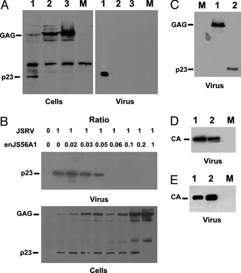
enJS56A1 Is Abundantly Expressed in the Cytoplasm of Transfected Cells and Blocks JSRV Exit. enJS56A1 does not release viral particles in the supernatant of transfected 293T cells, despite the presence of an intact gag gene and the use of the strong CMV promoter (7). It seemed possible that Gag was not expressed efficiently because of some transcriptional or translational defect. However, Western blotting analysis of lysates of 293T cells transfected with pCMV2en56A1 revealed abundant quantities of immature Gag (≈70 kDa) (Fig. 1A). As expected, lysates obtained from pCMV2JS21-transfected cells showed both the immature and the mature form of Gag, whereas only the latter was detectable in the supernatant.
Fig. 1.
enJS56A1 specifically inhibits JSRV viral particle release. (A) Western blot analysis of cell lysates (Left) and supernatants (Right) of 293T transfected or cotransfected with enJS56A1 and JSRV expression plasmids. Gag was detected with a JSRV p23 antiserum. Lanes: 1, JSRV; 2, enJS56A1; 3, enJS56A1 + JSRV; M, mock. (B) Western blot analysis of cell lysates and supernatants of 293T cells cotransfected with fixed amounts (14 μg) of JSRV expression plasmid and decreasing amounts of enJS56A1 expression plasmid. (C) JS21Δpro is able to release viral particles in the supernatant of transfected cells. Lanes: M, mock; 1, JS21Δpro; 2, JSRV. (D) enJS56A1 does not inhibit MPMV exit in cotransfection assays. Lanes: 1, MPMV; 2, MPMV+enJS56A1; M, mock. Gag was detected with anti-MPMV CA serum. (E) enJS56A1 does not inhibit M-MLV viral exit. Lanes: 1, M-MLV; 2, M-MLV+enJS56A1; M, mock. Gag was detected with anti-MLV CA serum.
We then determined whether enJS56A1 was able to influence the capacity of JSRV to exit the cells in cotransfection assays. Analysis of cell lysates from 293T cells cotransfected with pCMV2JS21 and pCMVen56A1 indicated the presence of abundant immature Gag protein in cell lysates, but no viral particles were detected in the supernatant (Fig. 1 A, lane 3). Titration of the amount of pCMV2enJS56A1 cotransfected with a fixed amount of pCMV2JS21 showed that enJS56A1 can inhibit JSRV particle release even at a 1:15 ratio (enJS56A1:JSRV plasmid DNA) (Fig. 1B). Quantitative analysis by RT assays of particles (data not shown) released in the supernatant of cells transfected with pCMV2JS21 or pCMV2enJS56A1 or cotransfected with an equal mixture of both plasmids revealed that the endogenous enJS56A1 reduces JSRV exit by ≈440-fold (geometric mean of the degree of inhibition in five independent experiments) to a level virtually indistinguishable from the background value seen with cells transfected with empty plasmid. Thus, the RT results are fully consistent with the Western blotting data, confirming the ability of enJS56A1 to block virus particle formation or exit by JSRV. As shown in Fig. 1 A, lane 2, only the uncleaved Gag polyprotein is seen in lysates of enJS56A1-transfected cells. It seemed possible that the failure of maturation cleavage in this virus was responsible for the defect in particle release. To test this possibility, we deleted nucleotides 2116–2147 in the protease-coding region of pJS21m (pJS21m derives from pCMV2JS21 by removal of the multiple cloning site after the 3′ long terminal repeat and introduction of an XbaI and a SalI site in gag by silent mutagenesis). This mutation prevented cleavage of pJS21 Gag but had no effect on particle production (Fig. 1C); thus, inhibition of Gag cleavage cannot explain the block in viral exit caused by enJS56A1. We also tested the ability of enJS56A1 to interfere with virus production by MPMV (another betaretrovirus) or M-MLV (a gammaretrovirus). As shown in Fig. 1 D and E, enJS56A1 had no detectable effect on these viruses.
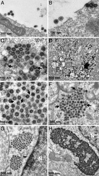
enJS56A1 Forms Intracytoplasmic Viral Particles. enJS56A1 is abundantly expressed in transfected 293T cells (Fig. 1). Consequently, we performed electron microscopy analysis to determine whether enJS56A1 was able to form viral particles. Cells transfected with JSRV expression plasmid showed, as expected, complete extracellular particles with envelope (Fig. 2A), particles approaching and budding from the cell membrane (Fig. 2B), and intracytoplasmic core particles (Fig. 2 C and D) typical of betaretroviruses. Cells transfected with enJS56A1 expression plasmid also showed intracytoplasmic viral particles more often visible in the perinuclear region (Fig. 2 E and F), but no budding or extracellular particles were observed, in agreement with Western blotting and RT analysis. A difference noted between the intracytoplasmic particles of JSRV and enJS56A1 was that the latter were in large agglomerations of tightly packed particles. The presence of incomplete particles in these clusters suggests that these are the sites of enJS56A1 assembly. However, the incomplete particles may simply represent particles that have been sectioned during sample preparation. In addition, we noticed that enJS56A1 particles appear to have projections from the viral core that at times seem to join particles together. These projections could be part of the enJS56A1 Gag or could be cellular structures linked to the enJS56A1 particles. Viral particles in cells cotransfected with both JSRV and enJS56A1 plasmids showed the same phenotype of enJS56A1 particles with large clusters of viral particles (Fig. 2 G and H).
Fig. 2.
enJS56A1 assembles intracytoplasmic viral particles. 293T cells were transfected with JSRV (A–D) or enJS56A1 (E and F) or cotransfected (G and H) with both expression plasmids and analyzed by electron microscopy. (A and B) Extracellular complete JSRV particles and particles approaching and budding from the membrane. (C and D) Intracytoplasmic JSRV. (E and F) enJS56A1 particles. (C and F) Arrows, incomplete viral particles. In E, arrows point to apparent projections from the enJS56A1 particles. (G and H) Large clusters of perinuclear viral particles in enJS56A1+JSRV cotransfected cells. N, nucleus.
Thus, enJS56A1 is able to induce the formation of viral particles that assemble in the cytoplasm, but they are not able to reach and bud from the cell membrane. Furthermore, expression of this endogenous genome prevents JSRV from producing virus into the culture supernatant.
Determinants of the enJS56A1 Defect in Viral Exit and Interference Map in Different Amino Acid Residues of the Amino-Terminal Gag. The enJS56A1 and JSRV Gag are 94% identical. The major differences are localized in two regions (VR1 and VR2) that are rich in proline residues (in JSRV) and localized in Gag between the putative MA and CA proteins (7, 13), probably in p23 (the exact boundaries of the JSRV Gag proteins are not known with certainty). To map the determinants of the defect of viral exit and interference of enJS56A1, a panel of chimeras between JSRV and enJS56A1 was constructed, and their ability to exit transfected cells and interfere with JSRV was determined.
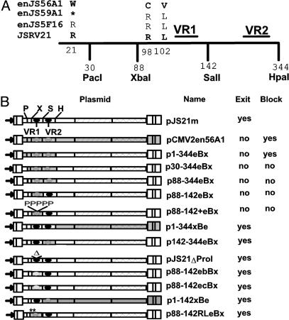
The schematic representation of the chimeras, the relative position of the restriction sites used for the molecular cloning, and the results obtained in transfection assays are shown in Fig. 3. The results of these tests can be summarized briefly as follows. The defect in virus production of enJS56A1, and its ability to interfere with JSRV virus production, can be attributed to amino acids in Gag proximal to residue 142. The VR1 per se is not responsible for the defect of enJS56A1 because chimeras p88–142ebBx and p88–142ecBx, containing, respectively, the VR1 from loci enJS5F16 and enJS59A1, were able to exit transfected cells (data not shown). However, a chimera with residues 88–142 from enJS56A1 (p88–142eBx) was defective, but the defect could be reversed by the two mutations C98R and V102L. Interestingly, p88–142eBx can be rescued by WT JSRV, and it can complement a protease-defective JSRV, leading to the production of particles with Gag cleavage products (data not shown). Thus, C98 and/or V102 of enJS56A1 block its ability to produce virus into the supernatant, but this defect is recessive in the presence of WT Gag (Fig. 3).
Fig. 3.
Determinants of enJS56A1 defect in viral exit and interference. (A) Schematization of the first 344 Gag amino acid residues. The restriction sites used for the construction of the chimeras, the VR1–VR2 regions, and the amino acid residues found to be critical for the enJS56A1 defect are indicated. enJS59A1 has a premature stop codon in gag upstream of position 88; amino acid residue at position 21 is indicated by *. (B) Schematic representation of the chimeras constructed in this study and results obtained in transfection and cotransfection assays.
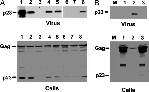
To further define the residues responsible for the defects in enJS56A1, we constructed a series of point-mutants of JSRV (Table 1). We found that mutants pJSR98C and pJSR98C-L102V did not release particles but did not interfere with WT JSRV production. In contrast, mutant pJSR21W replicated the phenotype of enJS56A1, failing to produce virus and interfering with JSRV production (Fig. 4A). Tests on these and other mutants lead to the conclusion that the critical mutation of the arginine residue at position 21 of JSRV into a tryptophan in enJS56A1 determines the dominant negative functions of the endogenous Gag. Mutations R98C and L102V are also responsible for additional defects in viral exit that by themselves are not dominant, although they decrease the amount of viral particles released by the cotransfected JSRV.
Table 1. Phenotypes of JSRV mutants.
| Mutant | Exit | Interference |
|---|---|---|
| pJSL102V | Yes | n.a. |
| pJSR98C | No | No |
| pJS21R21W | No | Yes |
| pJSG2A | No | No |
| pJSR98C-L102V | No | No |
| pJSR21W-R98C-L102V | No | Yes |
| pJSG2A-R98C-L102V | No | No |
| pJSG2V-R21W-R98C-L102V | No | Yes |
n.a., not applicable.
Fig. 4.
Western blotting analysis of JSRV and enJS56A1 mutants. Supernatants (Upper) and cell lysates (Lower) of 293T cells cotransfected with JSRV and critical mutant expression plasmids were analyzed by Western blotting for the presence of Gag proteins. (A) Lanes: 1, JSRV; 2, JSRV + JSG2A; 3, JSRV+JSR21W; 4, JSRV + JSR98C-L102V; 5, JSRV + JSG2A-R98C-L102V; 6, JSRV + JSR21W-R98C-L102V; 7, JSRV + JSG2V-R21W-R98C-L102V; 8, JSRV + JSR98C. (B) Restoration of enJS56A1 viral exit. Lanes: M, mock; 1, en56W21R-V102L; 2, en56W21R-C98R; 3, en56W21R. Note that the p23 of the endogenous Gag has a slightly lower molecular weight with respect to the homologous JSRV protein, probably because of the lack of proline residues in the VR1 and VR2 regions of the endogenous locus.
We also tested whether by affecting the myristoylation signal we would create mutants that are unable to exit and are transdominant over WT JSRV. Both single mutant pJS21G2A and triple mutant pJS21G2A-R98C-L102V were unable to release viral particles in the supernatant as expected; however, they did not interfere significantly with the exit of JSRV (Fig. 4). This finding suggests that R21 does not simply affect the efficacy of membrane targeting by the myristoylation signal.
Correction of enJS56A1 Defect in Viral Particle Release. Endogenous mutants pen56W21R and pen56W21R-V102L did not render enJS56A1 capable of releasing viral particles in the supernatant of transfected cells. However, double mutant pen56W21R-C98R did restore the capacity of enJS56A1 to exit transfected cells. The level of expression of en56W21R-C98R was in general lower than that of the WT JSRV (Fig. 4B).
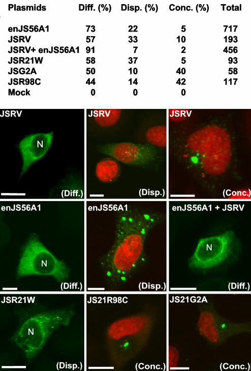
Confocal Microscopy. We next analyzed the pattern of expression of JSRV, enJS56A1, and the relevant mutants by confocal microscopy using a JSRV p23 antiserum (Fig. 5). We classified Gag-positive cells into three basic phenotypes: (i) diffuse, (ii) dispersed, and (iii) concentrated. Cells with a diffuse phenotype had a uniform cytoplasmic staining, usually more intense in the perinuclear region. In the dispersed phenotype, “dots” of fluorescence were visible and scattered in the cytoplasm, whereas in the concentrated phenotype, there was an area of intense staining in the vicinity of the nucleus. These terms are in accord with recent descriptions of cells expressing the betaretrovirus MPMV, which is assembled in the pericentriolar region of the cell (15). Most often, cells presented a mixed diffused-dispersed phenotype that we counted as dispersed. There were no significant differences in the distribution of the three phenotypes among enJS56A1, JSRV, and JSR21W (Fig. 5) in three independent experiments. However, cells transfected with enJS56A1 always presented the highest number of Gag-positive cells and the most intense staining, consistent with the inability of this Gag to be released from the cell. In addition, the “dispersed” foci of fluorescence in cells expressing enJS56A1 were much larger than those observed with JSRV or any other mutant. We expected cells cotransfected with JSRV and enJS56A1 expression plasmids to resemble enJS56A1-positive cells, given the dominant-negative property of its Gag; however, they showed mainly a diffuse phenotype with intense perinuclear staining. JS21R98C and JS21G2A consistently showed the highest proportion of cells with a concentrated phenotype. The fact that the phenotypes of the R98C and R21W mutants are different suggests that W21 and C98 contribute in different ways to the defects in enJS56A1.
Fig. 5.
Confocal microscopy of HeLa cells expressing or coexpressing enJS56A1, JSRV, and critical mutants. Positive cells have been quantified according to three phenotypes: diffuse (Diff.), dispersed (Disp.), and concentrated (Conc.). At the top is the quantification of a representative experiment. Values indicate relative percentage of each phenotype, and the last column indicates the total number of cells counted for each transfection. Photomicrographs are representative examples of cells expressing the indicated viruses and mutants. Note that the “dispersed” phenotype for enJS56A1-transfected cells consists of larger foci with a more intense fluorescence than in other samples. Gag staining is in green, and nuclei are in red or with the letter N.
Confocal microscopy confirms what we have seen by electron microscopy: enJS56A1 (and defective mutants) do not accumulate at the cell membrane. As shown by electron microscopy, the enJS56A1 particles seem to be present mainly in the perinuclear region, suggesting that the cytoplasmic staining in the dispersed and diffuse phenotype observed by confocal microscopy probably derives from nonparticulate Gag proteins.
Overall, these data suggest that the defect possessed by enJS56A1 seems to be at the level of viral particle trafficking between the cytoplasm and the cell membrane.
enJS56A1 Is Expressed in the Sheep Uterus. enJSRVs RNA and proteins are highly expressed in the sheep uterus (5–7). However, there are ≈20 copies of enJSRV loci, and no information is available on their relative expression levels. The critical R21W mutation of enJS56A1 is not present in the other two published full-length enJSRVs sequences, enJS5F16 and enJS59A1 (7). We performed RT-PCR on sheep endometrium by amplifying a region between the 5′ leader and the VR2 region of Gag. Primers were chosen in regions highly conserved between enJSRVs and JSRV sequences to amplify as many enJSRV loci as possible. Four independent PCRs were cloned into a PCR cloning vector, and 34 independent clones were fully sequenced. Sequence analysis revealed that 12 sequences of the 34 clones had some defect in the gag gene that would preclude production of functional protein (e.g., premature stop codons, frame shifts, or mutations in the first ATG). Of the remaining 22 clones with an intact gag ORF, 4 had the critical W21, C98, and V102 mutations characteristic of the enJS56A1 clone. These data suggest that enJS56A1 (or enJS56A1-like loci) is expressed in the sheep endometrium.
Discussion
In this paper, we have described an endogenous retrovirus of sheep (enJS56A1) with a dominant negative Gag protein that interferes with its exogenous counterpart (JSRV) at a postassembly level. This blockade represents a previously uncharacterized mechanism of retroviral interference. The other known ERV-mediated blocks are all at early stages of the retroviral replication cycle such as entry (by receptor competition) or at a preintegration step (Fv-1) (1).
JSRV/enJSRVs are betaretroviruses, and thus they assemble in the cytoplasm. Cells expressing enJS56A1 (or coexpressing enJS56A1 and JSRV) display large clusters of tightly packed particles in the cytoplasm. The almost regular organization of the enJS56A1 particles, and the presence in these groupings of incomplete cores, suggests that assembled particles are not able to traffic properly to the cell membrane.
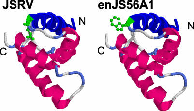
The tryptophan residue at position 21 of the enJS56A1 MA (replacing an arginine in JSRV) is the main determinant for the block induced by enJS56A1, although C98 and V102 contribute to the overall dominant negative activity of the endogenous Gag. The mutant JSR21W shows a dominant negative phenotype analogous to enJS56A1. Interestingly, R21 (and a glycine residue immediately after) is conserved in all betaretroviruses, including MPMV and related simian betaretroviruses, mouse mammary tumor virus, and human endogenous retrovirus-K. The JSRV/enJS56A1 MAs and MPMV MA are 39% identical and 64% similar, and this level of similarity allows for structural modeling on the basis of the homologous MPMV protein (16, 17) (Fig. 6). R21 falls at the end of the first α-helix of MA. Thus, the R to W mutation present in the enJS56A1 introduces a hydrophobic amino acid in an exposed portion of the protein. This nonconservative mutation could alter the ability of Gag to interact with cellular factors necessary for the trafficking of viral particles, although it is also possible that the mutation exerts its effects by altering the structure or assembly properties of Gag. The admixture of mutant Gag protein blocks the transport of assembled particles containing WT Gag; thus, it appears that the normal trafficking of WT particles to the plasma membrane depends on the concerted action of many WT Gag molecules. The myristoylation signal plays a critical role in targeting Gag and viral particles to the cell membrane (18, 19). Mutation of the myristoylation signal did not confer a dominant negative phenotype to the resulting mutant (JSG2A) but did block viral exit. However, a polybasic region downstream of the myristoylation signal in other retroviruses also has been found to be an important signal for membrane targeting of Gag (20). It is possible that the R21W mutation impairs membrane-targeting of JSRV Gag. Another class of mutants that interferes with release of assembled virions is the “late (L) domain” mutants. However, it seems unlikely that enJS56A1 is equivalent to an L domain mutant because such mutants of MPMV accumulate at the cell membrane (21, 22) and enJS56A1 still contains intact PSAP and PPXY motifs in its VR2 region.
Fig. 6.
Modeling of the putative JSRV and enJS56A1 MA proteins. The tertiary structures of the matrix proteins of enJS56A1 and JSRV were modeled by using the Swiss Model Server (http://swissmodel.expasy.org). The matrix protein of MPMV (Protein Data Bank ID code 1BAX) was used as a template. As the available MPMV matrix protein structure at the Protein Data Bank is a carbon trace structure, a tertiary structure including side chains was generated by using the MaxSprout database algorithm (www.ebi.ac.uk/maxsprout). Models are displayed in ribbons, and the W21 and R21 are displayed in green in a ball-and-stick format. The N and C terminals are indicated. The first α-helix is shown in blue. Both models were derived from the amino-terminal 92-aa residues of the JSRV/enJS56A1 Gag.
The agglomerates of viral particles visible in JSRV+enJS56A1 transfected cells resemble “viral factories” of large DNA viruses such as poxviruses, iridoviruses, and asfaviruses (23). These viral factories have been shown to have similar properties to the aggresomes, perinuclear cellular sites where misfolded or unassembled proteins are transported and sequestered by the cell. Aggresomes are localized in the vicinity of the centrosomes and enclosed in a characteristic vimentin cage (24). It will be interesting to determine whether JSRV and/or enJS56A1 assembly takes place in these cellular structures.
The exact biological role of the enJS56A1 locus in the evolutionary interplay with its exogenous counterpart and its host is difficult to establish. By RT-PCR, we determined that enJS56A1 (or similar loci containing W21, C98, and V102) are expressed in the sheep endometrium. We have previously shown that enJSRVs also can interfere with JSRV entry by receptor competition (9). Thus, enJSRVs have developed a two-step strategy and are capable of interfering in the replication cycle of related exogenous viruses at both early and late stages. Alternatively, or additionally, enJS56A1 might have been used by the host to “control” the replication of other enJSRVs with some damaging effect.
In conclusion, these studies have shown previously uncharacterized interfering properties of an endogenous retroviral Gag. Understanding the molecular mechanisms underlying the enJS56A1-induced block could spark new strategies for obtaining antiretroviral drugs. This study provides a previously uncharacterized example of an endogenous retrovirus with a dominant negative Gag protein that acts at a late step of the viral replication cycle. HIV Gag mutants with dominant negative effects have been derived in the late 1980s and early 1990s (14, 25), along with the hope of an effective “intracellular immunization” (14). Our results suggest that this phenomenon has, in fact, arisen in sheep as a result of natural selection. The exogenous counterpart of enJS56A1, JSRV, causes a major infectious disease of sheep with no available vaccine. The recent development of animal cloning technologies raises the possibility that a transgenic sheep in which enJS56A1 expression is directed to the differentiated epithelial cells of the lungs, the cells' target for JSRV transformation, would be resistant to JSRV-induced disease. Thus, the JSRV-enJS56A1 system could furnish a unique model to test the intracellular immunization theory in a naturally occurring retroviral infection.
Acknowledgments
We thank J. Urbauer, J. Shields, and M. Varela for help with molecular modeling and confocal microscopy; K. Dunlap (Texas A&M University) for providing sheep uterine RNA preparations; E. Hunter for reagents; and L. A. Jones and C. Pretto for technical help. This work was funded in part by a Georgia Cancer Coalition grant and National Institutes of Health Grant CA95706-01.
This paper was submitted directly (Track II) to the PNAS office.
Abbreviations: CMV, cytomegalovirus; enJSRVs, endogenous sheep retroviruses related to JSRV; ERV, endogenous retrovirus; JSRV, Jaagsiekte sheep retrovirus; MPMV, Mason–Pfizer monkey virus; M-MLV, Moloney murine leukemia virus; RT, reverse transcriptase.
References
- 1.Boeke, J. D. & Stoye, J. P. (1997) in Retroviruses, eds. Coffin, J. M., Hughes, S. H. & Varmus, H. E. (Cold Spring Harbor Lab. Press, Plainview, NY), pp. 343–436. [PubMed]
- 2.Palmarini, M., Mura, M. & Spencer, T. E. (2004) J. Gen. Virol. 85, 1–13. [DOI] [PubMed] [Google Scholar]
- 3.Palmarini, M., Sharp, J. M., de las Heras, M. & Fan, H. (1999) J. Virol. 73, 6964–6972. [DOI] [PMC free article] [PubMed] [Google Scholar]
- 4.Palmarini, M. & Fan, H. (2001) J. Natl. Cancer Inst. 93, 1603–1614. [DOI] [PubMed] [Google Scholar]
- 5.Spencer, T. E., Stagg, A. G., Joyce, M. M., Jenster, G., Wood, C. G., Bazer, F. W., Wiley, A. A. & Bartol, F. F. (1999) Endocrinology 140, 4070–4080. [DOI] [PubMed] [Google Scholar]
- 6.Palmarini, M., Gray, C. A., Carpenter, K., Fan, H., Bazer, F. W. & Spencer, T. (2001) J. Virol. 75, 11319–11327. [DOI] [PMC free article] [PubMed] [Google Scholar]
- 7.Palmarini, M., Hallwirth, C., York, D., Murgia, C., de Oliveira, T., Spencer, T. & Fan, H. (2000) J. Virol. 74, 8065–8076. [DOI] [PMC free article] [PubMed] [Google Scholar]
- 8.Palmarini, M., Datta, S., Omid, R., Murgia, C. & Fan, H. (2000) J. Virol. 74, 5776–5787. [DOI] [PMC free article] [PubMed] [Google Scholar]
- 9.Spencer, T. E., Mura, M., Gray, C. A., Griebel, P. J. & Palmarini, M. (2003) J. Virol. 77, 749–753. [DOI] [PMC free article] [PubMed] [Google Scholar]
- 10.Bieniasz, P. D. (2003) Trends Microbiol. 11, 286–291. [DOI] [PubMed] [Google Scholar]
- 11.Rhee, S. S., Hui, H. X. & Hunter, E. (1990) J. Virol. 64, 3844–3852. [DOI] [PMC free article] [PubMed] [Google Scholar]
- 12.Ausubel, F. M., Brent, R., Kingston, R. E., Moore, D. D., Seidman, J. G., Smith, J. A. & Struhl, K. (2000) Current Protocols in Molecular Biology (Wiley, New York).
- 13.Palmarini, M., Sharp, J. M., Lee, C. & Fan, H. (1999) J. Virol. 73, 10070–10078. [DOI] [PMC free article] [PubMed] [Google Scholar]
- 14.Trono, D., Feinberg, M. B. & Baltimore, D. (1989) Cell 59, 113–120. [DOI] [PubMed] [Google Scholar]
- 15.Sfakianos, J. N., LaCasse, R. A. & Hunter, E. (2003) Traffic 4, 660–670. [DOI] [PubMed] [Google Scholar]
- 16.Conte, M. R., Klikova, M., Hunter, E., Ruml, T. & Matthews, S. (1997) EMBO J. 16, 5819–5826. [DOI] [PMC free article] [PubMed] [Google Scholar]
- 17.Conte, M. R. & Matthews, S. (1998) Virology 246, 191–198. [DOI] [PubMed] [Google Scholar]
- 18.Rein, A., McClure, M. R., Rice, N. R., Luftig, R. B. & Schultz, A. M. (1986) Proc. Natl. Acad. Sci. USA 83, 7246–7250. [DOI] [PMC free article] [PubMed] [Google Scholar]
- 19.Rhee, S. S. & Hunter, E. (1987) J. Virol. 61, 1045–1053. [DOI] [PMC free article] [PubMed] [Google Scholar]
- 20.Freed, E. O. (1998) Virology 251, 1–15. [DOI] [PubMed] [Google Scholar]
- 21.Yasuda, J. & Hunter, E. (1998) J. Virol. 72, 4095–4103. [DOI] [PMC free article] [PubMed] [Google Scholar]
- 22.Gottwein, E., Bodem, J., Muller, B., Schmechel, A., Zentgraf, H. & Krausslich, H. G. (2003) J. Virol. 77, 9474–9485. [DOI] [PMC free article] [PubMed] [Google Scholar]
- 23.Andres, G., Simon-Mateo, C. & Vinuela, E. (1997) J. Virol. 71, 2331–2341. [DOI] [PMC free article] [PubMed] [Google Scholar]
- 24.Heath, C. M., Windsor, M. & Wileman, T. (2001) J. Cell Biol. 153, 449–455. [DOI] [PMC free article] [PubMed] [Google Scholar]
- 25.Mammano, F., Ohagen, A., Hoglund, S. & Gottlienger, H. G. (1994) J. Virol. 68, 4927–4936. [DOI] [PMC free article] [PubMed] [Google Scholar]


