Original citation: J. Clin. Invest. 114:280–290(2004). doi:10.1172/JCI200421583.
Citation for this Corrigendum: J. Clin. Invest. 114:142 (2004). doi:10.1172/JCI200421583E1.
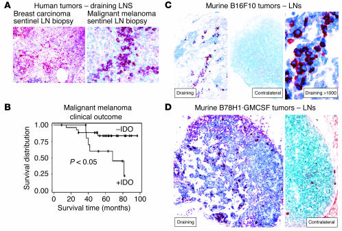
During the preparation of this manuscript for publication, an error was introduced into the panel labels of Figure 1. The correct figure appears below. We regret this error.
Figure 1.
Expression of IDO in human and murine TDLNs. (A) Sentinel (first draining) LN from patients with breast carcinoma (left, ×100) and malignant melanoma (right, ×400), showing an abnormal infiltration of IDO+ cells (red chromogen). (B) Kaplan-Meier survival plot of 40 patients with malignant melanoma, stratified into those with an abnormal accumulation of IDO+ cells in the sentinel LN (+IDO), versus a normal (negative) pattern. (C) Expression of IDO in murine B16F10 melanoma. Left: Draining inguinal LN from a mouse with a B16F10 tumor, day 12, stained for IDO (red, ×100). Middle: Contralateral inguinal LN from the same animal as at left, stained for IDO (red, ×100). Right: High-power view of IDO+ cells shown in the left panel (×1,000). Controls for staining (anti-IDO antibody neutralized with the immunizing peptide) showed a negative pattern similar to that seen in the contralateral LN (not shown). (D) Draining and contralateral LNs from a mouse with B78H1–GM-CSF tumor, day 12, stained for IDO (red, both ×200).


