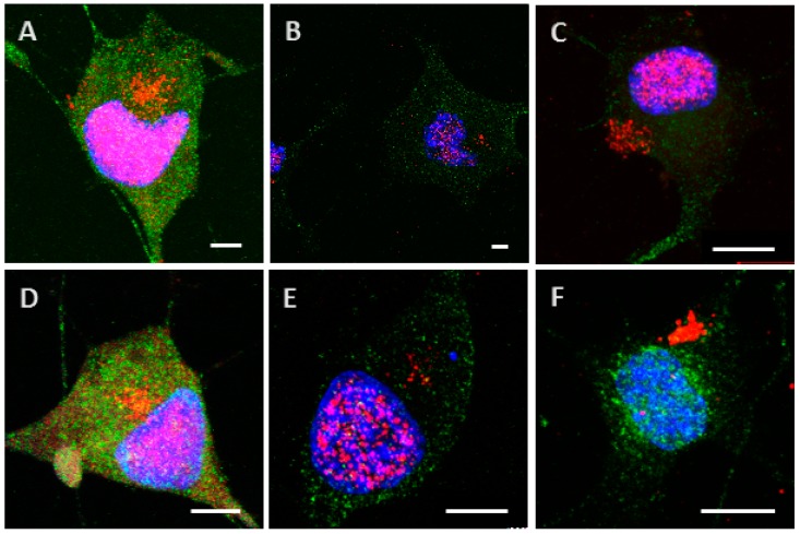
Figure A1.
Internalization and processing of the Tau protein (15 µg; A–C) and its highly phosphorylated form (1.5 µg; D–F) by Neuro2A cells. Following 24 h (A,D), or 48 h (B,E) of incubation with protein, cells were immunostained with a mouse anti-His-tag antibody targeted by an AlexaFluor® 594 antibody (red) and a rabbit anti-MAP2 antibody targeted by an AlexaFluor® 488 antibody (green). In other experiments, cells were placed in contact with protein for 8 h, whereupon the medium was replaced by one devoid of protein and maintained for 48 h (C,F). Nuclei were stained with DAPI. Different digital zooms were applied to the 60× optical zoom: A: no zoom, B: 1.2×, C: 2×, D: 1.6×, E: 2.1×, F: 2.3×. All scale bars correspond to 5 µm.

