Abstract
Pseudomonas aeruginosa defies eradication by antibiotics and is responsible for acute and chronic human infections due to a wide variety of virulence factors. Currently, it is believed that MvfR (PqsR) controls the expression of many of these factors indirectly via the pqs and phnAB operons. Here we provide strong evidence that MvfR may also bind and directly regulate the expression of additional 35 loci across the P. aeruginosa genome, including major regulators and virulence factors, such as the quorum sensing (QS) regulators lasR and rhlR, and genes involved in protein secretion, translation, and response to oxidative stress. We show that these anti-oxidant systems, AhpC-F, AhpB-TrxB2 and Dps, are critical for P. aeruginosa survival to reactive oxygen species and antibiotic tolerance. Considering that MvfR regulated compounds generate reactive oxygen species, this indicates a tightly regulated QS self-defense anti-poisoning system. These findings also challenge the current hierarchical regulation model of P. aeruginosa QS systems by revealing new interconnections between them that suggest a circular model. Moreover, they uncover a novel role for MvfR in self-defense that favors antibiotic tolerance and cell survival, further demonstrating MvfR as a highly desirable anti-virulence target.
Pseudomonas aeruginosa is a major nosocomial pathogen representing a critical threat for human health1,2 because of its tolerance and rapid development of resistance towards almost all current antimicrobial therapies3,4,5,6,7. P. aeruginosa acute and chronic infections are facilitated by a wide array of virulence factors, including toxins, small molecules and secondary metabolites as well as defense systems against host immunity and bacterial competitors. P. aeruginosa interactions with host and bacterial competitors generate environments with high levels of reactive oxygen species (ROS)8,9,10,11,12,13,14,15 that P. aeruginosa survives to by virtue of its multiple antioxidant systems16,17.
Most of P. aeruginosa’s virulence factors are controlled via the three major cell density dependent quorum sensing systems: LasR18, RhlR19,20 and MvfR (also known as PqsR)21,22,23,24. The current view is that these three systems are hierarchically connected with LasR positioned at the top of this hierarchy25,26,27. LasR and RhlR directly control the production of their respective activating inducers, acyl-homoserine lactones (HSL) 3-oxo-C12-HSL and C4-HSL encoded via the synthetases lasI and rhlI respectively18,28,29,30. LasR binds to 34 additional loci in P. aeruginosa genome, including mvfR and rhlR, and directly regulates the expression of multiple genes, including transcriptional regulators which ultimately result in the indirect modulation of more than 300 genes across the genome31. RhlR directly controls the expression of rhlAB and rhlC responsible for the biosynthesis of the rhamnolipid surfactants32,33 and also indirectly controls the expression of multiple genes34. MvfR also controls its own activity by binding and positively regulating the expression of pqsABCDE and phnAB operons that catalyze the biosynthesis of MvfR inducers and of ~60 distinct low-molecular-weight compounds21,22,23,35,36, including hydroxyquinolones (HAQs)37 and the non-HAQ molecule 2-AA38,39,40. Two of the most abundant HAQs (4-hydroxy-2-heptylquinoline [HHQ] and 3,4-dihydroxy-2-heptylquinoline [Pseudomonas Quinolone Signal-PQS]) bind and activate MvfR, leading to the induction of the many virulence factors that promote infection23,35,41,42,43. MvfR activity correlates with HHQ synthesis. Thus, an essential step of MvfR regulon activation by MvfR is the binding of MvfR protein to the pqsABCDE and phnAB operons23,35. So far, these were the only two operons to which MvfR was known to bind22,35,44 and the fact that MvfR is regulating the expression of 18% of P. aeruginosa genome45 was attributed to indirect effects.
The three QS systems appear to be interconnected in multiple and complex ways. RhlR and LasR QS systems both activate each other46. RhlR directly inhibits the expression of pqsA and mvfR by binding to their respective promoters35,44, and the MvfR regulon appears to be interconnected with RhlR via pqsE, the last gene of the pqs operon controlled by MvfR47. On the other hand LasR positively regulates MvfR, as it binds and induces mvfR expression during exponential phase27,35, with MvfR eventually becoming LasR-independent at the later stages of growth35. Another interconnection between the LasR and MvfR systems is that MvfR, via the pqs operon, controls the synthesis of the precursors of PQS and of the programmed cell death signal 2-n-heptyl-4-hydroxyquinoline-N-Oxide (HQNO)13, while LasR controls the enzymatic conversion of their precursors into these molecules by controlling the expression of pqsH and pqsL genes respectively26,37,48.
Here, our genome-wide analysis provides strong evidence that in addition to direct control of the pqsABCDE, phnAB and mvfR genes, MvfR may also bind to 34 additional loci across the genome of P. aeruginosa and fine-tune the expression of the associated genes. This work provides novel insights into the quorum sensing circuits in P. aeruginosa that are crucial for both pathogenesis and cell survival in deleterious environments, and its interconnection to the other P. aeruginosa QS systems, as well as its role in self-defense response that favors antibiotic tolerance.
Results
MvfR binds to and regulates the expression of multiple virulence-related loci in P. aeruginosa genome
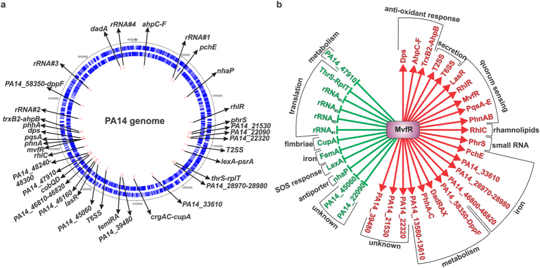
Previous studies reported that as cell density increases MvfR regulates more genes, reaching 18% of the P. aeruginosa genome at the onset of stationary phase45. To elucidate the mode of action of MvfR on the expression of QS-controlled genes, we utilized a genome-wide approach and performed chromatin immuno-precipitation sequencing (ChIPseq) coupled with RNA sequencing (RNAseq). To fully grasp the MvfR binding dynamics, we performed this analysis at four time points corresponding to different bacterial growth stages. We used cells from early (OD600nm 1.0), middle (OD600nm 2.0) and late (OD600nm 3.0) exponential phase as well as stationary phase (OD600nm 4.0) of growth. MvfR interacting DNA was immuno-precipitated and identified by Illumina sequencing. Table 1 and Fig. 1a show that MvfR binds to 37 loci across the PA14 genome. Amongst these 37 loci, we found the expected pqsA, phnA and mvfR promoters, thus validating our approach. MvfR binding was also validated in vivo (bacterial cultures) by ChIPqPCR on some of those key loci (Supplementary Figure S1).
Table 1. List of MvfR binding sites, their associated gene function and regulation.
| |
Regulated gene(s) | Function | MvfR binding (Input normalized coverage) | Locus | Regulation byMvfR | ||||
|---|---|---|---|---|---|---|---|---|---|
| Coordinates | OD 1 | OD 2 | OD 3 | OD 4 | |||||
| 155,548 | 157,602 | ahpC-ahpF | response to oxidative stress | 868 | 467 | over. | + | ||
| 733,577 | 738,665 | 16S, 23S, 5S & tRNA | translation (rRNA #1) | 748 | 504 | 503 | over. | − | |
| 788,364 | 789,806 | pyochelin operon | siderophore pyochelin | 375 | ins. | + | |||
| 1,170,619 | 1,171,875 | PA14_13580-13610 operonnhaP | ABC transporterNa+/H+ antiporter | 723 | prom. | + | |||
| − | |||||||||
| 1,651,474 | 1,652,473 | rhlR | rhlR QS system | 436 | end | + | |||
| 1,840,873 | 1,842,123 | phrS | small RNA | 643 | over. | + | |||
| 1,866,043 | 1,868,058 | PA14_21530 | unknown | 447 | 548 | over. | + | ||
| 1,923,884 | 1,925,012 | PA14_22090 | unknown | 586 | prom. | − | |||
| 1,943,865 | 1,944,935 | PA14_22320 | unknown | 381 | over. | + | |||
| 2,083,830 | 2,084,914 | T2SS operons | type 2 secretion system | 408 | over. | + | |||
| 2,201,076 | 2,202,455 | lexApsrA | SOS responsesigma factor | 497 | prom. | − | |||
| + | |||||||||
| 2,467,980 | 2,469,951 | infC operon | translation (initiation factor, tRNA and ribosomal proteins) | 821 | 277 | over. | − | ||
| 2,504,998 | 2,506,157 | PA14_28970 & 28980 | putative iron transporter | 107 | over. | + | |||
| 2,956,881 | 2,958,001 | pyoverdine operon | siderophore pyoverdine | 106 | ins. | + | |||
| 3,303,526 | 3,304,565 | crgAC & cupA operon | cupA fimbriae | 100 | end | − | |||
| 3,513,100 | 3,515,095 | PA14_39480 | unknown | 844 | 232 | ins. | + | ||
| 3,547,291 | 3,548,339 | femIRA | iron transpoter | 77 | prom. | + | |||
| 3,830,327 | 3,831,831 | T6SS locus 2 operons | type 6 secretion system | 545 | prom. | + | |||
| 4,019,224 | 4,020,417 | PA14_45060 | putative urea transporter | 94 | over. | − | |||
| 4,084,785 | 4,086,308 | lasR | lasR QS system | 490 | over. | + | |||
| 4,104,672 | 4,106,305 | PA14_46160 | unknown | 536 | 493 | over. | + | ||
| 4,167,308 | 4,168,950 | PA14_46810-46820 | putative iron transporter | 293 | 77 | over. | + | ||
| 4,246,274 | 4,247,621 | cobalamine operon | cobalamine biosynthesis | 86 | ins. | ? | |||
| 4,263,263 | 4,264,333 | PA14_47910 | ABC transporter (arabinose) | 99 | ins. | − | |||
| 4,294,736 | 4,295,982 | PA14_48240-48300 | putative antibiotic efflux pump | 103 | ins. | ? | |||
| 4,425,759 | 4,427,304 | PA14_49750 & rhlC | rhamnolipids biosynthesis | 119 | ins. | + | |||
| 4,561,261 | 4,564,718 | mvfR | MvfR QS system | 6,439 | 5,101 | 1,313 | over. | + | |
| 4,565,212 | 4,566,614 | phnAB operon | MvfR QS system | 378 | prom. | + | |||
| 4,570,486 | 4,572,591 | pqsABCDE operon | MvfR QS system | 1,083 | 2,118 | 647 | 254 | prom. | + |
| 4,602,850 | 4,604,494 | dps | response to oxidative stress | 596 | 526 | over. | + | ||
| 4,698,069 | 4,699,401 | phhABC operon | phenylalanine catabolism | 338 | 376 | over. | + | ||
| 4,722,947 | 4,725,071 | trxB2 & ahpB | response to oxidative stress | 497 | 736 | over. | + | ||
| 4,953,754 | 4,957,408 | 5S, 23S, tRNAs, 16S | translation (rRNA #2) | 516 | 606 | 422 | over. | − | |
| 5,200,388 | 5,201,551 | psdR | repressor of ABC transporter dppA-F (arginine) | 525 | prom. | − | |||
| 5,535,420 | 5,540,989 | 5S 23S tRNAs 16S | translation (rRNA #3) | 672 | 702 | 596 | over. | − | |
| 6,245,833 | 6,247,899 | dadA | D-alanine metabolism | 1,734 | 2,081 | 1,043 | over. | + | |
| 6,312,205 | 6,316,455 | 5S 23S tRNAs 16S | translation (rRNA #4) | 725 | 420 | over. | − | ||
over. = binding site is overlapping several genes, ins. = binding site is inside a gene, prom. = binding site is in a promoter region, end = binding site is at the end of a gene, + = positive regulation, − = negative regulation, ? = unclear regulation. For more details on gene regulation, see Supplementary Table S1.
Figure 1. MvfR binding sites.
(a) Localization of MvfR binding sites in PA14 genome. The first outer black circle represents the PA14 circular genome. Genes encoded on the positive strand (second circle) or negative strand (third circle) are shown in blue. Figure generated with IGV software102. MvfR binding sites are represented by the red rectangles (fourth circle). (b) Categorization of genes associated with MvfR binding sites. Positive regulation is indicated by a red arrow and negative regulation by a green flat bar.
To correlate MvfR binding with the targeted gene expression, RNAseq studies were carried out using the parental strain PA14 and the isogenic mvfR mutant. Gene expression analysis of the RNAseq studies indicates that MvfR regulates 95% of the genes associated with its binding sites, with 64% being induced and 31% repressed. MvfR action on the regulation of two remaining sites is unclear (Fig. 1b, Table 1 and Supplementary Table S1).
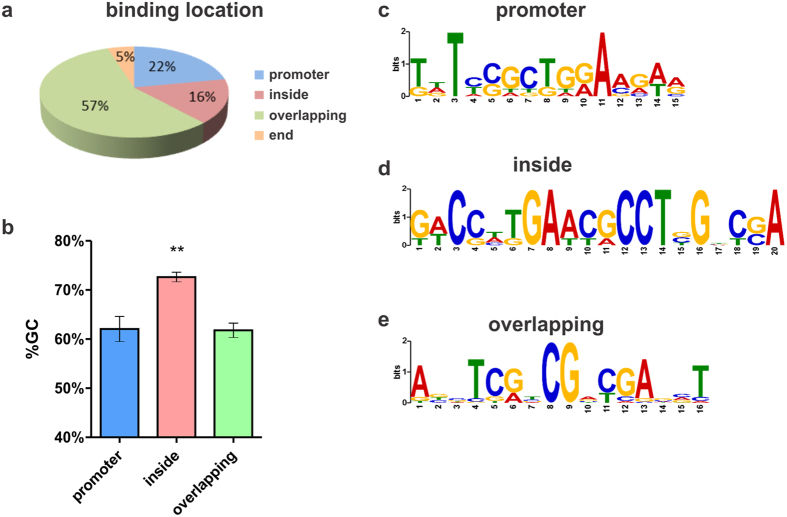
MvfR binding to the 37 loci is either at the promoter region (22%), extends over several genes (57%), within genes (16%), or at the end of genes (5%) (Fig. 2a). Interestingly, MvfR binding sites located within genes harbor a 72% GC content, which is significantly higher than all the other MvfR binding sites (Fig. 2b). These data suggest that MvfR may exhibit different binding patterns, and potentially recognize different DNA binding motifs, consistent with the ability of LysR Type of Transcriptional Regulators (LTTR) to bind at different gene regions49. In silico analysis using MEME suite50 indeed shows that the predicted consensus motifs differ based on MvfR binding location (Fig. 2c–e).
Figure 2. MvfR binding pattern.
(a) Frequency of MvfR binding sites based on location. (b) Percentage of GC content in the DNA sequence of MvfR binding sites according to the site location, either at the promoter region (blue), inside a gene (red) or overlapping several genes (green). Statistical significance was assessed using one way ANOVA + Dunnett’s post-test. (c–e) MvfR binding motif identified using MEME suite50 according to the site location, either at the promoter region (d), inside a gene (e) or overlapping several genes (f).
Functional categorization of the genes associated in 37 loci to which MvfR binds reveals that they are mainly involved in virulence related functions, including protein secretion, quorum sensing, rhamnolipids biosynthesis and iron acquisition but also in functions related to cell metabolism, transport of small molecules, translation and response to oxidative stress (Table 1 and Fig. 1b). The following sections focus on the MvfR binding and gene expression of some of these key virulence functions.
MvfR contributes to the induction of both RhlR and LasR QS systems
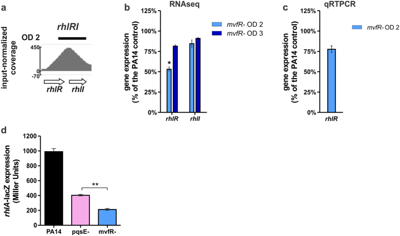
RhlR and LasR are the two other P. aeruginosa main QS systems, both directly and indirectly controlling multiple virulence genes, including the MvfR QS system. Figures 3a and S1 show that MvfR binds to the rhlR – rhlI locus at OD600nm 2.0. Accordingly, the expression of rhlR is significantly reduced in the mvfR mutant at OD600nm 2.0 (Fig. 3b,c), suggesting another level of regulation of the RhlR QS system aside from what was previously described via PqsE24,51,52. Indeed, as shown in Fig. 3d, the expression of rhlA – directly regulated by RhlR – is significantly lower in the mvfR mutant than in the pqsE mutant (p < 0.01) supporting the existence of an additional regulatory role of MvfR on the RhlR QS system independent of PqsE.
Figure 3. MvfR binds to and induces genes involved in the RhlR QS system.
(a) ChIPseq analysis reveals that MvfR binds to the rhlR-rhlI region. The black bar above the binding intensity plot represents the peak identified using SPP peak caller. (b) RNAseq analysis indicates that MvfR induces the expression of rhlR. Light blue bar = mvfR mutant at OD600nm 2, dark blue bar = mvfR mutant at OD600nm 3. (c) qRTPCR analysis validates that MvfR induces the expression of rhlR. Light blue bar = mvfR mutant at OD600nm 2. Data show the average +/− SEM of 3 independent replicates. (d) MvfR has an additional layer of control on rhlA expression in addition to PqsE as rhlA promoter activity is significantly lower in mvfR mutant than in pqsE mutant (p < 0.01, unpaired T test). Data show the average +/− SEM of at least 3 independent replicates.
The ChIPseq analysis also shows MvfR binding over the region containing lasR and rsaL genes (Figs 4a and S1). Consistently, lasI, rsaL and lasR expression is reduced in the mvfR mutant, indicating that MvfR acts as an activator for this region (Fig. 4b,c). MvfR binding on these genes occurs early (OD600nm 1.0) (Fig. 4a), which correlates with the early effect on the transcription of these genes (Fig. 4c).
Figure 4. MvfR binds to and fine tunes the expression of LasR QS system genes.
(a) ChIPseq analysis reveals that MvfR binds to rsaL-lasR region. The black bar above the binding intensity plots represents the peak identified using SPP peak caller. (b) RNAseq analysis indicates that MvfR induces the expression lasR, lasI and rsaL. Light blue bar = mvfR mutant at OD600nm 2, dark blue bar = mvfR mutant at OD600nm 3. (c) qRTPCR analysis validates that MvfR induces the expression of lasR. Faint blue bar = mvfR mutant at OD600nm 1, light blue bar = mvfR mutant at OD600nm 2. Data show the average +/− SEM of 3 independent replicates. Statistical significance was assessed using one way ANOVA + Dunnett’s post-test.
Taken together, these data suggest a direct control of MvfR on RhlR and LasR QS systems during early and mid-exponential phase. This finding provides an interesting alternative to our current view of the hierarchical regulation of the QS systems as it rather supports a circular, interconnected regulation of the three systems.
MvfR promotes a positive feedback loop via induction of the phrS small RNA
phrS is a small RNA known to positively regulate MvfR at the post-transcriptional level leading to increased PQS and pyocyanin production, ultimately promoting P. aeruginosa virulence53. As shown in Figs 5a and S1, MvfR binds to phrS gene region. Accordingly, phrS gene expression is significantly decreased in mvfR mutant relative to PA14 (Fig. 5b,c). Consistent with the MvfR binding pattern to phrS gene (Fig. 5a and Table 1), phrS expression is decreased at OD600nm 2.0 but not OD600nm 3.0 (Fig. 5b). These data indicate that MvfR promotes a positive feedback loop via the direct induction of the phrS small RNA.
Figure 5. MvfR generates a positive feedback loop by binding to and inducing the small RNA PhrS.
(a) ChIPseq analysis reveals that MvfR binds to phrS region. The black bar above the binding intensity plots represents the peak identified using SPP peak caller. (b) RNAseq analysis indicates that MvfR induces the expression of phrS. Light blue bar = mvfR mutant at OD600nm 2, dark blue bar = mvfR mutant at OD600nm 3. (c) qRTPCR analysis validates that MvfR induces the expression of phrS. Faint blue bar = mvfR mutant at OD600nm 1, light blue bar = mvfR mutant at OD600nm 2. Data show the average +/− SEM of 3 independent replicates. Statistical significance was assessed using one way ANOVA + Dunnett’s post-test.
MvfR induces type II and type VI protein secretion systems
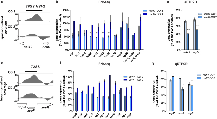
P. aeruginosa secretes important toxins (i.e. elastase, exotoxins, phospholipases) via the type II secretion system (T2SS) into the extracellular environment54. The type VI secretion system (T6SS) is mostly involved in antagonistic interactions with bacterial competitors55. We have shown previously that MvfR impacts the transcription of T6SS42 and T2SS and the level of secreted exoproducts of the T2SS22,45. MvfR positively regulated the transcription of T6SS HSI-II genes, while surprisingly they were reported to be unchanged in a pqsE mutant42, suggesting that MvfR is the major contributor for this regulation. This notion is supported by the fact that MvfR binds over hsiA2 and hcpD of T6SS HSI-II (Figs 6a and S1). Gene expression studies in the mvfR mutant reveal that MvfR induces the expression of most T6S HSI-II genes at OD600nm 2.0 but not at OD600nm 3.0 (Fig. 6b,c). As such, they are consistent with the impact of MvfR binding during early exponential phase and the absence of MvfR binding later (Fig. 6a and Table 1), and imply a direct role for MvfR in the expression of this locus at the early and mid-exponential stage of growth.
Figure 6. MvfR binds to and fine tunes the expression of T6SS and T2SS operons.
(a,d) ChIPseq analysis reveals that MvfR binds to hsiA2-hcpD (T6SS HSI-II) and xcpQ-xcpP-xcpR (T2SS) regions. The black bar above the binding intensity plots represents the peak identified using SPP peak caller. (b,e) RNAseq analysis indicates that MvfR induces the expression of most T6SS and T2SS genes. Light blue bar = mvfR mutant at OD600nm 2, dark blue bar = mvfR mutant at OD600nm 3. (c,f) qRTPCR analysis validates that MvfR induces the expression of T6SS and T2SS genes. Faint blue bar = mvfR mutant at OD600nm 1, light blue bar = mvfR mutant at OD600nm 2. Data show the average +/− SEM of 3 independent replicates. Statistical significance was assessed using one way ANOVA + Dunnett’s post-test.
Figures 6d and S1 show that MvfR also binds to T2SS genes xcpQ, xcpP and xcpR, key genes that confer functionality to this system for the secretion of several virulence exoproducts54,56. Accordingly, the expression of most T2SS genes is reduced in mvfR mutant at OD600nm 2.0 but no reduced expression of these genes is observed at OD600nm = 3, consistent with the absence of detectable MvfR binding at these later ODs (Fig. 6e,f and Table 1). Together, these data suggest that MvfR directly contributes to the regulation of key genes of T6SS HSI-II and T2SS at the early stages of P. aeruginosa growth.
MvfR binds to five translation-related loci and negatively impacts P. aeruginosa translation
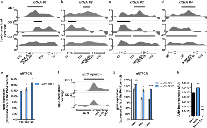
We reported previously that the MvfR negatively modulates the expression of P. aeruginosa protein translation genes6. However, the underlying molecular mechanism of this negative regulation is unclear. Figure 7 shows that MvfR binds to and controls the expression of five translation related loci. Binding of MvfR extends along an entire region comprised of genes encoding 5S, 16S, 23S rRNAs and two tRNAs. MvfR binding is present during OD600nm 1.0–3.0 in all four repeats of this region throughout the PA14 genome (Fig. 7a–d). In addition, MvfR binds to the translation initiation factor IF-3 encoding gene, infC, which is the second gene of an operon also encoding the translation-related genes, thrS, rpmL and rplT (Fig. 7f). Expanding on what we previously reported6, we show here that the expression of 16S, 23S and 5S rRNAs is increased in the mvfR mutant (Fig. 7e). Moreover, the expression of thrS, infC and rpmI genes is also significantly increased (Fig. 7g), indicating that MvfR acts as a negative regulator of this set of translation-related genes. Consistently, P. aeruginosa translational activity, measured by the incorporation of L-azidohomoalanine into proteins57, is significantly increased in the mvfR mutant compared to PA14 (Fig. 7h), validating the role of MvfR in translation inhibition. Overall, these findings imply a direct negative fine-tuning action of QS on P. aeruginosa translation machinery via MvfR binding to rRNA genes and infC operon.
Figure 7. MvfR slows down translation activity by binding to and negatively regulating translation related genes.
(a–d,f) ChIPseq analysis reveals that MvfR binds to the 4 rRNA regions – rRNA #1 (a), rRNA #2 (b), rRNA #3 (c) and rRNA #4 (d) – as well as the infC operon (f). The black bar above the binding intensity plots represents the peak identified using SPP peak caller. (e,g) qRTPCR analysis indicates that MvfR represses the expression of 16S, 23S and 5S rRNAs as well as thrS, infC, and rpmI. Faint blue bar = mvfR mutant at OD600nm 1, light blue bar = mvfR mutant at OD600nm 2. Data show the average +/− SEM of 3 independent replicates. Statistical significance was assessed using unpaired T test for rRNA genes and one way ANOVA + Dunnett’s post-test for infC operon genes. (h) MvfR inhibits the translation activity of P. aeruginosa as reflected by the measurement of newly synthetized proteins using the labeled amino-acid alanine AHA (L-azidohomoalanine). The translation inhibitor chloramphenicol (15 mg/L) was used as a control. Data show the average +/− SEM of three independent replicates. Statistical significance was assessed using one way ANOVA + Dunnett’s post-test.
MvfR induction of antioxidant genes contributes to antibiotic tolerance
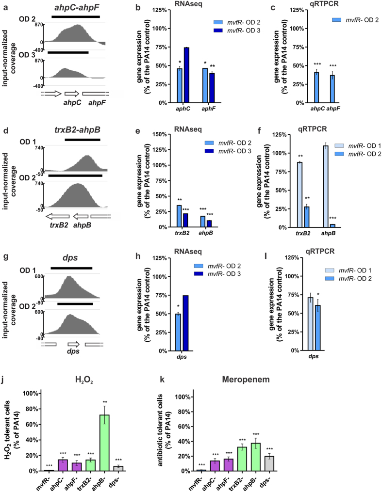
Perhaps one the most interesting sets of MvfR binding loci are those related to the oxidative stress response. As shown in Figs 8a,d,g and S1, MvfR binds to ahpC-ahpF, trxB2-ahpB and dps regions. Accordingly, the expression of all those genes is significantly reduced in the mvfR mutant (Fig. 8b,c,e–h). In corroboration, our previous microarray expression studies also showed that the expression of these genes is downregulated in the presence of MvfR QS system inhibitors42. The alkyl-hydroxyperoxidases AhpC, AhpF and AhpB, as well as the threonine reductase TrxB2, and the Dps protein are involved in the detoxification of hydrogen peroxide and organic peroxides17,58,59,60 and iron oxidation and sequestration. These proteins ultimately prevent the generation of lethal Fenton reaction derived hydroxyl radicals61,62. Consistently, we observed that AhpC, AhpF, TrxB2, AhpB and Dps allow tolerance to hydrogen peroxide given that mutants in those genes are 1.4 to 17 times more sensitive to H2O2 than PA14 (Fig. 8i). The mvfR mutant is also significantly more sensitive to H2O2 than PA14 (Fig. 8i), thereby supporting the relevance of MvfR in response to oxidative stress.
Figure 8. MvfR binds to and positively regulates genes involved in response to oxidative stress which leads to tolerance of H2O2 and antibiotic.
(a,d,g) ChIPseq analysis reveals that MvfR binds to ahpC-ahpF, trxB2-ahpB and dps regions. The black bar above the binding intensity plots represents the peak identified using SPP peak caller. (b,e,h) RNAseq analysis indicates that MvfR induces the expression of ahpC, ahpF, trxB2, ahpB and dps. Light blue bar = mvfR mutant at OD600nm 2, dark blue bar = mvfR mutant at OD600nm 3. (c,f,g) qRTPCR analysis validates that MvfR induces the expression of ahpC, ahpF, trxB2, ahpB and dps. Faint blue bar = mvfR mutant at OD600nm 1, light blue bar = mvfR mutant at OD600nm 2. Data show the average +/− SEM of 3 independent replicates. Statistical significance was assessed using one way ANOVA + Dunnett’s post-test. (i,j) mvfR mutant (red) as well as ahpC and ahpF mutants (purple), trxB2 and ahpB mutants (green), and dps mutant (grey) are more sensitive than PA14 to hydrogen peroxide (i) or the β-lactam antibiotic Meropenem (j). The survival fraction of PA14 control after H2O2 or Meropenem treatment is of 6.8 × 10⁻⁴ or 7.1 × 10⁻⁶ respectively. Data show the average +/− SEM of at least 3 independent replicates. Statistical significance was assessed using one way ANOVA + Dunnett’s post-test.
Importantly, antioxidant defenses are known to play a protective role against antibiotics via their ROS suppressing activities63,64,65,66,67,68,69. We previously described the importance of MvfR in antibiotic tolerance6,43,70, and here we asked whether the detoxification abilities of AhpC, AhpF, TrxB2, AhpB and Dps may contribute to this phenomenon. As shown in Fig. 8j, ahpC, ahpF, trxB2, ahpB and dps mutants are 2.7 to 7.7 times more sensitive to the β-lactam antibiotic Meropenem than the parental PA14 strain, indicating that direct MvfR control of AhpC-F, TrxB2-AhpB and Dps contributes to antibiotic tolerance.
Discussion
This study provides novel insights in the understanding of MvfR role in the complex regulation of QS and pathogenesis in P. aeruginosa. Indeed, our data strongly suggest that MvfR acts as a direct modulator of key P. aeruginosa virulence systems beyond the biosynthetic operons pqsABCDE and phnAB in line with its original nomenclature as Multiple Virulence Factor Regulator (MvfR)22. Moreover, this work permits us to better comprehend the regulation of QS and challenges the current hierarchical view that LasR is upstream of MvfR and that MvfR controls QS virulence only indirectly through the transcriptional control of the pqsABCDE operon24,51,52.
Previous studies have shown the broad impact of MvfR in the transcriptional regulation of up to 18% of P. aeruginosa genes45. However, the possibility of MvfR direct regulation for some of these genes has been overshadowed by the role of the HAQs and PqsE in virulence26,47,51,52,71,72. It is noteworthy that PQS is dispensable for virulence since pqsH mutation does not attenuate virulence in mice23. The suggested direct transcriptional regulation of MvfR on P. aeruginosa genes described here could account for the already established indirect regulation working via PqsE and HHQ/PQS26,47,51,52,71,72. Even though the 34 loci identified represent only a fraction of MvfR regulated genes, they are nonetheless relevant because they contain specific virulence factors as well as global regulators of virulence, including the two other major QS regulators LasR and RhlR. The increased number of genes regulated by MvfR at late exponential and stationary phases of growth most likely stems from direct MvfR regulation of key cellular, metabolic and QS regulatory functions at earlier stages of growth.
We noticed that MvfR exhibits different binding patterns and recognizes different DNA binding motifs. The predicted MvfR consensus binding motifs in both promoter regions and regions overlapping several genes share the classical LTTR TNxA palindrome structure49. Tomtom analysis73 indicates that the predicted consensus motif for sites located inside genes resembles the DNA binding motif of RutR, an E. coli transcriptional regulator known to bind specifically within genes74 which is consistent with this group of MvfR binding sites. Such intragenic binding and the ability of MvfR to act as negative and positive regulator have previously been described with other members of the LTTR family49,75. It is important to note that the crosslinking step inherent to all ChIP procedures may lead to the capture of protein-protein interactions and potentially to false-positive binding sites. Future in depth mechanistic studies focusing on MvfR binding patterns will allow us to conclusively demonstrate the MvfR direct regulation of the loci identified and obtain a better understanding of how and why MvfR interacts with DNA in this fashion.
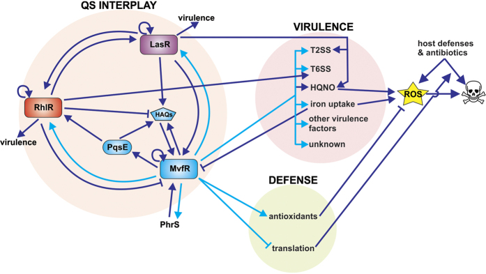
The longitudinal interrogation of MvfR binding during P. aeruginosa growth supports an interconnected regulation pathway for the three major QS systems, as MvfR is likely involved in the direct modulation of both LasR and RhlR QS systems, which were both previously shown to regulate the MvfR QS system27,35,44. The data presented here challenge the hierarchical regulation model of P. aeruginosa QS systems and introduce a circular regulation model (see proposed model in Fig. 9). Intriguingly, MvfR binding to and modulation of lasR and rhlR occurs at early and mid-exponential phase (OD600nm 1.0 and 2.0) raising the question of why this timing is important. At late exponential and stationary phase rhlR expression and C4-HSL levels max out45,46 and RhlR binds to mvfR and pqsA promoters acting as a direct repressor of the MvfR QS system35,44, promoting a tight negative auto-regulatory loop. In contrast, MvfR binding to lasR likely feeds back into the MvfR QS circuitry by inducing mvfR expression27,35, and the PqsH-mediated conversion of HHQ into PQS increases MvfR activity43,48. Two other suggested positive feedback loops occurring at the earlier stages of growth result from MvfR binding to itself and phrS small RNA, which was previously described as a post-transcriptional activator of MvfR53. Until now, phrS induction was reported to take place at the stationary growth phase as a result of its control by the transcriptional regulator ANR under hypoxic conditions53. Here, our data indicate that MvfR could directly induce phrS expression during early and mid-exponential phase, before ANR and hypoxia come into play. These two suggested positive feedback loops may benefit the MvfR QS system by rapidly increasing MvfR levels and generating functional intracellular levels of MvfR ligands before quorum levels are reached in the global cell population.
Figure 9. Proposed model of the role of MvfR direct regulation on QS interplay, virulence and defense.
This proposed model figure focuses on MvfR direct regulation and the systems that feedback to it either positively (arrow) or negatively (bar). Dark blue arrows/bars represent connections previously described in the literature whereas light blue arrows/bars represent new connections based on this study.
Another interesting regulation identified in this study is related to iron uptake and homeostasis. Indeed, we notice MvfR binding to and inducing the two siderophores, pyochelin and pyoverdin, and the putative iron transporters PA14_28970-28980 and PA14_46810-46820 (Table 1 and Supplementary Table S1). Iron is an essential cofactor for a wide variety of cellular processes but is especially scarce during host infection due to nutritional immunity76,77,78. However, excessive iron levels can lead to cellular damage and ultimately cell death as iron catalyzes ROS production via the Fenton reaction62,79. Therefore, iron homeostasis is critical. Once intracellular iron levels are high, uptake systems and their regulators, including MvfR, are repressed by the Fur transcriptional regulator80. This negative feedback loop also turns down the production of all other ROS-inducing systems under MvfR control (i.e. Pyocyanin, HQNO)8,13.
Even though the iron uptake and ROS producing systems can be deleterious for P. aeruginosa, they are nonetheless critical for survival, colonization and competition with other bacterial species8,9,10,81,82. Therefore, the ability of P. aeruginosa to survive when these systems are active offers an important selective advantage. Here we show that MvfR enhances protection against ROS that can be produced by these systems via binding to and inducing the expression of the anti-oxidant defense systems AhpC-F, AhpB-TrxB2 and Dps. These antioxidant proteins are known to limit ROS production by reducing the levels of hydrogen peroxide and iron available as substrates for the Fenton reaction59,61,62,83. As such MvfR modulation of these antioxidant genes may allow cells to survive the production of oxidative toxins (see proposed model in Fig. 9). This self-protective “toxin/anti-toxin” system is reminiscent of antibiotic producing bacteria that require a self-resistance mechanism to avoid committing suicide84,85. Protection against self-poisoning is not the only role of these antioxidants, as we show here that they contribute to antibiotic tolerance. MvfR’s ability to induce their expression provides an explanation for the previously unknown molecular mechanism of MvfR-mediated antibiotic tolerance. In addition to inducing antioxidant systems, we also show that MvfR binds to translation-related loci and represses translation. Since antibiotic tolerant cells are known to exhibit reduced protein synthesis and metabolic rates4,5,86,87, MvfR action on translation may also contribute to antibiotic tolerance (see proposed model in Fig. 9).
Overall, our data suggest that MvfR directly controls the expression of multiple virulence factors and plays a central role in the P. aeruginosa QS interplay as well as in antibiotic tolerance via the regulation of multiple antioxidant systems. This highlights the importance of MvfR as a critical virulence determinant and reinforces its potential as a highly desirable drug target candidate 42,43,88,89,90,91.
Methods
Bacterial strains, plasmids, growth conditions
UCBPP-PA14 (PA14) is a P. aeruginosa human clinical isolate92. All mutant strains including mvfR-22, pqsA-21 and pqsE-47 are isogenic to UCBPP-PA14. pECP60 plasmid containing the rhlA-lacZ reporter system was described previously45,93. Unless noted otherwise, all bacterial strains were grown in 5 mL LB Lenox medium (Fisher Scientific) at 37 °C under 200 rpm orbital shaking using glass tubes (VWR). To generate pJN-mvfR-FLAG plasmid expressing C-terminally-FLAG tagged MvfR, the 657-bp upstream region and mvfR coding region with C-terminal FLAG was amplified by PCR and then cloned into pJN105 treated with EcoR1 and Xho1 as described in94. The resulting plasmid was transformed into mvfR- cells. The mvfR- strain carrying pJN-mvfR-FLAG was grown in the presence of 15 μg/mL Gentamycin.
ChIPseq
5 ml cultures were inoculated at OD600nm 0.1, and grown at 37 °C 200 rpm in LB Lenox + 15 μg/mL Gentamycin. At OD600nm 1, 2, 3 and 4, cells were washed in fresh LB Lenox then pelleted and stored at −80 °C. The ChIP experiment was performed as described in43,95 using anti-FLAG M2 magnetic beads (Sigma). Library construction and Illumina DNA sequencing was performed by htSEQ (Seattle, WA). The sequence aligner BWA96 was used to map sequencing reads to a UCBPP-PA14 fasta reference genome file (RefSeq May 24, 2010). Peaks for samples at OD600nm 1, 2, 3 and 4 were identified using the ChipSeq peak caller SPP97 with OD3_input as the reference input. Peaks were filtered at a Z_score = 9 in order to limit the probability of false positives. It is noteworthy that pH changes over time during growth have the potential to affect iron bioavailability, and may also drive changes in the physiology of the microbe that could affect binding.
ChIP qPCR
We validated the ChIPseq findings via ChIP qPCR and not via EMSA studies because this approach allows us to interrogate MvfR binding in live cells where all biologically relevant components are present, and also to bypass the known difficulties to consistently purify full-length MvfR protein88,98. Samples were prepared as described in the ChIPseq section above. qPCR was then performed using primers designed to be in the middle of MvfR binding sites using the Primer 3 tool (http://bioinfo.ut.ee/primer3-0.4.0/). The primer sequences for each site are listed in Supplementary Table S2. Quantitative PCR was performed using Brilliant II SYBR Green QPCR Master Mix (Stratagene) as in43,47 with a Mx3005P qPCR machine (Stratagene). Data were analyzed using the percent input method and using rpoD as a negative control as in ref. 43.
RNAseq
PA14 and mvfR- cells were grown in LB Lenox at 37 °C 200 rpm until OD600nm 2 or OD600nm 3. Duplicates of each culture were processed for RNA extraction using the RNeasy kit (QIAGEN) and DNAse treatment was performed with the TURBO DNA-free kit (Thermo-Fisher). RNA samples were then subjected to rRNA depletion using RiboZero (Epicentre) followed by the construction of next-generation sequencing libraries using NEBNext Ultra Directional RNA Library Prep Kit (New England Biolabs). These libraries were sequenced on Illumina HiSeq 2500 instrument resulting in approximately 8.8 million reads per sample on average. Subsequent to alignment using BWA96, read counts for individual transcripts were produced with HTSEQ99, based on UCBPP-PA14 transcriptome annotation (NC_002516). The estimation of expression values and detection of differentially expressed transcripts was performed using EdgeR100. Statistical significance was assessed using false discovery rate.
qRT-PCR
RNA samples were prepared as described above for RNAseq. cDNA was generated by RT-PCR using Superscript III First-Strand kit (Invitrogen) according to manufacturer’s instructions. Specific primers were designed using Primer3. Primers used are described in Supplementary Table S2. RpoD expression was used as a reference gene as described in47,101. Quantitative PCR was performed using Brilliant II SYBR Green QPCR Master Mix (Stratagene) as in43,47 with a Mx3005P qPCR machine (Stratagene).
Translation activity assay
Translation activity was determined using Click chemistry as described in57 with modifications. PA14 and mvfR- cells were grown in LB Lenox at 37 °C 200 rpm overnight from a frozen stock then washed and resuspended in M9 media to OD600nm 0.1. Cells were grown for 5 h at 37 °C 200 rpm then 15 μg/mL chloramphenicol (or 10% ethanol vehicle) together with 1 μM Click-iT AHA L-azidohomoalanine (Life Technologies) were added for 45 minutes to the growing cultures. Cells were then pelleted and fixed in 4% PFA for 1 hour then washed in PBS. Cells were then lysed by sonication in 1% SDS 50 mM Tris-HCl pH8. Labelled solubilized proteins were subjected to reaction with Click-iT Protein Reaction Buffer kit (Life Technologies) containing 4 mM tetramethylrhodamine (TAMRA) alkyne (Life Technologies) according to manufacturer’s instructions. Proteins were then extracted by the methanol-chloroform method and washed twice in methanol as described in ref. 57. 2 μL of each extracted protein sample was finally dotted on a nitrocellulose membrane and imaged using 534 nm excitation (green)/607 nm emission (orange) filter in a FluorChem M imaging device (Protein Simple). Signal was then processed using ImageJ software in order to quantify AHA labeled proteins, representing nascent protein synthesis.
β-galactosidase gene reporter assay
PA14 and mvfR- cells containing pECP60 plasmid were grown in presence of 300 μg/mL Carbenicillin to maintain the plasmid. At OD600nm 2, 20 μL of cells were collected and incubated with 80 μL of permeabilization solution (100 mM Na2HPO4, 20 mM KCl, 2 mM MgSO4, 0.8 mg/mL CTAB, 0.4 mg/mL sodium deoxycholate and 5.4 μL/mL β-mercaptoethanol) for 30 min at 37 °C. Then 600 μL of substrate solution was added (60 mM Na2HPO4, 40 mM NaH2PO4, 1 mg/mL ONPG and 2.7 μL/mL β-mercaptoethanol) and incubated at 37 °C until yellow color was detected. 700 μL of Stop solution (1M Na2CO3) was finally added to block the reaction and OD420nm was measured. Miller units were calculated as follows: 1000 × OD420nm/(OD600nm × 0.02 mL × reaction time). More details on this protocol can be found at (http://openwetware.org/wiki/Beta-Galactosidase_Assay_%28A_better_Miller%29).
Tolerance to H2O2 or Meropenem
PA14, mvfR-, aphC-, ahpF-, trxB2-, ahpB- and dps- cells were grown at 37 °C 200 rpm in LB Lenox media until mid-exponential phase (OD600nm 2) then exposed to 300 mM hydrogen peroxide (H2O2) for 1 hour under the same incubating conditions. Before (t = 0) and after (t = 1 h) H2O2 addition, a 100 μL sample of each culture was collected, diluted and plated on LB agar plates to quantify the total number of bacteria (t = 0) and the surviving bacteria (t = 1 h). Colony forming units (CFUs) were counted after 24 h incubation at 37 °C. Tolerance to Meropenem was assessed the same way except the cells were grown in 1% TSB media and the killing was performed for 24 hours in presence of 10 μg/mL Meropenem (Sandoz, USA).
Statistical analyses
Statistical significance was assessed using unpaired T test or One Way ANOVA + Dunnett’s post-test in the case of multiple comparisons, as appropriate and indicated in the figure legends.
Additional Information
How to cite this article: Maura, D. et al. Evidence for Direct Control of Virulence and Defense Gene Circuits by the Pseudomonas aeruginosa Quorum Sensing Regulator, MvfR. Sci. Rep. 6, 34083; doi: 10.1038/srep34083 (2016).
Supplementary Material
Acknowledgments
We thank Anthony Anselmo and Ruslan Sadreyev from the MGH Department of Molecular Biology for their help on ChIPseq and RNAseq analysis. This work was supported by Shriners Hospital Postdoctoral Fellowship #84206 to D.M., Cystic Fibrosis Foundation fellowship #BALLOK15F0 to A.B. and by the research grants, Shriners #8770, Cystic Fibrosis Foundation #11P0, NIAID R33AI105902 and R56AI063433 to L.G.R.
Footnotes
Author Contributions D.M., R.H. and L.G.R. designed experiments. D.M., R.H., T.K. and A.B. performed experiments. D.M. and L.G.R. wrote the manuscript and prepared figures. All authors reviewed the manuscript.
References
- Gellatly S. L. & Hancock R. E. Pseudomonas aeruginosa: new insights into pathogenesis and host defenses. Pathog Dis 67, 159–173 (2013). [DOI] [PubMed] [Google Scholar]
- Kerr K. G. & Snelling A. M. Pseudomonas aeruginosa: a formidable and ever-present adversary. J Hosp Infect 73, 338–344 (2009). [DOI] [PubMed] [Google Scholar]
- Lister P. D., Wolter D. J. & Hanson N. D. Antibacterial-resistant Pseudomonas aeruginosa: clinical impact and complex regulation of chromosomally encoded resistance mechanisms. Clin Microbiol Rev 22, 582–610 (2009). [DOI] [PMC free article] [PubMed] [Google Scholar]
- Lewis K. Persister cells, dormancy and infectious disease. Nat Rev Microbiol 5, 48–56 (2007). [DOI] [PubMed] [Google Scholar]
- Lewis K. Persister Cells. Annual review of microbiology 64, 357–372 (2010). [DOI] [PubMed] [Google Scholar]
- Que Y. A. et al. A quorum sensing small volatile molecule promotes antibiotic tolerance in bacteria. PLoS One 8, e80140 (2013). [DOI] [PMC free article] [PubMed] [Google Scholar]
- Livermore D. M. Current epidemiology and growing resistance of gram-negative pathogens. Korean J Intern Med 27, 128–142 (2012). [DOI] [PMC free article] [PubMed] [Google Scholar]
- Lau G. W., Hassett D. J., Ran H. & Kong F. The role of pyocyanin in Pseudomonas aeruginosa infection. Trends Mol Med 10, 599–606 (2004). [DOI] [PubMed] [Google Scholar]
- Hassan H. M. & Fridovich I. Mechanism of the antibiotic action pyocyanine. J Bacteriol 141, 156–163 (1980). [DOI] [PMC free article] [PubMed] [Google Scholar]
- Baron S. S. & Rowe J. J. Antibiotic action of pyocyanin. Antimicrob Agents Chemother 20, 814–820 (1981). [DOI] [PMC free article] [PubMed] [Google Scholar]
- Machan Z. A., Taylor G. W., Pitt T. L., Cole P. J. & Wilson R. 2-Heptyl-4-hydroxyquinoline N-oxide, an antistaphylococcal agent produced by Pseudomonas aeruginosa. J Antimicrob Chemother 30, 615–623 (1992). [DOI] [PubMed] [Google Scholar]
- Van Ark G. & Berden J. A. Binding of HQNO to beef-heart sub-mitochondrial particles. Biochim Biophys Acta 459, 119–127 (1977). [DOI] [PubMed] [Google Scholar]
- Hazan R. et al. Auto Poisoning of the Respiratory Chain by a Quorum-Sensing-Regulated Molecule Favors Biofilm Formation and Antibiotic Tolerance. Curr Biol 26, 195–206 (2016). [DOI] [PMC free article] [PubMed] [Google Scholar]
- Paiva C. N. & Bozza M. T. Are reactive oxygen species always detrimental to pathogens? Antioxid Redox Signal 20, 1000–1037 (2014). [DOI] [PMC free article] [PubMed] [Google Scholar]
- Spooner R. & Yilmaz O. The role of reactive-oxygen-species in microbial persistence and inflammation. Int J Mol Sci 12, 334–352 (2011). [DOI] [PMC free article] [PubMed] [Google Scholar]
- Hassett D. J., Charniga L., Bean K., Ohman D. E. & Cohen M. S. Response of Pseudomonas aeruginosa to pyocyanin: mechanisms of resistance, antioxidant defenses, and demonstration of a manganese-cofactored superoxide dismutase. Infect Immun 60, 328–336 (1992). [DOI] [PMC free article] [PubMed] [Google Scholar]
- Ochsner U. A., Vasil M. L., Alsabbagh E., Parvatiyar K. & Hassett D. J. Role of the Pseudomonas aeruginosa oxyR-recG operon in oxidative stress defense and DNA repair: OxyR-dependent regulation of katB-ankB, ahpB, and ahpC-ahpF. J Bacteriol 182, 4533–4544 (2000). [DOI] [PMC free article] [PubMed] [Google Scholar]
- Gambello M. J. & Iglewski B. H. Cloning and characterization of the Pseudomonas aeruginosa lasR gene, a transcriptional activator of elastase expression. J Bacteriol 173, 3000–3009 (1991). [DOI] [PMC free article] [PubMed] [Google Scholar]
- Schuster M. & Greenberg E. P. A network of networks: quorum-sensing gene regulation in Pseudomonas aeruginosa. International journal of medical microbiology: IJMM 296, 73–81 (2006). [DOI] [PubMed] [Google Scholar]
- Venturi V. Regulation of quorum sensing in Pseudomonas. FEMS microbiology reviews 30, 274–291 (2006). [DOI] [PubMed] [Google Scholar]
- Deziel E. et al. Analysis of Pseudomonas aeruginosa 4-hydroxy-2-alkylquinolines (HAQs) reveals a role for 4-hydroxy-2-heptylquinoline in cell-to-cell communication. Proc Natl Acad Sci USA 101, 1339–1344 (2004). [DOI] [PMC free article] [PubMed] [Google Scholar]
- Cao H. et al. A quorum sensing-associated virulence gene of Pseudomonas aeruginosa encodes a LysR-like transcription regulator with a unique self-regulatory mechanism. Proceedings of the National Academy of Sciences 98, 14613 (2001). [DOI] [PMC free article] [PubMed] [Google Scholar]
- Xiao G. et al. MvfR, a key Pseudomonas aeruginosa pathogenicity LTTR-class regulatory protein, has dual ligands. Mol Microbiol 62, 1689–1699 (2006). [DOI] [PubMed] [Google Scholar]
- Williams P. & Cámara M. Quorum sensing and environmental adaptation in Pseudomonas aeruginosa: a tale of regulatory networks and multifunctional signal molecules. Current opinion in microbiology 12, 182–191 (2009). [DOI] [PubMed] [Google Scholar]
- Heeb S., Fletcher M. P., Chhabra S. R., Diggle S. P., Williams P. & Camara M. Quinolones: from antibiotics to autoinducers. FEMS Microbiol. Rev. 35, 247–274 (2010). [DOI] [PMC free article] [PubMed] [Google Scholar]
- Pesci E. C. et al. Quinolone signaling in the cell-to-cell communication system of Pseudomonas aeruginosa. Proc Natl Acad Sci USA 96, 11229–11234 (1999). [DOI] [PMC free article] [PubMed] [Google Scholar]
- McGrath S., Wade D. S. & Pesci E. C. Dueling quorum sensing systems in Pseudomonas aeruginosa control the production of the Pseudomonas quinolone signal (PQS). FEMS Microbiol Lett 230, 27–34 (2004). [DOI] [PubMed] [Google Scholar]
- Passador L., Cook J. M., Gambello M. J., Rust L. & Iglewski B. H. Expression of Pseudomonas aeruginosa virulence genes requires cell-to-cell communication. Science 260, 1127–1130 (1993). [DOI] [PubMed] [Google Scholar]
- Ochsner U. A., Koch A. K., Fiechter A. & Reiser J. Isolation and characterization of a regulatory gene affecting rhamnolipid biosurfactant synthesis in Pseudomonas aeruginosa. J Bacteriol 176, 2044–2054 (1994). [DOI] [PMC free article] [PubMed] [Google Scholar]
- Latifi A. et al. Multiple homologues of LuxR and LuxI control expression of virulence determinants and secondary metabolites through quorum sensing in Pseudomonas aeruginosa PAO1. Mol Microbiol 17, 333–343 (1995). [DOI] [PubMed] [Google Scholar]
- Gilbert K. B., Kim T. H., Gupta R., Greenberg E. P. & Schuster M. Global position analysis of the Pseudomonas aeruginosa quorum-sensing transcription factor LasR. Mol Microbiol 73, 1072–1085 (2009). [DOI] [PMC free article] [PubMed] [Google Scholar]
- Abdel-Mawgoud A. M., Lepine F. & Deziel E. Rhamnolipids: diversity of structures, microbial origins and roles. Appl Microbiol Biotechnol 86, 1323–1336 (2010). [DOI] [PMC free article] [PubMed] [Google Scholar]
- Reis R. S., Pereira A. G., Neves B. C. & Freire D. M. Gene regulation of rhamnolipid production in Pseudomonas aeruginosa–a review. Bioresour Technol 102, 6377–6384 (2011). [DOI] [PubMed] [Google Scholar]
- Brint J. M. & Ohman D. E. Synthesis of multiple exoproducts in Pseudomonas aeruginosa is under the control of RhlR-RhlI, another set of regulators in strain PAO1 with homology to the autoinducer-responsive LuxR-LuxI family. J Bacteriol 177, 7155–7163 (1995). [DOI] [PMC free article] [PubMed] [Google Scholar]
- Xiao G., He J. & Rahme L. G. Mutation analysis of the Pseudomonas aeruginosa mvfR and pqsABCDE gene promoters demonstrates complex quorum-sensing circuitry. Microbiology 152, 1679–1686 (2006). [DOI] [PubMed] [Google Scholar]
- Lepine F. et al. PqsA is required for the biosynthesis of 2,4-dihydroxyquinoline (DHQ), a newly identified metabolite produced by Pseudomonas aeruginosa and Burkholderia thailandensis. Biol Chem 388, 839–845 (2007). [DOI] [PMC free article] [PubMed] [Google Scholar]
- Lepine F., Milot S., Deziel E., He J. & Rahme L. G. Electrospray/mass spectrometric identification and analysis of 4-hydroxy-2-alkylquinolines (HAQs) produced by Pseudomonas aeruginosa. J Am Soc Mass Spectrom 15, 862–869 (2004). [DOI] [PubMed] [Google Scholar]
- Scott-Thomas A. J. et al. 2-Aminoacetophenone as a potential breath biomarker for Pseudomonas aeruginosa in the cystic fibrosis lung. BMC Pulm Med 10, 56 (2010). [DOI] [PMC free article] [PubMed] [Google Scholar]
- Kesarwani M. et al. A Quorum Sensing Regulated Small Volatile Molecule Reduces Acute Virulence and Promotes Chronic Infection Phenotypes. PLoS Pathogens 7, e1002192 (2011). [DOI] [PMC free article] [PubMed] [Google Scholar]
- Bandyopadhaya A. et al. The quorum sensing volatile molecule 2-amino acetophenon modulates host immune responses in a manner that promotes life with unwanted guests. PLoS Pathog 8, e1003024 (2012). [DOI] [PMC free article] [PubMed] [Google Scholar]
- Déziel E. et al. The contribution of MvfR to Pseudomonas aeruginosa pathogenesis and quorum sensing circuitry regulation: multiple quorum sensing-regulated genes are modulated without affecting lasRI, rhlRI or the production of N-acyl-L-homoserine lactones. Mol Microbiol 55, 998–1014 (2005). [DOI] [PubMed] [Google Scholar]
- Lesic B. et al. Inhibitors of pathogen intercellular signals as selective anti-infective compounds. PLoS Pathog 3, 1229–1239 (2007). [DOI] [PMC free article] [PubMed] [Google Scholar]
- Starkey M. et al. Identification of anti-virulence compounds that disrupt quorum-sensing regulated acute and persistent pathogenicity. PLoS Pathog 10, e1004321 (2014). [DOI] [PMC free article] [PubMed] [Google Scholar]
- Wade D. S. et al. Regulation of Pseudomonas quinolone signal synthesis in Pseudomonas aeruginosa. J Bacteriol 187, 4372–4380 (2005). [DOI] [PMC free article] [PubMed] [Google Scholar]
- Déziel E. et al. The contribution of MvfR to Pseudomonas aeruginosa pathogenesis and quorum sensing circuitry regulation: multiple quorum sensing-regulated genes are modulated without affecting lasRI, rhlRI or the production of N-acyl-L-homoserine lactones. Molecular microbiology 55, 998–1014 (2005). [DOI] [PubMed] [Google Scholar]
- Dekimpe V. & Deziel E. Revisiting the quorum-sensing hierarchy in Pseudomonas aeruginosa: the transcriptional regulator RhlR regulates LasR-specific factors. Microbiology 155, 712 (2009). [DOI] [PubMed] [Google Scholar]
- Hazan R. et al. Homeostatic interplay between bacterial cell-cell signaling and iron in virulence. PLoS Pathogens 6, e1000810 (2010). [DOI] [PMC free article] [PubMed] [Google Scholar]
- Gallagher L. A., McKnight S. L., Kuznetsova M. S., Pesci E. C. & Manoil C. Functions required for extracellular quinolone signaling by Pseudomonas aeruginosa. Journal of bacteriology 184, 6472 (2002). [DOI] [PMC free article] [PubMed] [Google Scholar]
- Maddocks S. E. & Oyston P. C. Structure and function of the LysR-type transcriptional regulator (LTTR) family proteins. Microbiology 154, 3609–3623 (2008). [DOI] [PubMed] [Google Scholar]
- Bailey T. L. & Elkan C. Fitting a mixture model by expectation maximization to discover motifs in biopolymers. Proc Int Conf Intell Syst Mol Biol 2, 28–36 (1994). [PubMed] [Google Scholar]
- McKnight S. L., Iglewski B. H. & Pesci E. C. The Pseudomonas quinolone signal regulates rhl quorum sensing in Pseudomonas aeruginosa. J Bacteriol 182, 2702–2708 (2000). [DOI] [PMC free article] [PubMed] [Google Scholar]
- Farrow J. M. et al. PqsE functions independently of PqsR-Pseudomonas quinolone signal and enhances the rhl quorum-sensing system. Journal of bacteriology 190, 7043–7051 (2008). [DOI] [PMC free article] [PubMed] [Google Scholar]
- Sonnleitner E. et al. The small RNA PhrS stimulates synthesis of the Pseudomonas aeruginosa quinolone signal. Mol Microbiol 80, 868–885 (2011). [DOI] [PubMed] [Google Scholar]
- Cianciotto N. P. Type II secretion: a protein secretion system for all seasons. Trends Microbiol 13, 581–588 (2005). [DOI] [PubMed] [Google Scholar]
- Russell A. B., Peterson S. B. & Mougous J. D. Type VI secretion system effectors: poisons with a purpose. Nat Rev Microbiol 12, 137–148 (2014). [DOI] [PMC free article] [PubMed] [Google Scholar]
- Douzi B., Filloux A. & Voulhoux R. On the path to uncover the bacterial type II secretion system. Philos Trans R Soc Lond B Biol Sci 367, 1059–1072 (2012). [DOI] [PMC free article] [PubMed] [Google Scholar]
- Hatzenpichler R. et al. In situ visualization of newly synthesized proteins in environmental microbes using amino acid tagging and click chemistry. Environ Microbiol 16, 2568–2590 (2014). [DOI] [PMC free article] [PubMed] [Google Scholar]
- Lu J. & Holmgren A. The thioredoxin antioxidant system. Free Radic Biol Med 66, 75–87 (2014). [DOI] [PubMed] [Google Scholar]
- Dubbs J. M. & Mongkolsuk S. Peroxiredoxins in bacterial antioxidant defense. Subcell Biochem 44, 143–193 (2007). [DOI] [PubMed] [Google Scholar]
- Wei Q. et al. Global regulation of gene expression by OxyR in an important human opportunistic pathogen. Nucleic Acids Res 40, 4320–4333 (2012). [DOI] [PMC free article] [PubMed] [Google Scholar]
- Chiancone E. & Ceci P. The multifaceted capacity of Dps proteins to combat bacterial stress conditions: Detoxification of iron and hydrogen peroxide and DNA binding. Biochim Biophys Acta 1800, 798–805 (2010). [DOI] [PubMed] [Google Scholar]
- Calhoun L. N. & Kwon Y. M. Structure, function and regulation of the DNA-binding protein Dps and its role in acid and oxidative stress resistance in Escherichia coli: a review. J Appl Microbiol 110, 375–386 (2011). [DOI] [PubMed] [Google Scholar]
- Wu N. et al. Ranking of persister genes in the same Escherichia coli genetic background demonstrates varying importance of individual persister genes in tolerance to different antibiotics. Front Microbiol 6, 1003 (2015). [DOI] [PMC free article] [PubMed] [Google Scholar]
- Vega N. M., Allison K. R., Khalil A. S. & Collins J. J. Signaling-mediated bacterial persister formation. Nat Chem Biol 8, 431–433 (2012). [DOI] [PMC free article] [PubMed] [Google Scholar]
- Goswami M., Mangoli S. H. & Jawali N. Involvement of reactive oxygen species in the action of ciprofloxacin against Escherichia coli. Antimicrob Agents Chemother 50, 949–954 (2006). [DOI] [PMC free article] [PubMed] [Google Scholar]
- Yeom J., Imlay J. A. & Park W. Iron homeostasis affects antibiotic-mediated cell death in Pseudomonas species. J Biol Chem 285, 22689–22695 (2010). [DOI] [PMC free article] [PubMed] [Google Scholar]
- Calhoun L. N. & Kwon Y. M. The ferritin-like protein Dps protects Salmonella enterica serotype Enteritidis from the Fenton-mediated killing mechanism of bactericidal antibiotics. Int J Antimicrob Agents 37, 261–265 (2011). [DOI] [PubMed] [Google Scholar]
- Dwyer D. J., Collins J. J. & Walker G. C. Unraveling the physiological complexities of antibiotic lethality. Annu Rev Pharmacol Toxicol 55, 313–332 (2015). [DOI] [PubMed] [Google Scholar]
- Khakimova M., Ahlgren H. G., Harrison J. J., English A. M. & Nguyen D. The stringent response controls catalases in Pseudomonas aeruginosa and is required for hydrogen peroxide and antibiotic tolerance. J Bacteriol 195, 2011–2020 (2013). [DOI] [PMC free article] [PubMed] [Google Scholar]
- Hazan R., Que Y. A., Maura D. & Rahme L. G. A method for high throughput determination of viable bacteria cell counts in 96-well plates. BMC Microbiol 12, 259 (2012). [DOI] [PMC free article] [PubMed] [Google Scholar]
- Mashburn L. M. & Whiteley M. Membrane vesicles traffic signals and facilitate group activities in a prokaryote. Nature 437, 422–425 (2005). [DOI] [PubMed] [Google Scholar]
- Häussler S. & Becker T. The pseudomonas quinolone signal (PQS) balances life and death in Pseudomonas aeruginosa populations. PLoS pathogens 4, e1000166 (2008). [DOI] [PMC free article] [PubMed] [Google Scholar]
- Gupta S., Stamatoyannopoulos J. A., Bailey T. L. & Noble W. S. Quantifying similarity between motifs. Genome Biol 8, R24 (2007). [DOI] [PMC free article] [PubMed] [Google Scholar]
- Shimada T., Ishihama A., Busby S. J. & Grainger D. C. The Escherichia coli RutR transcription factor binds at targets within genes as well as intergenic regions. Nucleic Acids Res 36, 3950–3955 (2008). [DOI] [PMC free article] [PubMed] [Google Scholar]
- Viswanathan P., Ueki T., Inouye S. & Kroos L. Combinatorial regulation of genes essential for Myxococcus xanthus development involves a response regulator and a LysR-type regulator. Proc Natl Acad Sci USA 104, 7969–7974 (2007). [DOI] [PMC free article] [PubMed] [Google Scholar]
- Cornelis P. Iron uptake and metabolism in pseudomonads. Appl Microbiol Biotechnol 86, 1637–1645 (2010). [DOI] [PubMed] [Google Scholar]
- Cornelis P., Matthijs S. & Van Oeffelen L. Iron uptake regulation in Pseudomonas aeruginosa. Biometals 22, 15–22 (2009). [DOI] [PubMed] [Google Scholar]
- Soares M. P. & Weiss G. The Iron age of host-microbe interactions. EMBO Rep 16, 1482–1500 (2015). [DOI] [PMC free article] [PubMed] [Google Scholar]
- Imlay J. A., Chin S. M. & Linn S. Toxic DNA damage by hydrogen peroxide through the Fenton reaction in vivo and in vitro. Science 240, 640–642 (1988). [DOI] [PubMed] [Google Scholar]
- Troxell B. & Hassan H. M. Transcriptional regulation by Ferric Uptake Regulator (Fur) in pathogenic bacteria. Front Cell Infect Microbiol 3, 59 (2013). [DOI] [PMC free article] [PubMed] [Google Scholar]
- Machan Z., Taylor G., Pitt T., Coke P. & Wilson R. 2-Heptyl-4-hydroxyquinoline N-oxide, an antistaphylococcal agent produced by Pseudomonas aeruginosa. Journal of Antimicrobial Chemotherapy 30, 615–623 (1992). [DOI] [PubMed] [Google Scholar]
- Van Ark G. & Berden J. A. Binding of HQNO to Beef-Heart sub mitochondrial particles. Biochimica et Biophysica Acta 459, 119–137 (1977). [DOI] [PubMed] [Google Scholar]
- Poole L. B. Bacterial defenses against oxidants: mechanistic features of cysteine-based peroxidases and their flavoprotein reductases. Arch Biochem Biophys 433, 240–254 (2005). [DOI] [PubMed] [Google Scholar]
- Cundliffe E. & Demain A. L. Avoidance of suicide in antibiotic-producing microbes. J Ind Microbiol Biotechnol 37, 643–672 (2010). [DOI] [PubMed] [Google Scholar]
- Hopwood D. A. How do antibiotic-producing bacteria ensure their self-resistance before antibiotic biosynthesis incapacitates them? Mol Microbiol 63, 937–940 (2007). [DOI] [PubMed] [Google Scholar]
- Shah D. et al. Persisters: a distinct physiological state of E. coli. BMC Microbiol 6, 53 (2006). [DOI] [PMC free article] [PubMed] [Google Scholar]
- Cho J. et al. Escherichia coli persister cells suppress translation by selectively disassembling and degrading their ribosomes. Mol Microbiol 95, 352–364 (2015). [DOI] [PubMed] [Google Scholar]
- Ilangovan A. et al. Structural basis for native agonist and synthetic inhibitor recognition by the Pseudomonas aeruginosa quorum sensing regulator PqsR (MvfR). PLoS Pathog 9, e1003508 (2013). [DOI] [PMC free article] [PubMed] [Google Scholar]
- Storz M. P. et al. Validation of PqsD as an anti-biofilm target in Pseudomonas aeruginosa by development of small-molecule inhibitors. J Am Chem Soc 134, 16143–16146 (2012). [DOI] [PubMed] [Google Scholar]
- Lu C. et al. Discovery of antagonists of PqsR, a key player in 2-alkyl-4-quinolone-dependent quorum sensing in Pseudomonas aeruginosa. Chem Biol 19, 381–390 (2012). [DOI] [PubMed] [Google Scholar]
- Klein T. et al. Identification of small-molecule antagonists of the Pseudomonas aeruginosa transcriptional regulator PqsR: biophysically guided hit discovery and optimization. ACS Chem Biol 7, 1496–1501 (2012). [DOI] [PubMed] [Google Scholar]
- Rahme L. G. et al. Common virulence factors for bacterial pathogenicity in plants and animals. Science (New York, NY) 268, 1899–1902 (1995). [DOI] [PubMed] [Google Scholar]
- Pesci E. C., Pearson J. P., Seed P. C. & Iglewski B. H. Regulation of las and rhl quorum sensing in Pseudomonas aeruginosa. J Bacteriol 179, 3127–3132 (1997). [DOI] [PMC free article] [PubMed] [Google Scholar]
- Wilder C. N., Diggle S. P. & Schuster M. Cooperation and cheating in Pseudomonas aeruginosa: the roles of the las, rhl and pqs quorum-sensing systems. ISME J 5, 1332–1343 (2011). [DOI] [PMC free article] [PubMed] [Google Scholar]
- Castang S., McManus H. R., Turner K. H. & Dove S. L. H-NS family members function coordinately in an opportunistic pathogen. Proceedings of the National Academy of Sciences of the United States of America 105, 18947–18952 (2008). [DOI] [PMC free article] [PubMed] [Google Scholar]
- Li H. & Durbin R. Fast and accurate short read alignment with Burrows-Wheeler transform. Bioinformatics 25, 1754–1760 (2009). [DOI] [PMC free article] [PubMed] [Google Scholar]
- Kharchenko P. V., Tolstorukov M. Y. & Park P. J. Design and analysis of ChIP-seq experiments for DNA-binding proteins. Nat Biotechnol 26, 1351–1359 (2008). [DOI] [PMC free article] [PubMed] [Google Scholar]
- Kefala K. et al. Purification, crystallization and preliminary X-ray diffraction analysis of the C-terminal fragment of the MvfR protein from Pseudomonas aeruginosa. Acta Crystallogr Sect F Struct Biol Cryst Commun 68, 695–697 (2012). [DOI] [PMC free article] [PubMed] [Google Scholar]
- Anders S., Pyl P. T. & Huber W. HTSeq–a Python framework to work with high-throughput sequencing data. Bioinformatics 31, 166–169 (2015). [DOI] [PMC free article] [PubMed] [Google Scholar]
- Robinson M. D., McCarthy D. J. & Smyth G. K. edgeR: a Bioconductor package for differential expression analysis of digital gene expression data. Bioinformatics 26, 139–140 (2010). [DOI] [PMC free article] [PubMed] [Google Scholar]
- Savli H. Expression stability of six housekeeping genes: a proposal for resistance gene quantification studies of Pseudomonas aeruginosa by real-time quantitative RT-PCR. Journal of Medical Microbiology 52, 403–408 (2003). [DOI] [PubMed] [Google Scholar]
- Robinson J. T. et al. Integrative genomics viewer. Nat Biotechnol 29, 24–26 (2011). [DOI] [PMC free article] [PubMed] [Google Scholar]
Associated Data
This section collects any data citations, data availability statements, or supplementary materials included in this article.


