Abstract
Background:
Musculoskeletal injuries stemming from forceful muscular contractions during seizures have been documented in the literature. Reports of multiple seizure-induced spinal fractures, in the absence of external trauma and without risk factors for fracture, are rare.
Case Presentation:
A 28-year-old male, newly diagnosed with epilepsy, presented to a chiropractic clinic with the complaint of mid-thoracic pain beginning after a tonic-clonic seizure with no associated external trauma. Radiographs revealed the impression of five new vertebral compression fractures from T4 to T8.
Discussion:
This report highlights the importance of a complete history and examination of patients with a history of tonic-clonic seizures and back pain, especially when considering spinal adjustments.
Summary:
This case report presents an argument that a tonic-clonic seizure, in the absence of external trauma or significant risk factors for fracture, resulted in multiple vertebral compression fractures.
Keywords: chiropractic, seizure, back pain, thoracic, compression fracture, spinal manipulation
Abstract
Contexte:
Des études sur les blessures musculosquelettiques résultant de contractions musculaires forcées pendant les crises épileptiques ont déjà été publiées dans les revues scientifiques. Les rapports de fractures vertébrales multiples causées par des crises épileptiques, en l’absence de traumatismes externes et sans facteurs de risque de fracture, sont rares.
Exposé de cas:
Un homme de 28 ans, qui a reçu un diagnostic récent d’épilepsie, s’est présenté à une clinique de chiropratique se plaignant d’une douleur mi-dorsale débutant après une crise de grand mal sans traumatisme externe associé. Les radiographies révèlent l’impression de cinq nouvelles fractures vertébrales par compression de T4 à T8.
Discussion:
Ce rapport souligne l’importance d’un historique complet et de l’examen des patients ayant des antécédents d’une crise de grand mal et de douleurs dorsales, en particulier si l’on envisage des ajustements vertébraux.
Résumé:
Ce rapport de cas présente l’argument qu’une crise de grand mal, en l’absence de traumatismes externes ou de facteurs de risque significatifs pour fracture, a donné lieu à de multiples fractures vertébrales par compression.
Keywords: chiropratique, crise épileptique, douleur dorsale, thoracique, fracture par compression, manipulation vertébrale
Background
Musculoskeletal injuries stemming from forceful muscular contraction during seizures and associated external trauma (e.g. due to falls), have been documented in the literature.1–8 Early electroconvulsive therapy studies documented spinal fractures secondary to induced convulsions.9,10 However, epileptic seizure-induced fractures of the spine, in the absence of external trauma, are rarely reported in the literature.11–14
Powerful seizure-related abdominal and paraspinal muscle contractions create compressive flexion forces that induce significant vertebral axial loading15, which can lead to vertebral compression fractures9,16. These compression fractures are most commonly found in the upper and middle thoracic spine, compared to external trauma-induced compression fractures, which are more often found in the lower thoracic and lumbar spine.9,16 Although very rare, seizure-induced burst fractures resulting in neurological compromise (e.g. cauda equina compression) have also been reported, highlighting the extent to which muscular contractions can induce injury.17,18 The following report describes a case of seizure-induced vertebral compression fractures from T4 to T8, in the absence of external trauma or known risk factors for vertebral fracture.
Case Presentation
Clinical History
A 28-year-old, otherwise healthy male, presented to a chiropractic clinic with a ten-day history of mid-thoracic discomfort the onset of which was immediately following a tonic-clonic seizure. This was his second known seizure. His first was 15 months prior, with no known associated injury. Investigation (brain computed tomography (CT), magnetic resonance imaging (MRI), and electroencephalogram (EEG)) after his first seizure led to treatment for primary generalized epilepsy with lamotrigine, which has not been found to significantly affect bone mineral density in the short term.19,20 No other risk factors for osteopenia/osteoporosis were identified, including excessive alcohol consumption or tobacco use. The more recent seizure was observed by his wife, who described the convulsion posture as trunk forward flexion with rotation while sitting on a couch. There was no associated fall or other external trauma. Since this seizure he reported suffering mild to moderate back discomfort aggravated with prolonged lying and standing, taking deep breaths, and coughing. Night pain was also reported. Relieving factors included: ice, heat, and sitting in a chair with lumbar support. There were no radicular symptoms or signs identified.
Examination
Tenderness was present over the mid-thoracic spinous processes. Spasm of the thoracic paraspinal musculature was noted, especially on the left. All thoracic spine active ranges of motion (ROM) were mildly uncomfortable and described by the patient as “tight”, especially on the left. Specifically, active thoracic spine ROM was decreased especially in right rotation and right lateral flexion, due to increased left thoracic pain. Right and left active thoracic spine extension combined with ipsilateral rotation and lateral flexion produced mild mid-thoracic discomfort. Prone posterior-anterior compression over the costotransverse joints was unremarkable. When the patient transitioned on and off the examination table he exhibited significant guarding, beyond what was expected in a patient with typical mechanical back pain.
Imaging
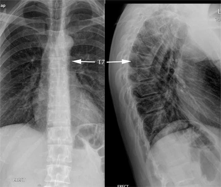
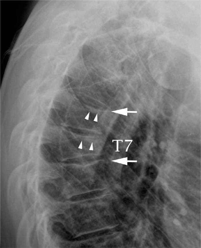
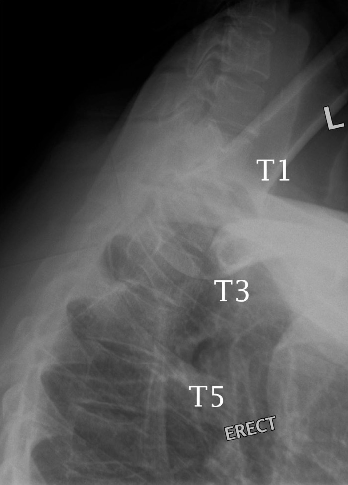
Based on the clinical assessment, the differential diagnosis included thoracic spine fracture(s). Plain radiographic examination was conducted. The AP and lateral radiographs of the thoracic spine (Figure 1a) revealed a 50% anterior compression fracture at T7 with a slight convex posterior vertebral body margin. Accentuated superior endplate concavities (white arrows) with 20% loss of central vertebral body heights of T6 and T8 are depicted on the close-up lateral view (Figure 1b). Hazy zones of condensed trabeculae (arrowheads) were noted subjacent to the T6 and T7 superior endplates (Figure 1b). Anterior wedging and slight superior endplate concavity were demonstrated at T4 and T5 on the swimmer’s view (Figure 2).
Figure 1a.
AP and lateral views of the thoracic spine demonstrated moderate anterior wedged deformity of T7 vertebral body with approximately 50% loss of vertebral body height.
Figure 1b.
Close-up lateral view of the upper thoracic spine revealed the convex deformity of the posterior vertebral body of T7 as well as the accentuated superior endplate concavities of the T6 and T8 (white arrows). Zones of impacted trabeculae (arrowheads) were evident subjacent to the T6 and T7 superior endplates.
Figure 2.
Swimmer’s view revealed the accentuated superior endplate concavities at T4, T5 and T6 when compared to the T3 superior endplate.
Diagnosis and Management
The clinical impression was that of acute seizure-induced mid-to upper thoracic spine compression fractures (T4–T8). While there are clinical practice guidelines for the management of osteoporotic vertebral compression fractures21, only a small body of low-level evidence was available focusing on the management of younger individuals presenting with seizure-induced spinal fractures11–14. The patient was referred to his family medical doctor for further investigations and to discuss the utility of bracing and pain medications. Dual-energy x-ray absorptiometry (DEXA) was found to be normal and the patient did not feel the need to pursue bracing or invasive treatment options. Conservative management was multimodal, including advice regarding: relative rest, cryotherapy, and gentle thoracic extension exercises to be performed at home. During a follow-up approximately six-weeks post-seizure, he stated that he was “doing very well”. During a three-month post-seizure follow-up, he noted a gradual reduction in his discomfort over time and was only occasionally uncomfortable when standing for long periods. During a fourteen-month post-seizure follow-up, he noted that he has continued to do very well and rarely feels any back discomfort.
Discussion
Compression fractures from T4 to T8 are suggestive of a muscular-induced mechanism, such as an epileptic seizure or electroconvulsive therapy.22 Seizure-induced thoracic compression fractures in our case are differentiated from Scheuermann’s disease based on the location in the upper thoracic vertebrae. Scheuermann’s disease occurs in the middle thoracic and thoracolumbar spines commonly and not in the upper thoracic spine.22 Furthermore, compression fractures do not usually demonstrate the characteristic irregular endplates/Schmorl’s nodes and disc narrowing found in Scheuermann’s disease. Upper thoracic compression fractures can be found in the elderly population with osteoporotic spines. Our patient is young with a normal bone density imaging finding. These upper thoracic compression fractures are easily differentiated from old healed compression fractures by the appearance of impacted zone of trabeculae subjacent to the deformed endplates. The compression fractures in this case are also differentiated from pathological vertebral compression fractures, which can have decreased anterior and posterior vertebral body heights (uniform collapse), which should prompt further investigation.22
The literature on compression fractures is focused on the osteoporotic population, as poor bone density is a significant risk factor for vertebral compression fractures.23 Although most osteoporotic vertebral compression fractures are stable and managed conservatively23, they may not always be obvious during the clinical encounter or on plain radiographs. Evidence suggests this is also true of seizure-induced vertebral fractures.15 This is important, as a delay in diagnosis and the delivery of inappropriate treatments may be detrimental to the patient. Youssef et al.15 describe the case of a tonic-clonic seizure-induced burst fracture in a 35 year old male who initially presented with mild paraspinal pain and a normal neurological exam. This seizure occurred while he was sitting on a couch and similar to our case, suffered no external trauma. This man was treated using a Risser cast; however, this was not successful. He subsequently underwent posterior spine fusion with Cotrel-Dubousset segmental instrumentation with iliac crest bone grafting.15 The potential for subtle spinal fractures to be mistaken for a simple musculoskeletal sprain/strain, in the absence of external trauma, in patients who have suffered a tonic-clonic seizure, is highlighted in this previous case report.15
The potential for further complications may arise, as discussed by Youssef et al.15 in patients presenting in a post-ictal state with this requiring the attending health care practitioner (likely an emergency department physician) to perform a careful examination of the shoulders, hips, pelvic girdle, as well as the entire spine14. Youssef et al.15 suggest that the presence of mild paraspinal pain after a seizure should still provoke suspicion of spinal fractures and prompt radiographic evaluation. Plain radiographs can have a significant false-negative rate when attempting to detect spinal fractures, especially unstable spinal fractures (e.g. burst fractures), suggesting the need for advanced imaging such as CT or MRI to provide an accurate diagnosis and optimal patient management.24–26 In particular, advanced imaging (MRI) is required to rule out spinal cord or nerve root compromise.23 MRI can also help confirm the age of compression fractures, as it can show bony edema in acute fractures.23
The current case could have been misdiagnosed as acute mechanical back pain originating from a sprain/strain injury where spinal imaging may not be indicated.27 Mechanical back pain is often treated by chiropractors and other manual health care providers with spinal mobilization and/or adjustments. However, these interventions are considered to be absolute contraindications in the presence of new spinal compression fracture(s), highlighting the potential risk of missed spinal fracture(s).28,29
Haldeman and Rubinstein30 published four cases where patients were noted to have compression fractures following chiropractic spinal manipulation. Although a causal relationship between the spinal manipulation and the fractures in these four cases was unclear, the authors noted that failure to identify compression fractures with the subsequent application of spinal adjustments into the fractured area can increase pain and prolong disability.30 Furthermore, the potential to cause iatrogenic spinal cord injury is of great concern.
Summary
This case highlights the possibility that the compression fractures identified were likely the result of a recent seizure. This suggests that severe muscular contractions secondary to tonic-clonic seizures, in the absence of secondary trauma/external forces, can result in spinal fractures. The report highlights the importance of obtaining an appropriate history and undertaking a musculoskeletal and neurological examination, as indicated, in patients presenting with spinal pain following a tonic-clonic seizure. The symptoms and signs of epileptic seizure-induced spinal fractures may be mild and resemble simple acute mechanical back pain. Health care providers should consider radiographic evaluation when a tonic-clonic seizure results in spinal pain, especially before delivering spinal mobilization or adjustments.
References:
- 1.Shaw JL. Bilateral posterior fracture-dislocation of the shoulder and other trauma caused by convulsive seizures. J Bone Joint Surg Am. 1971;53(7):1437–1440. [PubMed] [Google Scholar]
- 2.Brown RJ. Bilateral dislocation of the shoulders. Injury. 1984;15(4):267–273. doi: 10.1016/0020-1383(84)90012-3. [DOI] [PubMed] [Google Scholar]
- 3.Kristiansen B, Christensen S. Fractures of the proximal end of the humerus caused by convulsive seizures. Injury. 1984;16(2):108–109. doi: 10.1016/s0020-1383(84)80009-1. [DOI] [PubMed] [Google Scholar]
- 4.Dastgeer GM, Mikolich DJ. Fracture-dislocation of manubriosternal joint: an unusual complication of seizures. J Trauma. 1987;27(1):91–93. doi: 10.1097/00005373-198701000-00020. [DOI] [PubMed] [Google Scholar]
- 5.Hepburn DA, Steel JM, Frier BM. Hypoglycemic convulsions cause serious musculoskeletal injuries in patients with IDDM. Diabetes Care. 1989;12(1):32–34. doi: 10.2337/diacare.12.1.32. [DOI] [PubMed] [Google Scholar]
- 6.Finelli PF, Cardi JK. Seizure as a cause of fracture. Neurology. 1989;39(6):858–860. doi: 10.1212/wnl.39.6.858. [DOI] [PubMed] [Google Scholar]
- 7.Marty B, Simmen HP, Käch K, Trentz O. [Bilateral anterior shoulder dislocation fracture after an epileptic seizure. A case report] Unfallchirurg. 1994;97(7):382–384. [PubMed] [Google Scholar]
- 8.Engel T, Lill H, Korner J, Josten C. [Bilateral posterior fracture-dislocation of the shoulder caused by an epileptic seizure – diagnostic, treatment and result] Unfallchirurg. 1999;102(11):897–901. doi: 10.1007/s001130050499. [DOI] [PubMed] [Google Scholar]
- 9.Kelly JP. Fractures complicating electro-convulsive therapy and chronic epilepsy. J Bone Joint Surg Br. 1954;36-B(1):70–79. doi: 10.1302/0301-620X.36B1.70. [DOI] [PubMed] [Google Scholar]
- 10.Lingley JR, Robbins LL. Fractures following electroshock therapy. Radiology. 1947;48(2):124–128. doi: 10.1148/48.2.124. [DOI] [PubMed] [Google Scholar]
- 11.Takahashi T, Tominaga T, Shamoto H, et al. Seizure-induced thoracic spine compression fracture: case report. Surg Neurol. 2002;58(3–4):214–216. doi: 10.1016/s0090-3019(02)00837-6. [DOI] [PubMed] [Google Scholar]
- 12.Gnanalingham K, Macanovic M, Joshi S, et al. Non-traumatic compression fractures of the thoracic spine following a seizure – treatment by percutaneous kyphoplasty. Minim Invasive Neurosurg. 2004;47(4):256–257. doi: 10.1055/s-2004-818521. [DOI] [PubMed] [Google Scholar]
- 13.Ghayem Hasankhani E, Omidi-Kashani F. Multiple lumbar vertebral fractures following a single idiopathic seizure in an otherwise healthy patient: a case report. Med J Islam Repub Iran. 2013;27(4):233–235. [PMC free article] [PubMed] [Google Scholar]
- 14.Uvaraj NR, Gopinath NR, Bosco A. Non-traumatic vertebral fractures: An uncommon complication following the first episode of a convulsive seizure. Internatl J Case Reports Images. 2014;5(2):135–139. [Google Scholar]
- 15.Youssef JA, Mccullen GM, Brown CC. Seizure-induced lumbar burst fracture. Spine. 1995;20(11):1301–1303. doi: 10.1097/00007632-199506000-00020. [DOI] [PubMed] [Google Scholar]
- 16.Vasconcelos D. Compression fractures of the vertebrae during major epileptic seizures. Epilepsia. 1973;14(3):323–328. doi: 10.1111/j.1528-1157.1973.tb03967.x. [DOI] [PubMed] [Google Scholar]
- 17.Sharma A, Avery L, Novelline R. Seizure-induced lumbar burst fracture associated with conus medullaris-cauda equina compression. Diagn Interv Radiol. 2011;17(3):199–204. doi: 10.4261/1305-3825.DIR.3638-10.2. [DOI] [PubMed] [Google Scholar]
- 18.Napier RJ, Nolan PC. Diagnosis of vertebral fractures in post-ictal patients. Emerg Med J. 2011;28(2):169–170. doi: 10.1136/emj.2009.088021. [DOI] [PubMed] [Google Scholar]
- 19.Beniczky SA, Viken J, Jensen LT, et al. Bone mineral density in adult patients treated with various antiepileptic drugs. Seizure. 2012;21(6):471–472. doi: 10.1016/j.seizure.2012.04.002. [DOI] [PubMed] [Google Scholar]
- 20.Miziak B, Błaszczyk B, Chrościńska-Krawczyk M, et al. The problem of osteoporosis in epileptic patients taking antiepileptic drugs. Expert Opin Drug Saf. 2014;13(7):935–946. doi: 10.1517/14740338.2014.919255. [DOI] [PubMed] [Google Scholar]
- 21.Esses SI, Mcguire R, Jenkins J, et al. The treatment of symptomatic osteoporotic spinal compression fractures. J Am Acad Orthop Surg. 2011;19(3):176–182. doi: 10.5435/00124635-201103000-00007. [DOI] [PubMed] [Google Scholar]
- 22.Yochum TR, Rowe LJ. Yochum and Rowe’s Essentials of Skeletal Radiology. 3rd.ed. Philadelphia, Pa: Lippincott Williams & Wilkins; 2005. [Google Scholar]
- 23.Wong CC, Mcgirt MJ. Vertebral compression fractures: a review of current management and multimodal therapy. J Multidiscip Healthc. 2013;6:205–214. doi: 10.2147/JMDH.S31659. [DOI] [PMC free article] [PubMed] [Google Scholar]
- 24.Ballock RT, Mackersie R, Abitbol JJ, Cervilla V, Resnick D, Garfin SR. Can burst fractures be predicted from plain radiographs? J Bone Joint Surg Br. 1992;74(1):147–150. doi: 10.1302/0301-620X.74B1.1732246. [DOI] [PubMed] [Google Scholar]
- 25.Dai LY. Imaging diagnosis of thoracolumbar burst fractures. Chin Med Sci J. 2004;19(2):142–144. [PubMed] [Google Scholar]
- 26.Dai LY, Wang XY, Jiang LS, Jiang SD, Xu HZ. Plain radiography versus computed tomography scans in the diagnosis and management of thoracolumbar burst fractures. Spine. 2008;33(16):E548–52. doi: 10.1097/BRS.0b013e31817d6dee. [DOI] [PubMed] [Google Scholar]
- 27.Chou R, Qaseem A, Snow V, et al. Diagnosis and treatment of low back pain: a joint clinical practice guideline from the American College of Physicians and the American Pain Society. Ann Intern Med. 2007;147(7):478–491. doi: 10.7326/0003-4819-147-7-200710020-00006. [DOI] [PubMed] [Google Scholar]
- 28.Gibbons P, Tehan P. Patient positioning and spinal locking for lumbar spine rotation manipulation. Man Ther. 2001;6(3):130–138. doi: 10.1054/math.2001.0404. [DOI] [PubMed] [Google Scholar]
- 29.Reid DC, Henderson R, Saboe L, et al. Etiology and clinical course of missed spine fractures. J Trauma. 1987;27(9):980–986. doi: 10.1097/00005373-198709000-00005. [DOI] [PubMed] [Google Scholar]
- 30.Haldeman S, Rubinstein SM. Compression fractures in patients undergoing spinal manipulative therapy. J Manipulative Physiol Ther. 1992;15(7):450–454. [PubMed] [Google Scholar]


