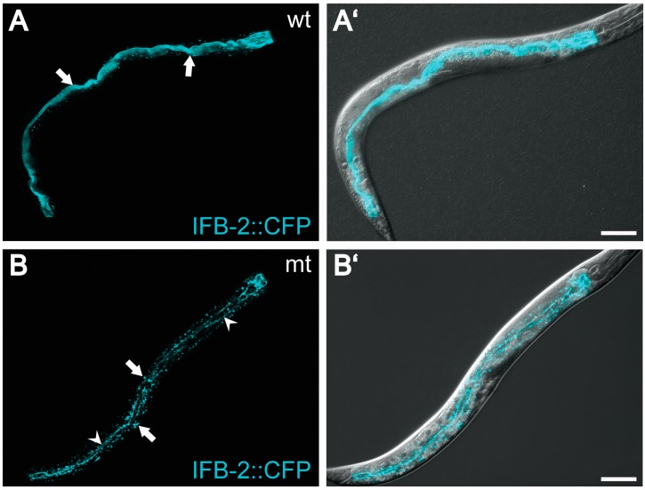
Figure 3.
Mutation of the intestinal filament organizer IFO-1 leads to a collapse of the intermediate filament network onto the C. elegans apical junction (CeAJ). (A) The confocal fluorescence micrographs (projection views) show the distribution of the fluorescent transgene product IFB-2: CFP in a wild type background (wt; strain BJ49; see [58]); Note the exclusive fluorescence in the intestine (anterior to the right upper corner; overlay image with Nomarski optics in (A')); IFB-2: CFP is concentrated in the evenly shaped endotube (arrows), which demarcates the ovoid intestinal lumen; (B) shows the distribution of IFB-2: CFP in ifo-1 mutant strain BJ133 (mt; [58]); Note that the intermediate filament network of the endotube has collapsed completely into aggregates decorating the C. elegans apical junction (fine lines marked by arrowheads) and formed cytoplasmatic granules (arrows). Overlay image with Nomarski optics in (B'). Scale bars = 50 μm.

