

Abstract
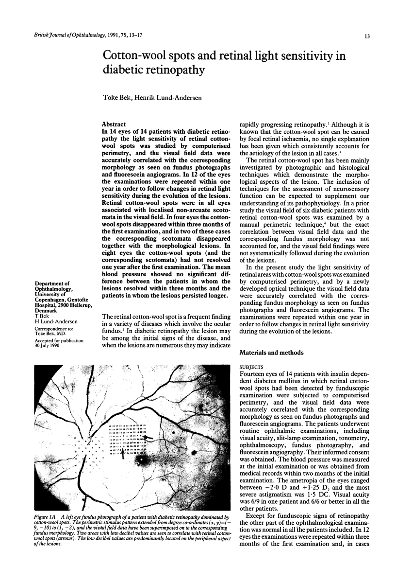
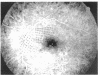
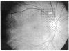
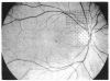
In 14 eyes of 14 patients with diabetic retinopathy the light sensitivity of retinal cotton-wool spots was studied by computerised perimetry, and the visual field data were accurately correlated with the corresponding morphology as seen on fundus photographs and fluorescein angiograms. In 12 of the eyes the examinations were repeated within one year in order to follow changes in retinal light sensitivity during the evolution of the lesions. Retinal cotton-wool spots were in all eyes associated with localised non-arcuate scotomata in the visual field. In four eyes the cotton-wool spots disappeared within three months of the first examination, and in two of these cases the corresponding scotomata disappeared together with the morphological lesions. In eight eyes the cotton-wool spots (and the corresponding scotomata) had not resolved one year after the first examination. The mean blood pressure showed no significant difference between the patients in whom the lesions resolved within three months and the patients in whom the lesions persisted longer.
Full text
PDF


Images in this article


Selected References
These references are in PubMed. This may not be the complete list of references from this article.
- Ashton N. Pathophysiology of retinal cotton-wool spots. Br Med Bull. 1970 May;26(2):143–150. doi: 10.1093/oxfordjournals.bmb.a070766. [DOI] [PubMed] [Google Scholar]
- Bek T., Lund-Andersen H. Accurate superimposition of perimetry data onto fundus photographs. Acta Ophthalmol (Copenh) 1990 Feb;68(1):11–18. doi: 10.1111/j.1755-3768.1990.tb01642.x. [DOI] [PubMed] [Google Scholar]
- Bek T., Lund-Andersen H. The influence of stimulus size on perimetric detection of small scotomata. Graefes Arch Clin Exp Ophthalmol. 1989;227(6):531–534. doi: 10.1007/BF02169446. [DOI] [PubMed] [Google Scholar]
- Brown G. C., Brown M. M., Hiller T., Fischer D., Benson W. E., Magargal L. E. Cotton-wool spots. Retina. 1985 Fall-Winter;5(4):206–214. doi: 10.1097/00006982-198500540-00003. [DOI] [PubMed] [Google Scholar]
- DIEZEL P. B., WILLERT H. G. [Morphology and histochemistry of hard and soft exudates of the retina in diabetes mellitus and essential hypertension]. Klin Monbl Augenheilkd Augenarztl Fortbild. 1961 Nov;139:475–491. [PubMed] [Google Scholar]
- ESMANN V., LUNDBAEK K., MADSEN P. H. TYPES OF EXUDATES IN DIABETIC RETINOPATHY. Acta Med Scand. 1963 Sep;174:375–384. doi: 10.1111/j.0954-6820.1963.tb07936.x. [DOI] [PubMed] [Google Scholar]
- HODGE J. V., DOLLERY C. T. RETINAL SOFT EXUDATES. A CLINICAL STUDY BY COLOUR AND FLUORESCENCE PHOTOGRAPHY. Q J Med. 1964 Jan;33:117–131. [PubMed] [Google Scholar]
- Kohner E. M., Dollery C. T., Bulpitt C. J. Cotton-wool spots in diabetic retinopathy. Diabetes. 1969 Oct;18(10):691–704. doi: 10.2337/diab.18.10.691. [DOI] [PubMed] [Google Scholar]
- McLeod D., Marshall J., Kohner E. M., Bird A. C. The role of axoplasmic transport in the pathogenesis of retinal cotton-wool spots. Br J Ophthalmol. 1977 Mar;61(3):177–191. doi: 10.1136/bjo.61.3.177. [DOI] [PMC free article] [PubMed] [Google Scholar]
- Roth J. A. Central visual field in diabetes. Br J Ophthalmol. 1969 Jan;53(1):16–25. doi: 10.1136/bjo.53.1.16. [DOI] [PMC free article] [PubMed] [Google Scholar]
- Roy M. S., Rick M. E., Higgins K. E., McCulloch J. C. Retinal cotton-wool spots: an early finding in diabetic retinopathy? Br J Ophthalmol. 1986 Oct;70(10):772–778. doi: 10.1136/bjo.70.10.772. [DOI] [PMC free article] [PubMed] [Google Scholar]
- Williams D. K., Drance S. M., Harris G. S., Fairclough M. Diabetic cotton-wool spots: an evaluation using perimetric and angiographic techniques. Can J Ophthalmol. 1970 Jan;5(1):68–77. [PubMed] [Google Scholar]