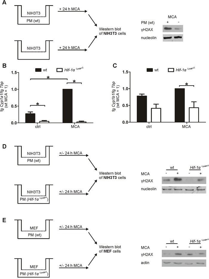
Figure 3. Impaired Cyp1a1 and Cyp1b1 expression in Hif-1αLysM−/− macrophages and fibroblast DNA damage in an in vitro coculture.

A. Schematic illustration of the NIH3T3/peritoneal macrophages (PMs) coculture set up, stimulated with MCA for 24 h. A representative Western blot of phosphorylated γH2AX in NIH3T3 cells cocultured with or without PMs is shown. Nucleolin served as a loading control. B. Cyp1a1 and C. Cyp1b1 mRNA expression of PMs that were isolated from wt and Hif-1αLysM−/− mice and stimulated with MCA or dimethylsulfoxide (DMSO: ctrl) for 8 h assessed by qPCR. mRNA levels were normalized to Tbp. The ratio of Cyp1a1 or Cyp1b1 and Tbp in wt mice after stimulation with MCA was set to 1. Mean values ± SEM of n = 4 are presented (each n contains 3 mice/genotype). * P < 0.05. D. Illustration of the cocultures of NIH3T3 cells with PMs isolated from wt or Hif-1αLysM−/− mice E. and the cocultures of MEF cells with PMs isolated from wt or Hif-1αLysM−/− mice, stimulated with MCA or DMSO for 24 h. Representative Western analysis of phosphorylated H2AX in NIH3T3 is shown. Nucleolin or actin served as loading controls.
